

Paper Menu >>
Journal Menu >>
 Vol.2, No.8, 445-447 (2013) Case Reports in Clinical Medicine http://dx.doi.org/10.4236/crcm.2013.28116 Successful left-sided accessory pathway ablation without reference catheter in patient with atresia of coronary sinus and thin persistent left superior vena cava* Qingxing Chen, Ye Xu, Kuang Cheng, Wenqing Zhu# Department of Cardiology, Zhongshan Hospital, The Shanghai Institute of Cardiovascular Diseases, Fudan University, Shanghai, China; #Corresponding Author: [email protected] Received 7 September 2013; revised 1 October 2013; accepted 29 October 2013 Copyright © 2013 Qingxing Chen et al. This is an open access article distributed under the Creative Commons Attribution License, which permits unrestricted use, distribution, and reproduction in any medium, provided the original work is properly cited. ABSTRACT We report a case of atrioventricular reentrant tachycardia (AVRT) with ostial atresia of the coronary sinus (CS). Without the anatomic an- giography, radiofrequency (RF) energy was ap- plied at the mitral valve annulus and the bypass tract was eliminated. After the therapy proce- dure, by CS angiography, we knew the persis- tent left superior vena cava (PLSVC), and the coronary sinus was connected with vena cava superior, very thin in a diameter. The therapy procedure was successful. The patient has re- mained completely symptom free. Keyw ords: Ablation; Accessory Pathway; Atresia of the Coronary Sinus 1. INTRODUCTION Coronary sinus (CS) atresia is a rare cardiac anatomic variant which causes failure to cannulate CS and consid- erable challenges in affecting cure. CS ostial atresia was associated with a few persistent left superior vena cavas (PLSVC), an unroofed coronary sinus, or was a postop- erative complication [1-5]. Patients with CS ostial atresia have been reported to have atrioventricular reentrant tachycardia [3-5], atrial flutter [4], and atrioventricular nodal reentrant tachycardia [6]. We report a case of CS ostial atresia with anomalous venous drainage into PLSVC with a left-sided accessory pathway (AP) that was successfully eliminated by cathe- ter ablation. 2. CASE REPORT A 57-year-old man with recurrent paroxysmal su- praventricular tachycardia (PSVT) effective to propa- fenone was referred to our hospital for a cardiac electro- physiologicstudy (EPS) and ablation one month ago. A 12-lead electrocardiogram (ECG) done during sinus rhythm exhibited no delta waves. A 12-lead ECG done during the palpitation revealed a regular, narrow QRS tachycardia at a rate of 170 bpm with P’-waves in leads II and aVF, QRS-P’ interval was 86ms; this suggested that the patient had an atrioventricular reentrant tachy- cardia (AVRT) through a concealed accessory pathway (AP). The physical examination, chest x-ray, echocar- diograms and serological examinations were normal. After written informed consent was obtained from the patient, EPS was done after the withdrawal of all antiar- rhythmic drugs for five elimination half-lives. One quadripolar electrode catheter was cannulated from Vena cava inferior, positioned in the right ventricu- lar apex. One multipolar electrode catheter was cannu- lated from Subclavian Vein. Attempts to cannulate the coronary sinus (CS) were unsuccessful. Without CS an- giography, we set a proximal electrode of multipolar catheter on the ostial of CS. Baseline intervals during sinus were as follows: sinus cycle length 660 ms, atrial His (AH) interval 79 ms, and His-ventricular (HV) interval 64 ms. There was no ven- tricular preexcitation during sinus rhythm and decre- mental ventricular pacing down to 350 ms. A regular, narrow QRS tachycardia with a cycle length of 350 ms was repeatly induced. During tachycardia, proximal mul- tipolar electrode was revealed V-A interval 86 ms, mid- dle electrode was revealed V-A interval 145 ms, distal *Conflict of Interest: None. Copyright © 2013 SciRes. OPEN A CCESS 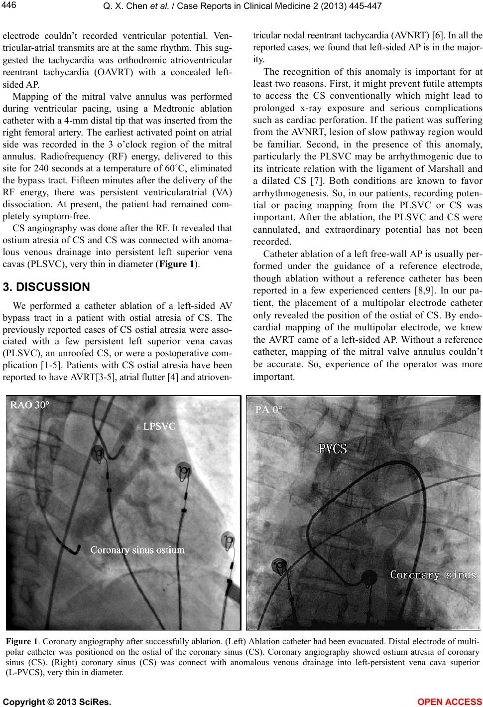 Q. X. Chen et al. / Case Reports in Clinical Medicine 2 (2013) 445-447 446 electrode couldn’t recorded ventricular potential. Ven- tricular-atrial transmits are at the same rhythm. This sug- gested the tachycardia was orthodromic atrioventricular reentrant tachycardia (OAVRT) with a concealed left- sided AP. Mapping of the mitral valve annulus was performed during ventricular pacing, using a Medtronic ablation catheter with a 4-mm distal tip that was inserted from the right femoral artery. The earliest activated point on atrial side was recorded in the 3 o’clock region of the mitral annulus. Radiofrequency (RF) energy, delivered to this site for 240 seconds at a temperature of 60˚C, eliminated the bypass tract. Fifteen minutes after the delivery of the RF energy, there was persistent ventricularatrial (VA) dissociation. At present, the patient had remained com- pletely symptom-free. CS angiography was done after the RF. It revealed that ostium atresia of CS and CS was connected with anoma- lous venous drainage into persistent left superior vena cavas (PLSVC), very thin i n diameter (Figure 1). 3. DISCUSSION We performed a catheter ablation of a left-sided AV bypass tract in a patient with ostial atresia of CS. The previously reported cases of CS ostial atresia were asso- ciated with a few persistent left superior vena cavas (PLSV C), an unroofed CS, or were a posto perative com- plication [1-5]. Patients with CS ostial atresia have been repor ted to have AVRT[3-5], atrial flutter [4] and atrioven- tricular nodal reentrant tachycardia (AVNRT) [6]. In all the reported cases, we found that left-sided AP is in the major- ity. The recognition of this anomaly is important for at least two re asons. First, i t might prevent fu tile attemp ts to access the CS conventionally which might lead to prolonged x-ray exposure and serious complications such as cardiac perforation. If the patient was suffering from the AVNRT, lesion of slow pathway region would be familiar. Second, in the presence of this anomaly, particularly the PLSVC may be arrhythmogenic due to its intricate relation with the ligament of Marshall and a dilated CS [7]. Both conditions are known to favor arrhythmogenesis. So, in our patients, recording poten- tial or pacing mapping from the PLSVC or CS was important. After the ablation, the PLSVC and CS were cannulated, and extraordinary potential has not been recorded. Cathe ter ab la t ion o f a le f t fr ee -wal l A P is usuall y p er- formed under the guidance of a reference electrode, though ablation without a reference catheter has been reported in a few experienced centers [8,9]. In our pa- tient, the placement of a multipolar electrode catheter only revealed the position of the ostial of CS. By endo- cardial mapping of the multipolar electrode, we knew the AVRT came of a left-sided AP. Without a reference catheter, mapping of the mitral valve annulus couldn’t be accurate. So, experience of the operator was more important. Figure 1. Coronary angiography after successfully ablation. (Left) Ablation catheter had been evacuated. Distal electrode of multi- polar catheter was positioned on the ostial of the coronary sinus (CS). Coronary angiography showed ostium atresia of coronary sinus (CS). (Right) coronary sinus (CS) was connect with anomalous venous drainage into left-persistent vena cava superior (L-PVCS), very thin in diameter. Copyright © 2013 SciRes. OPEN A CCESS  Q. X. Chen et al. / Case Reports in Clinical Medicine 2 (2013) 445-447 447 In conclusion, we report a case of CS ostial atresia with anomalous venous drainage into PLSVC with a left-sided AP that was successfully eliminated by catheter ablation. REFERENCES [1] Santoscoy, R., Walters, H.L., Ross, R.D., Lyons, J.M. and Hakimi, M. (1996) Coronary sinus ostial atresia with per- sistent left superior vena cava. The Annals of Thoracic Surgery, 61, 879-882. http://dx.doi.org/10.1016/0003-4975(95)01137-4 [2] Adatia, I. and de Gittenberger-de Groot, A.C. (1995) Un- roofed coronary sinus and coronary sinus orifice atresia. Implications for management of complex congenital heart disease. American College of Cardiology, 25, 948-953. http://dx.doi.org/10.1016/0735-1097(94)00475-6 [3] Takatsuki, S., Mitamura, H., Ieda, M. and Ogawa, S. (2001) Accessory pathway associated with an anomalous coronary vein in a patient with Wolff-Parkinson-White syndrome. Journal of Cardiovascular Electrophysiology, 12, 1080-1082. http://dx.doi.org/10.1046/j.1540-8167.2001.01080.x [4] Luik, A., Deisenhofer, I., Estner, H., Ndrepepa, P.A., Zrenner, B. and Schmitt, C. (2006) Atresia of the coro- nary sinus in patients with supraventricular tachycardia. Pacing and Clinical Electrophysiology, 29, 171-174. http://dx.doi.or g/10.1111/j.1540-8159.2006.00312.x [5] Khairy, P., Triedman, J.K., Juraszek, A. and Cecchin, F. (2005) Inability to cannulate the coronary sinus in pa- tients with supraventricular arrhythmias: Congenital and acquired coronary sinus atresia. Journal of Interventional Cardiac Electrophysiology, 12, 123-127. http://dx.doi.org/10.1007/s10840-005-6547-1 [6] Okuyama, Y., Oka, T., Mizuno, H., Sakai, T., Hirayama, A. and Kodama, K. (2005) A case of atrioventricular nodal reentrant tachycardia with atresia of the coronary sinus ostium. International Heart Journal, 46, 899-902. http://dx.doi.org/10.1536/ihj.46.899 [7] Jah, N.K., Gogna, A., Tan, T.H., Wong, K.Y. and Shankar, S. (2003) Atresia of coronary sinus with retrograde drain- age via persistent left superior vena cava. The Annals of Thoracic Surgery, 76, 2091-2092. http://dx.doi.org/10.1016/S0003-4975(03)01040-3 [8] Kuck, K.H. and Schluter, M. (1991) Single-catheter ap- proach to radiofrequency current ablation of left-sided accessory pathways in patients with Wolff-Parkinson- White syndrome. Circulation, 84, 2366-2375. http://dx.doi.org/10.1161/01.CIR.84.6.2366 [9] Brugada, J., Garcia -Bola o, I. , Figue ire do, M., Puigfe l, M., Matas, M. and Navarro-Lopez, F. (1997) Radiofrequency ablation of concealed left free wall accessory pathways without coronary sinus catheterization: Results in 100 consecutive patients. Journal of Cardiovascular Electro- physiology, 8, 249-253. http://dx.doi.or g/10.1111/j.1540-8167.1997.tb00787.x Copyright © 2013 SciRes. OPEN ACCESS |