

Paper Menu >>
Journal Menu >>
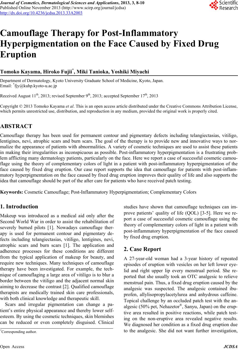 Journal of Cosmetics, Dermatological Sciences and Applications, 2013, 3, 8-10 Published Online November 2013 (http://www.scirp.org/journal/jcdsa) http://dx.doi.org/10.4236/jcdsa.2013.33A2003 Open Access JCDSA Camouflage Therapy for Post-Inflammatory Hyperpigmentation on the Face Caused by Fixed Drug Eruption Tomoko Kayama, Hiroko Fujii*, Miki Tanioka, Yoshiki Miyachi Department of Dermatology, Kyoto University Graduate School of Medicine, Kyoto, Japan. Email: *[email protected] Received August 11th, 2013; revised September 9th, 2013; accepted September 17th, 2013 Copyright © 2013 Tomoko Kayama et al. This is an open access article distributed under the Creative Commons Attribution License, which permits unrestricted use, distribution, and reproduction in any medium, provided the original work is properly cited. ABSTRACT Camouflage therapy has been used for permanent contour and pigmentary defects including telangiectasias, vitiligo, lentigines, nevi, atrophic scars and burn scars. The goal of the therapy is to provide new and innovative ways to nor- malize the appearance of patients with abnormalities. A variety of cosmetic techn iques are used to assist these patients in making their irregularities as inconspicuous as possible. Post-inflammatory hyperpigmentation is a frustrating prob- lem afflicting many dermatology patients, particularly on the face. Here we report a case of successful cosmetic camou- flage using the theory of complementary colors of light in a patient with post-inflammatory hyperpigmentation of the face caused by fixed drug eruption. Our case report supports the idea that camouflage for patients with post-inflam- matory hyperpigmentation on the face caused by fixed drug eruption improves their quality of life and also supports the idea that camouflage should be part of the after care for patients who have received patch testing. Keywords: Cosmetic Camouflage; Post-Inflammato ry Hyperpigmentation; Complementary Colors 1. Introduction Makeup was introduced as a medical aid only after the Second World War in order to assist the rehab ilitation of severely burned pilots [1]. Nowadays camouflage ther- apy is used for permanent contour and pigmentary de- fects including telangiectasias, vitiligo, lentigines, nevi, atrophic scars and burn scars [1]. The application and adherence processes for these conditions are different from the typical application of makeup for beauty, and require new techniques. Many techniques of camouflage therapy have been investigated. For example, the tech- nique of camouflaging a large area of vitilig o is to blur a border between the vitiligo and the adjacent normal skin aiming to decrease the contrast [2]. Qualified camouflage therapists are medically trained skin care professionals, with both clinical knowledge and therapeutic skill. Scars and irregular pigmentation can change a pa- tient’s entire physical appearance and thereby lower self- esteem. By using the cosmetic techniques, skin blemishes can be reduced or even completely disguised. Clinical studies have shown that camouflage techniques can im- prove patients’ quality of life (QOL) [3-5]. Here we re- port a case of successful cosmetic camouflage using the theory of complementary colors of light in a patient with post-inflammatory hyperpigmentation of the face caused by fixed drug eruption. 2. Case Report A 27-year-old woman had a 3-year history of repeated episodes of eruption with vesicles on her left lower eye- lid and right upper lip every menstrual period. She re- ported that she usually took an OTC analgesic to relieve menstrual pain. Thus, a fixed drug eruption caused by the analgesic was suspected. The analgesic contained ibu- profen, allylisopropylacetylurea and anhydrous caffeine. Topical challenge by an occluded patch test with the an- algesic (50% pet, Nehazetor®, Sanyu, Japan) on the erup- tive area resulted in positive reactions, while patch test- ing on the non-eruptive area revealed negative results. We diagnosed her co ndition as a fixed drug eruption due to the analgesic. She did not want further investigation, *Corresponding author. 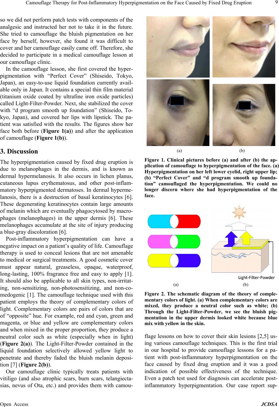 Camouflage Therapy for Post-Inflammatory Hyperpigmentation on the Face Caused by Fixed Drug Eruption 9 so we did not perform patch tests with compon ents of the analgesic and instructed her not to take it in the future. She tried to camouflage the bluish pigmentation on her face by herself, however, she found it was difficult to cover and her camouflage easily came off. Therefore, she decided to participate in a medical camouflage lesson at our camouflage clinic. In the camouflage lesson, she first covered the hyper- pigmentation with “Perfect Cover” (Shiseido, Tokyo, Japan), an easy-to-use liquid foundation currently avail- able only in Japan. It con tains a special thin film material (titanium oxide coated by ultrafine iron oxide particles) called Light-Filter-Powder. Next, she stabilized the co ver with “d program smooth up foundation” (Shiseido, To- kyo, Japan), and covered her lips with lipstick. The pa- tient was satisfied with the results. The figures show her face both before (Figure 1(a)) and after the application of camouflage (Figure 1(b)). 3. Discussion The hyperpigmentation caused by fixed drug eruption is due to melanophages in the dermis, and is known as dermal hypermelanosis. It also occurs in lichen planus, cutaneous lupus erythematosus, and other post-inflam- matory hyperpigmented dermatoses. In dermal hyperme- lanosis, there is a destruction of basal keratinocytes [6]. These degenerating keratinocytes contain large amounts of melanin which are eventually phagocytosed by macro- phages (melanophages) in the upper dermis [6]. These melanophages accumulate at the site of injury producing a blue-gray discoloration [6]. Post-inflammatory hyperpigmentation can have a negative impact on a patient’s qu a lity of life. Camouflage therapy is used to conceal lesions that are not amenable to medical or surgical treatments. A good cosmetic cover must appear natural, greaseless, opaque, waterproof, long-lasting, 100% fragrance free and easy to apply [1]. It should also be applicable to all skin types, non-irritat- ing, non-sensitizing, non-photosensitizing, and non-co- medogenic [1]. The camouflage technique used with this patient employs the theory of complementary colors of light. Complementary colors are pairs of colors that are of “opposite” hue. For example, red and cyan, green and magenta, or blue and yellow are complementary colors and when mixed in the proper propor tion, they produce a neutral color such as white (especially when in light) (Figure 2(a)). The Light-Filter-Powder contained in the liquid foundation selectively allowed yellow light to penetrate and thereby faded the bluish melanin deposi- tion [7] (Figure 2(b)). Our camouflage clinic typically treats patients with vitiligo (and also atrophic scars, burn scars, telangiecta- sias, nevus of Ota, etc.) and provides them with camou- (a) (b) Figure 1. Clinical pictures before (a) and after (b) the ap- plication of camouflage to hyperpigmentation of the face. (a) Hyperpigmentation on her left lower eye lid, right upper lip; (b) “Perfect Cover” and “d program smooth up founda- tion” camouflaged the hyperpigmentation. We could no longer discern where she had hyperpigmentation of the face. (a) (b) Figure 2. The schematic diagram of the theory of comple- mentary colors of light. (a) When complementary colors are mixed, they produce a neutral color such as white; (b) Through the Light-Filter-Powder, we see the bluish pig- mentation in the upper dermis looked white because blue mix with yellow in the skin. flage lessons on how to cover their skin lesions [2,5] us- ing various camouflage techniques. This is the first trial in our hospital to provide camouflage lessons for a pa- tient with post-inflammatory hyperpigmentation on the face caused by fixed drug eruption and it was a good indication of possible effectiveness of the technique. Even a patch test used for diagnosis can accelerate post- inflammatory hyperpigmentation. Our case report sup- Open Access JCDSA  Camouflage Therapy for Post-Inflammatory Hyperpigmentation on the Face Caused by Fixed Drug Eruption Open Access JCDSA 10 ports the idea that camouflage for patients with post- inflammatory hyperpigmentation on the face caused by fixed drug eruption improves their quality of life, and that camouflage should be part of the after care for pa- tients who have received patch testing. REFERENCES [1] C. Antoniou and C. Stefanaki, “Cosmetic Camouflage,” Journal of Cosmetic Dermatology, Vol. 5, No. 4, 2006, pp. 297-301. http://dx.doi.org/10.1111/j.1473-2165.2006.00274.x [2] M. Tanioka and Y. Miyachi, “Camouflage for Vitiligo,” Dermatologic Therapy, Vol. 22, No. 1, 2009, pp. 90-93. http://dx.doi.org/10.1111/j.1529-8019.2008.01220.x [3] B. Hell, F. Frangillo-Engler, E. Heissler, et al., “Camou- flage in Head and Neck Region—A Non-Invasive Option for Skin Lesions, ” International Journal of Oral and Ma x- illofacial Surgery, Vol. 28, No. 2, 1999, pp. 90-94. http://dx.doi.org/10.1016/S0901-5027(99)80199-4 [4] Y. Morita, T. Kondo, A. Iijima and K. Usuda, “Covering Foundation Improves Quality of Life in Patients with Melasma,” Skin Research, Vol. 8, No. 4, 2009, pp. 435- 439. [5] M. Tanioka, Y. Yamamoto, M. Kato and Y. Miyachi, “Camouflage for Patients with Vitiligo Vulgaris Improved Their Quality of Life,” Journal of Cosmetic Dermatology, Vol. 9, No. 1, 2010, pp. 72-75. http://dx.doi.org/10.1111/j.1473-2165.2010.00479.x [6] V. D. Callender, S. St Surin-Lord, E. C. Davis and M. Maclin, “Postinflammatory Hyperpigmentation: Etiologic and Therapeutic Considerations,” American Journal of Clinical Dermatology, Vol. 12, No. 2, 2011, pp. 87-99. http://dx.doi.org/10.2165/11536930-000000000-00000 [7] Z. K. Draelos, “Cosmetics in the Postsurgical Patient,” Dermatologic Clinics, Vol. 13, No. 2, 1995, pp. 461-465. |