

Paper Menu >>
Journal Menu >>
 Advances in Bioscience and Biotechnology, 2013, 4, 974-978 ABB http://dx.doi.org/10.4236/abb.2013.411129 Published Online November 2013 (http://www.scirp.org/journal/abb/) Biochemical variability between two Egyptian Stenodactylus species (Reptilia: Gekkonidae) inhabiting North Sinai Mohamed A. M. Kadry1*, Sayed A. M. Amer1,2 1Department of Zoology, Faculty of Science, Cairo University, Cairo, Egypt 2Department of Biology, Faculty of Science, Taif University, Taif, Saudi Arabia Email: *[email protected] Received 24 August 2013; revised 24 September 2013; accepted 15 October 2013 Copyright © 2013 Mohamed A. M. Kadry, Sayed A. M. Amer. This is an open access article distributed under the Creative Commons Attribution License, which permits unrestricted use, distribution, and reproduction in any medium, provided the original work is properly cited. ABSTRACT Polyacrylamide gel electrophoreses for malate dehy- drogenase (Mdh) and beta-esterase (β-Est) isoenzymes were conducted for biochemical differentiation be- tween two Stenodactylus gekkonid species inhabiting North Sinai of Egypt. Total lipids and proteins of liver and muscle tissues in both species were also analyzed. A total of three Mdh isoforms were re- corded in the analysis, in which the activity of Mdh-2 and Mdh-3 seemed to be higher in S. petrii than in S. sthenodactylus. This high activity could be supported by the significant increase in the total lipids and pro- teins in liver and muscle tissues of the species. It may thus be reasonable to suppose that S. petrii is more active, energetic and adaptable in the desert habitat than S. sthenodactylus. β-Est showed six fractions in S. petrii and only one fraction in S. sthenodactylus. It is therefore noticeable that β-Est is more highly ex- pressed in S. petrii than in S. sthenodactylus. Keywords: Electrophoreses; Physiological Ecology; Geckos; Isoenzymes; Lipids; Proteins 1. INTRODUCTION Gekkonidae are among the lizards known for their strik- ing range of morphological characteristics, ecological habitats and body sizes. In Egypt, most of the gekkonid species are found in and around human habitation. Some, however, live in Egyptian deserts [1]. Many studies determined the relationships among members of the family Gekkonidae on the basis of mor- phological and environmental characteristics [2,3], kar- yotyping [4,5], and biochemical [6-8] and molecular variations [9-19]. The genus Stenodactylus contains 13 recognized species. The species Stenodactylus stheno- dactylus and S. petrii are distributed in Egypt, Iran, Iraq, Syria, Jordan and Arabian Peninsula [1], and areas from Sudan to Mauritania [20]. Isoenzymes are multiple forms of a single enzyme. The forms are often marked by different isoelectric points and hence separable by electrophoresis. Malate dehydrogenase (Mdh) is considered as one of the most extensively studied isozyme systems [21]. This enzyme with lactate dehydrogenase constitutes a very suitable system for studying several metabolic, genetic, ecologi- cal features, and they are very useful in systematic stud- ies. As a homodimeric enzyme, Mdh is well known for the many cell compartment-specific isoenzymes that cha- racterize various organisms. There is a mitochondrial Mdh functioning in the tricarboxylic acid cycle which is usually NAD+-dependent. Most eukaryotes that have been studied also have a cytosolic Mdh isoform. The cytosolic Mdh, also known as NADP-malic enzyme (ME), catalyzes the NADP dependent oxidative decar- boxylation of malate into pyruvate and carbon dioxide to generate NADPH. ME is thought to be a key enzyme in lipid biosynthesis [22,23]. Esterase isoenzymes (Est)—as one kind of the lipid-hydrolyzing enzymes—possess high significance in genetics and toxicology [24]. The present study aims to investigate the patterns of the inter- specific biochemical variations between two common gekkonid species (S. petrii and S. sthenodactylus) inhab- iting the Sinai desert of Egypt. 2. MARERIALS AND METHODS 2.1. Taxon Sampling and Study Area Collected for this project were a total of 12 individuals *Corresponding author. OPEN ACCESS 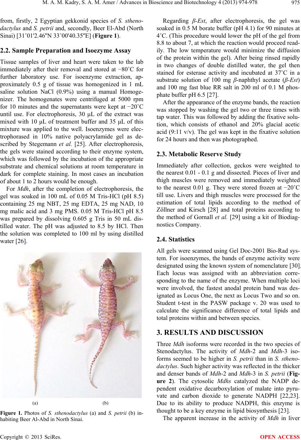 M. A. M. Kadry, S. A. M. Amer / Advances in Bioscience and Biotechnology 4 (2013) 974-978 975 from, firstly, 2 Egyptian gekkonid species of S. stheno- dactylus and S. petrii and, secondly, Beer El-Abd (North Sinai) [31˚01'2.46''N 33˚00'40.35''E] (Figure 1). 2.2. Sample Preparation and Isoezyme Assay Tissue samples of liver and heart were taken to the lab immediately after their removal and stored at −80˚C for further laboratory use. For isoenzyme extraction, ap- proximately 0.5 g of tissue was homogenized in 1 mL saline solution NaCl (0.9%) using a manual Homoge- nizer. The homogenates were centrifuged at 5000 rpm for 10 minutes and the supernatants were kept at −20˚C until use. For electrophoresis, 30 μL of the extract was mixed with 10 μL of treatment buffer and 35 μL of this mixture was applied to the well. Isoenzymes were elec- trophorased in 10% native polyacrylamide gel as de- scribed by Stegemann et al. [25]. After electrophoresis, the gels were stained according to their enzyme system, which was followed by the incubation of the appropriate substrate and chemical solutions at room temperature in dark for complete staining. In most cases an incubation of about 1 to 2 hours would be enough. For Mdh, after the completion of electrophoresis, the gel was soaked in 100 mL of 0.05 M Tris-HCl (pH 8.5) containing 25 mg NBT, 25 mg EDTA, 25 mg NAD, 10 mg malic acid and 3 mg PMS. 0.05 M Tris-HCl pH 8.5 was prepared by dissolving 0.605 g Tris in 50 mL dis- tilled water. The pH was adjusted to 8.5 by HCl. Then the solution was completed to 100 ml by using distilled water [26]. (a) (b) Figure 1. Photos of S. sthenodactylus (a) and S. petrii (b) in- habiting Beer Al-Abd in North Sinai. Regarding β-Est, after electrophoresis, the gel was soaked in 0.5 M borate buffer (pH 4.1) for 90 minutes at 4˚C. (This procedure would lower the pH of the gel from 8.8 to about 7, at which the reaction would proceed read- ily. The low temperature would minimize the diffusion of the protein within the gel). After being rinsed rapidly in two changes of double distilled water, the gel then stained for esterase activity and incubated at 37˚C in a substrate solution of 100 mg β-naphthyl acetate (β-Est) and 100 mg fast blue RR salt in 200 ml of 0.1 M phos- phate buffer pH 6.5 [27]. After the appearance of the enzyme bands, the reaction was stopped by washing the gel two or three times with tap water. This was followed by adding the fixative solu- tion, which consists of ethanol and 20% glacial acetic acid (9:11 v/v). The gel was kept in the fixative solution for 24 hours and then was photographed. 2.3. Metabolic Reserve Study Immediately after collection, geckos were weighted to the nearest 0.01 - 0.1 g and dissected. Pieces of liver and thigh muscles were removed and immediately weighted to the nearest 0.01 g. They were stored frozen at −20˚C till use. Livers and thigh muscles were processed for the estimation of total lipids according to the method of Zöllner and Kirsch [28] and total proteins according to the method of Gornall et al. [29] using a kit of Biodiag- nostics Company. 2.4. Statistics All gels were scanned using Gel Doc-2001 Bio-Rad sys- tem. For isoenzymes, the bands of enzyme activity were designated using the known system of nomenclature [30]. Each locus was assigned with an abbreviation corre- sponding to the name of the enzyme. When multiple loci were involved, the fastest anodal protein band was des- ignated as Locus One, the next as Locus Two and so on. Student t-test in the PASW package v. 20 was used to calculate the significance difference of total lipids and total proteins within and between species. 3. RESULTS AND DISCUSSION Three Mdh isoforms were recorded in the two species of Stenodactylus. The activity of Mdh-2 and Mdh-3 iso- forms seemed to be higher in S. petrii than in S. stheno- dactylus. Such higher activity was reflected in the thicker and denser bands of Mdh-2 and Mdh-3 in S. petrii (Fig- ure 2). The cytosolic Mdhs catalyzed the NADP de- pendent oxidative decarboxylation of malate into pyru- vate and carbon dioxide to generate NADPH [22,23]. Due to its ability to produce NADPH, this enzyme is thought to be a key enzyme in lipid biosynthesis [23]. The apparent increase in the activity of Mdh in liver Copyright © 2013 SciRes. OPEN ACCESS 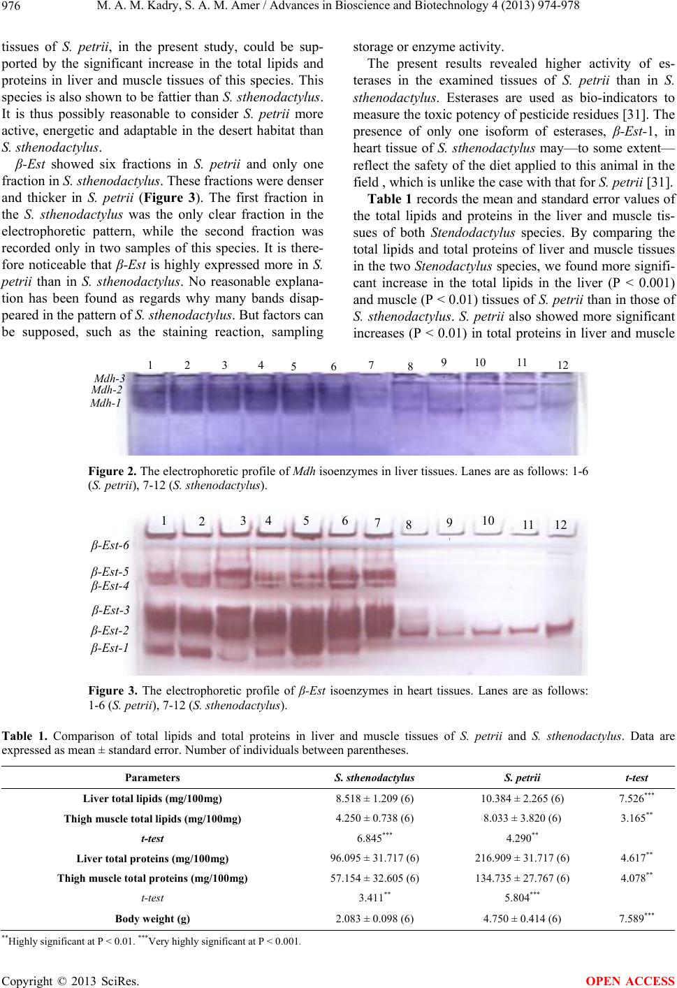 M. A. M. Kadry, S. A. M. Amer / Advances in Bioscience and Biotechnology 4 (2013) 974-978 Copyright © 2013 SciRes. 976 storage or enzyme activity. tissues of S. petrii, in the present study, could be sup- ported by the significant increase in the total lipids and proteins in liver and muscle tissues of this species. This species is also shown to be fattier than S. sthenodactylus. It is thus possibly reasonable to consider S. petrii more active, energetic and adaptable in the desert habitat than S. sthenodactylus. The present results revealed higher activity of es- terases in the examined tissues of S. petrii than in S. sthenodactylus. Esterases are used as bio-indicators to measure the toxic potency of pesticide residues [31]. The presence of only one isoform of esterases, β-Est-1, in heart tissue of S. sthenodactylus may—to some extent— reflect the safety of the diet applied to this animal in the field , which is unlike the case with that for S. petrii [31]. β-Est showed six fractions in S. petrii and only one fraction in S. sthenodactylus. These fractions were denser and thicker in S. petrii (Figure 3). The first fraction in the S. sthenodactylus was the only clear fraction in the electrophoretic pattern, while the second fraction was recorded only in two samples of this species. It is there- fore noticeable that β-Est is highly expressed more in S. petrii than in S. sthenodactylus. No reasonable explana- tion has been found as regards why many bands disap- peared in the pattern of S. sthenodactylus. But factors can be supposed, such as the staining reaction, sampling Table 1 records the mean and standard error values of the total lipids and proteins in the liver and muscle tis- sues of both Stendoda ctylus species. By comparing the total lipids and total proteins of liver and muscle tissues in the two Stenodactylus species, we found more signifi- cant increase in the total lipids in the liver (P < 0.001) and muscle (P < 0.01) tissues of S. petrii than in those of S. sthenodactylus. S. petrii also showed more significant increases (P < 0.01) in total proteins in liver and muscle 1 2 3 4 56 789 1 1011 12 M dh-1 M dh-2 M dh-3 Figure 2. The electrophoretic profile of Mdh isoenzymes in liver tissues. Lanes are as follows: 1-6 (S. petrii), 7-12 (S. sthenodactylus). 12 3 456789 1 10 11 12 β -Est-1 β -Est-2 β -Est-3 β -Est-4 β -Est-5 β-Est-6 Figure 3. The electrophoretic profile of β-Est isoenzymes in heart tissues. Lanes are as follows: 1-6 (S. petrii), 7-12 (S. sthenodactylus). Table 1. Comparison of total lipids and total proteins in liver and muscle tissues of S. petrii and S. sthenodactylus. Data are expressed as mean ± standard error. Number of individuals between parentheses. Parameters S. sthenodactylus S. petrii t-test Liver total lipids (mg/100mg) 8.518 ± 1.209 (6) 10.384 ± 2.265 (6) 7.526*** Thigh muscle total lipids (mg/100mg) 4.250 ± 0.738 (6) 8.033 ± 3.820 (6) 3.165** t-test 6.845*** 4.290** Liver total protei ns (mg/100mg) 96.095 ± 31.717 (6) 216.909 ± 31.717 (6) 4.617** Thigh muscle total proteins (mg/100mg) 57.154 ± 32.605 (6) 134.735 ± 27.767 (6) 4.078** t-test 3.411** 5.804*** Body weight (g) 2.083 ± 0.098 (6) 4.750 ± 0.414 (6) 7.589*** ** Highly significant at P < 0.01. ***Very highly significant at P < 0.001. OPEN ACCESS  M. A. M. Kadry, S. A. M. Amer / Advances in Bioscience and Biotechnology 4 (2013) 974-978 977 tissues than S. sthenodactylus. Within each species, total lipids and proteins were significantly higher in liver (P < 0.01, P < 0.001) tissues than in muscle tissues. 4. CONCLUSION In conclusion, S. petrii displayed higher physiological performance and activity than S. sthenodactylus, while isoenzyme expression was higher in the first species than in the second. The accumulation of total lipids and pro- teins was also significantly higher in the first species than in the second. Data analysis in the present research project does not support what is concluded in the study of Amer [6] for both species and within S. petrii. We therefore can affirm that the identification of S. petrii is incorrect in the study by Amer [6], while the research results point to the identity of other haplotypes of S. sthenodactylus than that of S. petrii. 5. ACKNOWLEDGMENTS We are grateful to Dr. Shawkat Ahmed at Ain Shams University of Egypt for his technical support in conducting the practical part of isoenzyme assay in this work. REFERENCES [1] Ali, R.A.M. (2012) Genetic variation among nine Egyp- tian gecko species (Reptilia: Gekkonidae) based on RAPD- PCR. Journal of Life Science, 9, 154-162. [2] Baha El Din, M. (1997) A new species of tarentola (Squamata: Gekkonidae) from the western desert of Egypt. African Journal of Herpetology, 46, 30-35. http://dx.doi.org/10.1080/21564574.1997.9649973 [3] Saleh, M.A. (1997) Amphibians and reptiles of Egypt. [Cairo]: Egyptian Environmental Affairs Agency. [4] Castiglia, R. (2004) First chromosomal analysis for the genus Lygodactylus (Gray, 1864): The karyotype of L. picturatus (Squamata, Gekkonidae, Gekkoninae). African Journal of Herpetology, 53, 95-97. http://dx.doi.org/10.1080/21564574.2004.9635502 [5] Kawai, A., Ishijima, J., Nishida, C., Kosaka, A., Ota, H., Kohno, S. and Matsuda, Y. (2009) The ZW sex chromo- somes of Gekkohokouensis (Gekkonidae, Squamata) re- present highly conserved homology with those of avian species. Chromosoma, 118, 43-51. http://dx.doi.org/10.1007/s00412-008-0176-2 [6] Amer, S.A.M. (1999) Morphometric, genetic and meta- bolic variations in relation to the taxonomic separation of gekkonids in Egypt (Gekkonidae: Reptilia). PhD Thesis, Cairo University, Egypt. [7] Macey, R., Ananjeva, B., Wang, Y. and Papenfuss, J. (2000) Phylogenetic relationships among Asian gekkonid lizards formerly of the genus Cyrtodactylus based on cladistic analyses of allozymic data: Monophyly of Cyr- topodion and Mediodactylus. Journal of Herpetology, 34, 258-265. http://dx.doi.org/10.2307/1565422 [8] Qin, X., Liang, Y. and Huang, X. (2006) Isozymes analy- sis on different tissues from three different populations of Gekko gecko. Guangxi Science, 13, 310-316. [9] Carranza, S., Arnold, N., Mateo, A. and Geniez, P. (2002) Relationships and evolution of the North African geckos, Geckonia and Tarentola (Reptilia: Gekkonidae), based on mitochondrial and nuclear DNA sequences. Molecular Phylogenetics and Evolution, 23, 244-256. http://dx.doi.org/10.1016/S1055-7903(02)00024-6 [10] Han, D., Zhou, K. and Bauer, M. (2004) Phylogenetic relationships among gekkotan lizards inferred from C- mos nuclear DNA sequences and anew classification of the Gekkota. Biological Journal of Linnean Society, 83, 353-368. h tt p: //d x. doi .org/10 .1111/j .1095-8312.2004.00393.x [11] Vences, M., Wanke, S., Vieites, R., Branch, R., Glaw, F. and Meyer, A. (2004) Natural colonization or introduc- tion? Phylogeographical relationships and morphological differentiation of house geckos (Hemidactylus) from Ma- dagascar. Biological Journal of Linnean Society, 83, 115- 130. h tt p:// dx.d oi. org/1 0. 1111/j. 1095-8312.2004.00370.x [12] Jesus, J., Brehm, A. and Harris, J. (2005) Phylogenetic relationships of Hemidactylus geckos from the Gulf of Guinea islands: Patterns of natural colonizations and an- thropogenic introductions estimated from mitochondrial and nuclear DNA sequences. Molecular Phylogenetics and Evolution, 34, 480-485. http://dx.doi.org/10.1016/j.ympev.2004.11.006 [13] Rocha, S., Carretero, M. and Harris, J. (2005) Diversity and phylogenetic relationships of Hemidactylus geckos from the Comoro Islands. Molecular Phylogenetics and Evolution, 35, 292-299. http://dx.doi.org/10.1016/j.ympev.2004.11.023 [14] Qin, X., Liang, Y., Huang, X. and Pang, G. (2005) RAPD analysis on genetic divergence and phylogenesis of Gekko gecko from different areas. Chinese Journal of Zoology, 40, 14. [15] Carranza, S. and Arnold, N. (2006) Systematics, bio- geography, and evolution of Hemidactylus geckos (Rep- tilia: Gekkonidae) elucidated using mitochondrial DNA sequences. Molecular Phylogenetics and Evolution, 38, 531-545. http://dx.doi.org/10.1016/j.ympev.2005.07.012 [16] Bansal, R. and Karanth, P. (2010) Molecular phylogeny of Hemidactylus geckos (Squamata: Gekkonidae) of the Indian subcontinent reveals a unique Indian radiation and an Indian origin of Asian house geckos. Molecular Phy- logenetics and Evolution, 57, 459-465. http://dx.doi.org/10.1016/j.ympev.2010.06.008 [17] Rato, C., Carranza, S., Pereira, A., Carretero, M. and Harris, J. (2010) Conflicting patterns of nucleotide diver- sity between mtDNA and nDNA in the Moorish gecko, Tarentola mauritanica. Molecular Phylogenetics and Evolution, 56, 962-971. http://dx.doi.org/10.1016/j.ympev.2010.04.033 [18] Busais, S. and Joger, U. (2011) Molecular phylogeny of the gecko genus Hemidactylus Oken, 1817 on the main- land of Yemen (Reptilia: Gekkonidae). Zoology in the Middle East, 53, 25-34. http://dx.doi.org/10.1080/09397140.2011.10648859 Copyright © 2013 SciRes. OPEN ACCESS  M. A. M. Kadry, S. A. M. Amer / Advances in Bioscience and Biotechnology 4 (2013) 974-978 978 [19] Fujita, K. and Papenfuss, J. (2011) Molecular systematics of Stenodactylus (Gekkonidae), an Afro-Arabian gecko species complex. Molecular Phylogenetics and Evolution, 58, 71-75. http://dx.doi.org/10.1016/j.ympev. 2010.10.014 [20] Baha El Din, M. (2006) A guide to reptiles and amphibi- ans of Egypt. Cairo: American University in Cairo Press. [21] Laganà, G., Bellocco, E., Mannucci, C., Leuzzi, U., Tel- lone, E., Kotyk1, A. and Galtieri, A. (2006) Enzymatic urea adaptation: Lactate and malate dehydrogenase in el- asmobranchs. Physiological Research, 55, 675-688. [22] Lo, A.S.Y., Liew, C.T., Ngai, S.M., Tsui, S.K.W., Fung, K.P., Lee, C.Y. and Waye, M.M.Y. (2005) Developmen- tal regulation and cellular distribution of human cytosolic malate dehydrogenase (MDH1). Journal of Cell Bio- chemistry, 94, 763-773. http://dx.doi.org/10.1002/jcb.20343 [23] Daniš, P. and Farkas, R. (2009) Hormone-dependant and hormone-independant control of metabolic and develop- mental functionals of malate dehydrogenase. Endocrine Regulations, 43, 39-52. http://dx.doi.org/10.4149/endo_2009_01_39 [24] Shahjahan, R.M., Karim, A., Begum, R.A., Alam, M.S. and Begum, A. (2008) Tissue specific esterase isozyme banding pattern in Nile Tilapia (Oreochromis niloticus). University Journal of Zoology (Rajshahi University), 27, 1-5. [25] Stegemann, H., Afify, A.M.R. and Hussein, K.R.F. (1985) Cultivar identification of dates (Phoenix dactylifera) by protein patterns. Second international symposium of bio- chemical approaches to identification of cultivars, Broun- schweig, Germany, 44. [26] Jonathan, F.W. and Wendel, N.F. (1990) Visualization and interpretation of plant isozymes. In: Soltis, D.E. and Soltis, P.S., Eds., Isozymes in Plant Biology, Champan and Hall, London, 5-45. [27] Scandaliojs, G. (1964) Tissue-specific isozyme variations in maize. Journal of Heredity, 55, 281-285. [28] Zöllner, N. and Kirsch, K. (1962) Colorimetric method for determination of total lipids. Journal of Experimental Medicine, 135, 545-550. http://dx.doi.org/10.1007/BF02045455 [29] Gornall, A.G., Bardawill, C.J. and David, M.M. (1949) Determination of serum proteins by means of the biuret reaction. Journal of Biological Chemistry, 177, 751-766. [30] Allendorff, W. and Utter, F.M. (1978) Population genet- ics. In: Hoaran, W.S. and Randalal, D.J., Eds., Fish Phy- siology 8, Academic Press, New York, 407-454. [31] Al-Harbi, M.S. and Amer, S.A.M. (2012) Comparison of energy-related isoenzymes between production and rac- ing Arabian camels. Advances in Bioscience and Bio- technology, 3, 1124-1128. Copyright © 2013 SciRes. OPEN ACCESS |