

Paper Menu >>
Journal Menu >>
 Engineering, 2013, 5, 363-367 http://dx.doi.org/10.4236/eng.2013.510B073 Published Online October 2013 (http://www.scirp.org/journal/eng) Copyright © 2013 SciRes. ENG Model-Based Analysis of Ventilation Inhomogeneity in Respiratory Mechanics Christoph Schranz1, Kévin Meffray1, Claudius Stahl2, Knut Möller1 1Institute of Technical Medicine, Furtwangen University, Villingen-Schwenningen, Germany 2Department of Anaesthesiology and Critical Care Medicine, University of Medical Center Freiburg, Germany Email: [email protected] Received June 2013 ABSTRACT Individualized models of respiratory mechanics help to reduce potential harmful effects of mechanical ventilation by supporting the evaluation of patient-specific lung protective ventilation strategies. Assessing ventilation inhomogenei- ties might be an important aspect in optimizing ventilator settings. The aim of this study is to capture and analyze ven- tilation inhomogeneity by a mathematical model using clinical data. The results show that the lung physiology of me- chanically ventilated patients without lung condition can be described by an inhomogeneity model revealing two alveo- lar compartments with median time constants of 0.4 an d 3.9 s. Thus, the IHM in combination with specific ventilation maneuver might be suitable to capture lung physiology for model-based optimization of ventilator settings but requires additional image-based investigations to further support the validity of the model. Keywords: Respiratory Mechanics; Inhomogeneity Model; Parameter Identification; Model-B ased Therapy 1. Introduction Non-adapted ventilator settings risks are severe side ef- fects in intensive care patients during mechanical venti- lation [1]. Optimized patient-specific settings can be ob- tained by individualizing physiological models using clinical data and p ar ameter identification methods. Indi- vidualized models provide insight into patient’s physiol- ogy that is not directly measurable. Thus, they offer sig- nificant potential to evaluate and guide personalized lung protective ventilator strategies on intensive care units [2-4]. The concept of model-based therapy applicable at the bedside of the patient requires models that are as simple as possible, while capturing all relevant dynamics and being identifiable with limited available measure- ment set. Relevant dynamics of lung mechanics are significantly affected by ventilation inhomogeneity [5]. Thus, inho- mogeneities in lung mechanics might provide useful in- formation on the lung tissue response to modified venti- lator settings. Currently, ventilation inhomogeneity can be captured by computed tomography (CT) [6] or by electro impedance tomography [7]. An alternative ap- proach involves the Inhomogeneity Model (IHM) of res- piratory mechanics [8], wh ich has been a strong force in the field of pulmonary physiology ev er since [5]. This paper presents the assessment and analysis of the inhomogeneity model in mechanically ventilated patients to evaluate its potential for model-based therapy. 2. Material & Methods 2.1. Models and Parameter Identification First Order Model (FOM): The FOM is the simplest re- presentation of lung mechanics and considers homoge- neous ventilation. The equation of motion is given in (1) and the electrical analog is shown in Figure 1. The res is- tive element R (cmH2O·s/L) corresponds to the resistance of the central and peripheral airways and the compliant compartment C (mL/cmH2O) represents the elasticity of alveolar tissue and the chest wall [5] defining the respi- ratory time constant τ = R·C. The patient-specific para- meters R and C are determined by Multiple Linear Re- gression using measured data samples of flow rate ( V ) as model input and airway pressure (paw) as model output [9]. pC represents the pressure in the elastic compartment. VRpp V C p Caw C += =1 (1) Inhomogeneity Model (I HM): The IHM is a two- compartment model representing two different alveolar regions by two compliances (C1, C2 in mL/cmH2O) with their own local airway (R1, R2 in cmH2O∙s/L) connected to the airway opening. This model assumes parallel ven- tilation inhomogeneity in the lungs described by the two 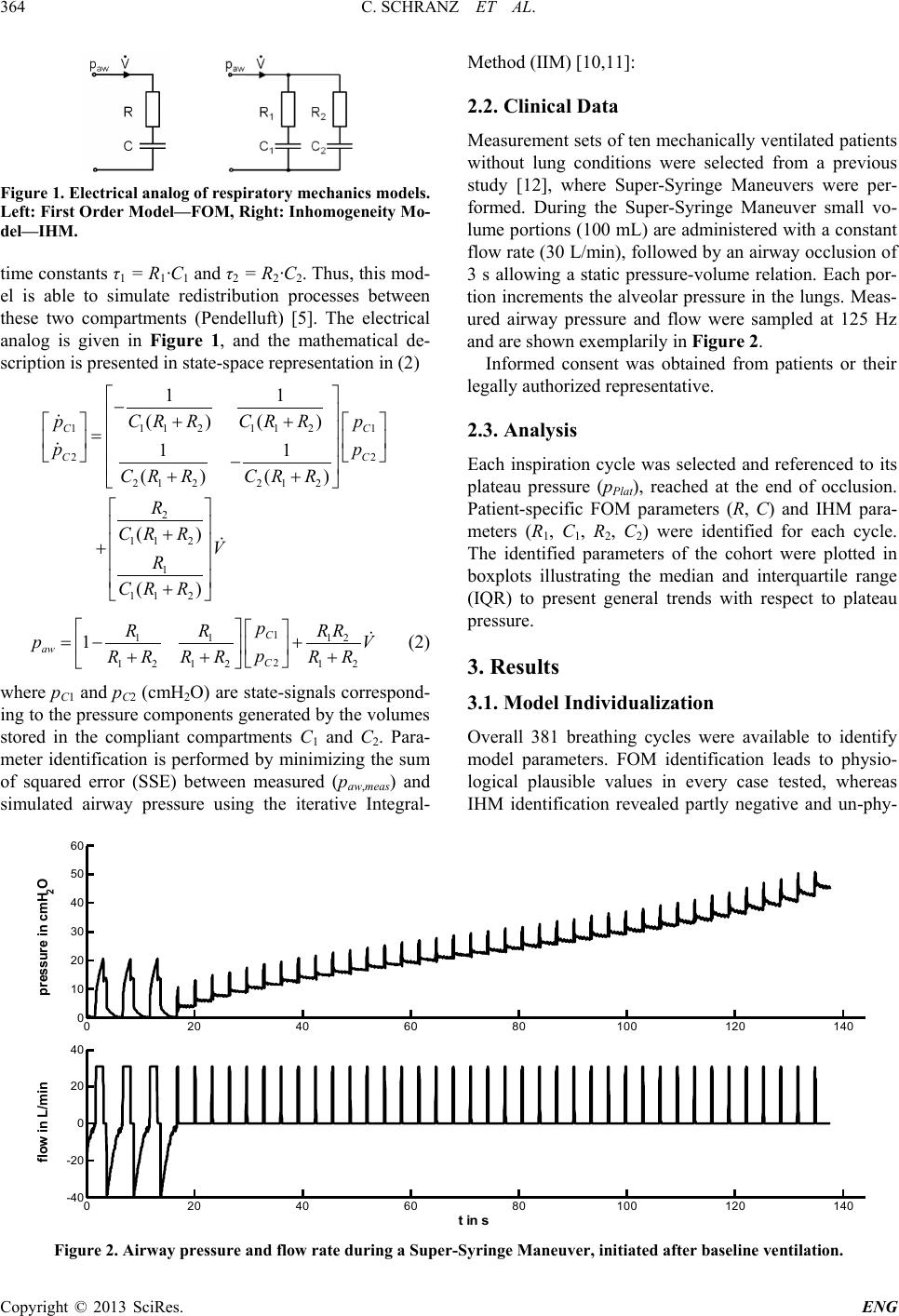 C. SCHRANZ ET AL. Copyright © 2013 SciRes. ENG 364 Figure 1. Electrical analog of respiratory mechanics models. Left: First Order Model—FOM, Right: Inho moge neity M o- del—IHM. time constants τ1 = R1·C1 and τ2 = R2·C2. Thus, this mod- el is able to simulate redistribution processes between these two compartments (Pendelluft) [5]. The electrical analog is given in Figure 1, and the mathematical de- scription is presented in state-space representation in (2) 11 211 2 11 22 21 221 2 2 11 2 1 11 2 11 ()() 11 () () () () CC CC CR RCR R pp pp CR RCR R R CR RV R CR R − ++ = − ++ + + + 1 1 112 2 12 1212 1 C aw C p R RRR pV p RR RRRR =−+ ++ + (2) where pC1 and pC2 (cmH2O) are state-signals correspond- ing to the pressure components gene rated by the volumes stored in the compliant compartments C1 and C2. Para- meter identification is performed by minimizing the sum of squared error (SSE) b etween measured (paw,meas) and simulated airway pressure using the iterative Integral- Method (IIM) [10,11]: 2.2. Clinical Data Measurement sets of ten mechanically ventilated patients without lung conditions were selected from a previous study [12], where Super-Syringe Maneuvers were per- formed. During the Super-Syringe Maneuver small vo- lume portions (100 mL) are administered with a constant flow rate (30 L/min), followed by an airway occlusion of 3 s allowing a static pressure-volume relation. Each por- tion increments the alveolar pressure in the lungs. Meas- ured airway pressure and flow were sampled at 125 Hz and are shown exemplarily in Figure 2. Informed consent was obtained from patients or their legally authorized representative. 2.3. Analysis Each inspiration cycle was selected and referenced to its plateau pressure (pPlat), reached at the end of occlusion. Patient-specific FOM parameters (R, C) and IHM para- meters (R1, C1, R2, C2) were identified for each cycle. The identified parameters of the cohort were plotted in boxplots illustrating the median and interquartile range (IQR) to present general trends with respect to plateau pressure. 3. Results 3.1. Model Individualization Overall 381 breathing cycles were available to identify model parameters. FOM identification leads to physio- logical plausible values in every case tested, whereas IHM identification revealed partly negative and un-phy- Figure 2. Airway pressure and fl ow rate during a Super-Syringe Maneuver, initiated after baseline ventilation. 020406080100 120 140 0 10 20 30 40 50 60 pressure in cmH 2 O 020 40 60 80100120140 -40 -20 0 20 40 t in s flow in L/min 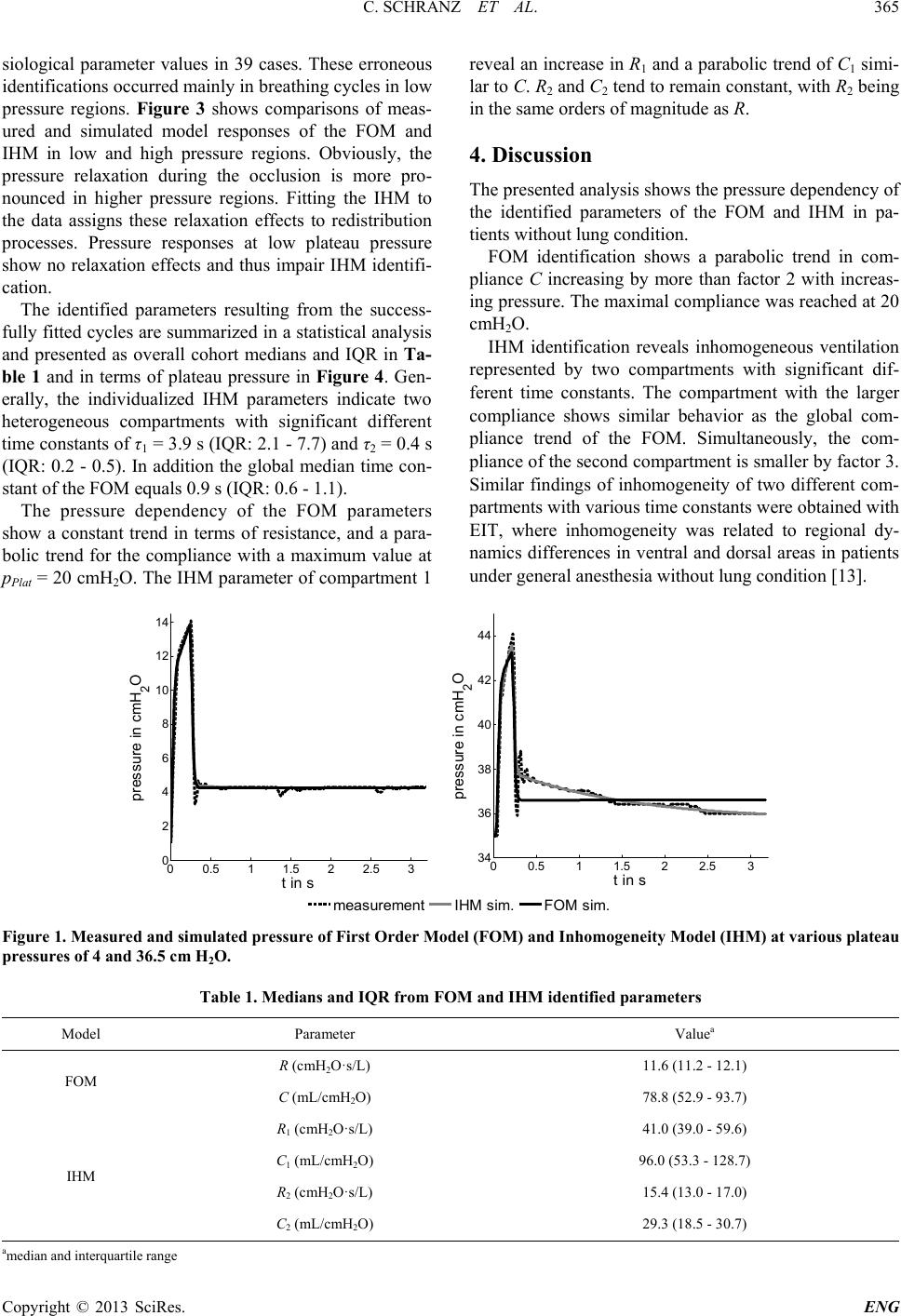 C. SCHRANZ ET AL. Copyright © 2013 SciRes. ENG 365 siological parameter values in 39 cases. These erroneous identifications occur r ed mainly in breathing cycles in low pressure regions. Figure 3 shows comparisons of meas- ured and simulated model responses of the FOM and IHM in low and high pressure regions. Obviously, the pressure relaxation during the occlusion is more pro- nounced in higher pressure regions. Fitting the IHM to the data assigns these relaxation effects to redistribution processes. Pressure responses at low plateau pressure show no relaxation effects and thus impair IHM identifi- cation. The identified parameters resulting from the success- fully fitted cycles are summarized in a statistical analysis and presented as overall cohort medians and IQR in Ta- ble 1 and in terms of plateau pressure in Figure 4. Gen- erally, the individualized IHM parameters indicate two heterogeneous compartments with significant different time constants of τ1 = 3.9 s (IQR: 2.1 - 7.7) and τ2 = 0.4 s (IQR: 0.2 - 0.5). In addition the global median time con- stant of the FOM equals 0.9 s (IQR : 0.6 - 1.1). The pressure dependency of the FOM parameters show a constant trend in terms of resistance, and a para- bolic trend for the compliance with a maximum value at pPlat = 20 cmH2O. The IHM parameter of compartment 1 reveal an increase in R1 and a parabolic trend of C1 simi- lar to C. R2 and C2 tend to remain constant, with R2 being in the same orders of magnitude as R. 4. Discussion The presented an alysis show s the pressur e depend ency of the identified parameters of the FOM and IHM in pa- tients without lung condition. FOM identification shows a parabolic trend in com- pliance C increasing by more than factor 2 with increas- ing pressure. The maximal compliance was reached at 20 cmH2O. IHM identification reveals inhomogeneous ventilation represented by two compartments with significant dif- ferent time constants. The compartment with the larger compliance shows similar behavior as the global com- pliance trend of the FOM. Simultaneously, the com- pliance of the second compartment is smaller by factor 3. Similar findings of inhomogeneity of two different com- partments with various time constants were obtained with EIT, where inhomogeneity was related to regional dy- namics differences in ventral and dorsal areas in patients under general ane s t he s i a wi thout lun g c onditi on [13]. Figure 1. Measured and simulated pressure of First Order Model (FOM) and Inhomogeneity Model (IHM) at various plateau pressures of 4 and 36.5 cm H2O. Table 1. Medi ans and I Q R f rom FOM and IHM i dentified param eters Model Parameter Valuea FOM R (cmH2O·s/L) 11.6 (11.2 - 12.1) C (mL/c mH2O) 78.8 (52.9 - 93.7) IHM R1 (cmH2O·s/L) 41.0 (39.0 - 59.6) C1 (mL/cmH2O) 96.0 (53.3 - 128.7) R2 (cmH2O·s/L) 15.4 (13.0 - 17.0) C2 (mL/cmH2O) 29.3 (18.5 - 30.7) amedian and interquartile range 00.5 11.5 22.5 3 0 2 4 6 8 10 12 14 t in s press ure in c m H2O measurement IHM sim.FOM sim. 00.5 11.5 22.5 3 34 36 38 40 42 44 t in s press ure in c m H2O 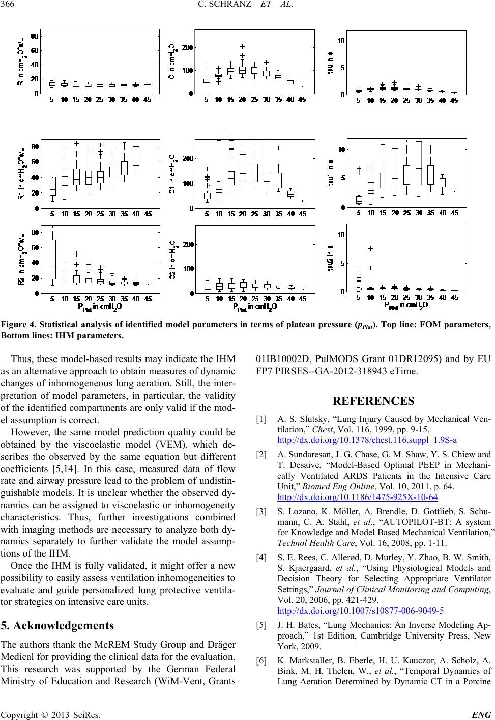 C. SCHRANZ ET AL. Copyright © 2013 SciRes. ENG 366 Figure 4. Statistical analysis of identified model parameters in terms of plateau pressure (pPlat). Top line: FOM parameters, Bottom lines: IHM parameters. Thus, these model-based results may indicate the IHM as an alternative approach to obtain measures of dynamic changes of inhomogeneous lung aeration. Still, the inter- pretation of model parameters, in particular, the validity of the identified compartments are only valid if the mod- el assumption is correct. However, the same model prediction quality could be obtained by the viscoelastic model (VEM), which de- scribes the observed by the same equation but different coefficients [5,14]. In this case, measured data of flow rate and airway pressure lead to the problem of undistin- guishable models. It is unclear whether the observed dy- namics can be assigned to viscoelastic or inhomogeneity characteristics. Thus, further investigations combined with imaging methods are necessary to analyze both dy- namics separately to further validate the model assump- tions of the IHM. Once the IHM is fully validated, it might offer a new possibility to easily assess ventilation inhomogeneities to evaluate and guide personalized lung protective ventila- tor strategies on intensive care units. 5. Acknowledgements The authors thank the McREM Study Group and Dräger Medical for providing the clinical data for the evaluation. This research was supported by the German Federal Ministry of Education and Research (WiM-Vent, Grants 01IB10002D, PulMODS Grant 01DR12095) and by EU FP7 PIRSES--GA-2012-318943 eTime. REFERENCES [1] A. S. Slutsky, “Lung Injury Caused by Mechanical Ven- tilation,” Chest, Vol. 116, 1999, pp. 9-15. http://dx.doi.org/10.1378/chest.116.suppl_1.9S-a [2] A. Sundaresan, J. G. Chase, G. M. Shaw, Y. S. Chiew and T. Desaive, “Model-Based Optimal PEEP in Mechani- cally Ventilated ARDS Patients in the Intensive Care Unit,” Biomed Eng Online, Vol. 10, 2011, p. 64. http://dx.doi.org/10.1186/1475-925X-10-64 [3] S. Lozano, K. Möller, A. Brendle, D. Gottlieb, S. Schu- mann, C. A. Stahl, et al., “AUTOPILOT-BT: A system for Knowledge and Model Based Mechanical Ventilation,” Technol Health Care, Vol. 16, 2008, pp. 1-11. [4] S. E. Rees, C. Allerød, D. Murley, Y. Zhao, B. W. Smith, S. Kjaergaard, et al., “Using Physiological Models and Decision Theory for Selecting Appropriate Ventilator Settings,” Journal of Clinical Monitoring and Computing, Vol. 20, 2006, pp. 421-429. http://dx.doi.org/10.1007/s10877-006-9049-5 [5] J. H. Bates, “Lung Mechanics: An Inverse Modeling Ap- proach,” 1st Edition, Cambridge University Press, New York, 2009. [6] K. Markstaller, B. Eberle, H. U. Kauczor, A. Scholz, A. Bink, M. H. Thelen, W., et al., “Temporal Dynamics of Lung Aeration Determined by Dynamic CT in a Porcine  C. SCHRANZ ET AL. Copyright © 2013 SciRes. ENG 367 Model of ARDS,” British Journal of Anaesthesia, Vol. 87, 2001, pp. 459-468. http://dx.doi.org/10.1093/bja/87.3.459 [7] Z. Zhao, K. Möller, D. Steinmann, I. Frerichs and J. Guttmann, “Evaluation of an Electrical Impedance To- mography-Based Global Inhomogeneity Index for Pul- monary Ventilation Distribution,” Intensive Care Medi- cine, Vol. 35, 2009, pp. 1900-1906. http://dx.doi.org/10.1007/s00134-009-1589-y [8] A. B. Otis, C. B. McKerrow, R. A. Bartlett, J. Mead, M. B. McIlroy, N. J. Selver-Stone, et al., “Mechanical Fac- tors in Distribution of Pulmonary Ventilation,” Journal of Applied Physiology, Vol. 8, 1956, pp. 427-443. [9] C. Schranz, C. Knöbel, J. Kretschmer, Z. Zhao and K. Möller, “Hierarchical Parameter Identification in Models of Respiratory Mechanics,” IEEE Transactions on Bio- medical Engineering, Vol. 58, 2011, pp. 3234-3241. http://dx.doi.org/10.1109/TBME.2011.2166398 [10] C. Schranz, P. D. Docherty, K. Möller and J. G. Chase, “Integral-Based Identification of an Inhomogeneity Mod- el in Respiratory Mechanics,” 6th International Confe- rence on Bioinformatics and Biomedical Engineering, Shanghai, 2012, pp. 575-578. http://dx.doi.org/10.1186/1475-925X-11-38 [11] C. Schranz, P. D. Docherty, Y. S. Chiew, K. Möller and J. G. Chase, “Iterative Integral Parameter Identification of a Respiratory Mechanics Model,” BioMedical Engineering OnLine, Vol. 11, 2012. [12] C. A. Stahl, K. Möller, S. Schumann, R. Kuhlen, M. Sy- dow, C. Putensen, et al., “Dynamic versus Static Respi- ratory Mechanics in Acute Lung Injury and Acute Respi- ratory Distress Syndrome,” Critical Care Medicine, Vol. 34, 2006, pp. 2090-2098. http://dx.doi.org/10.1097/01.CCM.0000227220.67613.0D [13] S. Pulletz, M. Kott, G. Elke, D. Schädler, B. Vogt, N. Weiler, et al., “Dynamics of Regional Lung Aeration De- termined by Electrical Impedance Tomography in Pa- tients with Acute Respiratory Distress Syndrome,” Multi- disciplinary Respiratory Medicine, Vol. 7, 2012, p. 44. http://dx.doi.org/10.1186/2049-6958-7-44 [14] G. Avanzolini and P. Barbini, “Comments on “Estimating Respiratory Mechanical Parameters in Parallel Compart- ment Models”, IEEE Transactions on Biomedical Engi- neering, Vol. 29, 1982, pp. 772-774. http://dx.doi.org/10.1109/TBME.1982.324873 |