

Paper Menu >>
Journal Menu >>
 Engineering, 2013, 5, 352-356 http://dx.doi.org/10.4236/eng.2013.510B071 Published Online Octob er 2013 (http://www.scirp.org/journal/eng) Copyright © 2013 SciRes. ENG Sonothromblysis in Combination w ith Thrombolytic Drugs in a Rabbit Model Using MRI-Guidance Christakis Damianou1, Nicos Mylonas2, Kleanthis Ioannides3 1Cyprus University of Technology, Cyprus 2Frederick University Cyprus, Limassol, Cyprus 3Polikliniki Ygia, Limassol, Cyprus Email: [email protected], [email protected] Received June 2013 ABSTRACT The potential of MRI-guided focused ultrasound (MRgFUS) combined with the thrombolytic drug recombinant tissue plas mino gen a ctiva to r (r t-PA), to d issolve clots in the carotid of a New Zealand rab bit in vivo is evaluated. A spherical- ly-focused tr a nsd uce r s of 5 cm diameter; focusing at 10 cm and operating at 1 MHz was used. A pulsed ultrasound pro- tocol was used that maintains a tissue temperature increase of less than 1˚C in the clot (called safe temperature). MRgFUS has t he potentials to dissolve clots t hat are injected in the carotid of rabbits in vivo. It was found that the ti me needed for opening the carotid artery using ultrasound and rt-PA was decreased compared to just using rt-P A. The pro- posed protocol was monitored using Magnetic Resonance Angiography (MRA) ever y 1 min. Keywords: Ultrasound; MRI; Stroke; Thrombolysis 1. Introduction The feasibility of ultrasound to enhance thrombolysis was reported in the mid 70s [1,2]. In the foll owin g ye ars, several in vitro studies (Kimura et al. 1994 [3] and Spengos K et al. 2000 [4]) have confirmed the above results. In the former studies, the range of intensity va- ried from 0.2 - 2.0 W/cm2 (spatial peak temporal average intensity) and the freque ncy from 20 kHz to 2 MHz using unfocused ultrasound. It is speculated that ultrasound accelerates enzymatic fibrinolysis primarily through mechanical mechanisms, by enhancing the effectiveness of thrombolytic drugs, possibly by exposing more binding sites on the fibrin to the r ecombi nant ti ssue p lasmi nogen a ctivato r (r t-PA) [5]. Other mechanical effects of ultrasound, such as cavita- tion and radiation force, are possibly influencing drug transport [6,7]. Acoustic cavitation plays a very signifi- cant r ole in ul trasound -accelerated fibrinolysis [6]. Other theories revolve around the fact that ultrasound promotes motion of fluid around the clot, an effect called micro- streaming [7]. Birnbaum et al. in 1998 [8], reported that in vivo ar- terial clot dissolution can be achieved with intravenous microbubbles and transcutaneous ultrasound delivery alone. Moreover, a study has shown the effectiveness of transcutaneous ultrasound and intravascular microbub- bles in lysing intracrania l c lot in pigs [9]. Administration of gaseous microspheres dramatically lowers the threshold for cavitation and increases the lytic activity of ultrasound (Holland CK and Aplef RE 1990[10]). Because the bubbles are destroyed in the process, they must be constantly injected for complete clot dissolution. Recent studies have demonstrated that microbubbles can be concentrated at the surface of clot by attaching a glycoprotein receptor antagonist, which increases their adherence to acute clot, to the bubbles [11]. The use of Focused ultrasound for destroying clots has received attention lately (Frenkel V et al. 2006 [12]; Shawa G et al. 2009 [13]; Hölscher T et al. 2011[14] and Laing S et al. 20 11[ 15]). In the stud y b y Frenke l V et al. 2006 [12], clots from humans in vitro were ablated using pulse ultrasound with a 1 MHz single element transducer in syne rg y with r t -PA. In t hi s stud y the c oncl us ion is t ha t by using rt-PA and focused ultrasound, improved clot destruction rates were achieved. In another study, using unfocused ultrasound at 120 KHz [12], showed that ul- trasound has the potential of destroying clots in vitro in combination with Liposomes loaded with rt-PA. Human whole blood clots were ablated though the skull using a hemispheric phased array transducer with 1,000 single piezo elements [13] using pulse ultrasound. The conclu- sion of this study is that ultrasound alone can be utilized to destro y clots. In addition to these in vitro studies, there are some in- 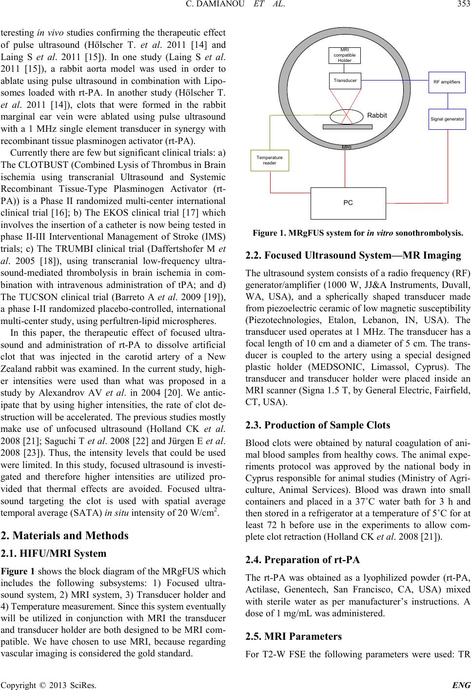 C. DAMIANOU ET AL. Copyright © 2013 SciRes. ENG 353 teresting in vivo studies confirming the therapeutic effect of pulse ultrasound (Hölscher T. et al. 2011 [14] and Laing S et al. 2011 [15]). In one study (Laing S et al. 2011 [15]), a rabbit aorta model was used in order to ablate using pulse ultrasound in combination with Lipo- somes loaded with rt-PA. In another study (Hölscher T. et al. 2011 [14]), clots that were formed in the rabbit marginal ear vein were ablated using pulse ultrasound with a 1 MHz single element transducer in synergy with recombinant tissue plasminog e n activator (rt-PA). Currently there are few but sig ni ficant c linical trials: a) The CLOTBUST (Combined Lysis of Thrombus in Brain ischemia using transcranial Ultrasound and Systemic Recombinant Tissue-Type Plasminogen Activator (rt- PA)) is a Phase II randomized multi-center international clinical trial [16]; b) The EKOS clinical trial [17] which involves the inser tion of a catheter is now being tested in phase II-III Interventional Management of Stroke (IMS) trials; c) The TRUMBI clinical trial (Daffertshofer M et al. 2005 [18]), using transcranial low-frequency ultra- sound-mediated thrombolysis in brain ischemia in com- bination with intravenous administration of tPA; and d) The TUCSON clinical trial (Barreto A et al. 2009 [19]), a phase I-II randomized placebo-controlled, international multi-center study, using perfultr en-lipid microsp heres. In this paper, the therapeutic effect of focused ultra- sound and administration of rt-PA to dissolve artificial clot that was injected in the carotid artery of a New Zealand rabbit was examined. In the current study, high- er intensities were used than what was proposed in a study by Alexandrov AV et al. in 2004 [20]. We antic- ipate that by using higher intensities, the rate of clot de- struction will be accelerated. The previous studies mostly make use of unfocused ultrasound (Holland CK et al. 2008 [21]; S aguchi T et al. 200 8 [22 ] and Jür gen E et al. 2008 [23]). Thus, the intensity levels that could be used were limited. I n this stud y, focused ultrasound is investi- gated and therefore higher intensities are utilized pro- vided that thermal effects are avoided. Focused ultra- sound targeting the clot is used with spatial average temporal average (SATA) in situ intensity of 20 W/cm2. 2. Materials and Methods 2.1. HIFU /MRI System Figure 1 shows the block diagram of the MRgFUS which includes the following subsystems: 1) Focused ultra- sound system, 2) MRI system, 3) Transducer holder and 4) Temperature measurement. Since thi s system e ventually will be utilized in conjunction with MRI the transducer and transducer holder are both designed to be MRI com- patible. We have chosen to use MRI, because regarding vascular imaging is considered the gold standard. MRI compatible Holder Transducer PC RF amplifiers Signal generator Temperature reader MRI Rabbit Figure 1 . MRgFUS system for in vitro sonothrombolysis. 2.2. Focused Ultrasound System—MR Imaging The ultra sound syste m co nsi sts o f a ra dio fr eque ncy (RF ) generator/amplifier (1000 W, JJ&A Instruments, Duvall, WA, USA), and a spherically shaped transducer made from piezoelectric ceramic of low magnetic susceptibility (Piezotechnologies, Etalon, Lebanon, IN, USA). The transducer used operates at 1 MHz. The transducer has a focal length of 10 cm and a diameter of 5 cm. The trans- ducer is coupled to the artery using a special designed plastic holder (MEDSONIC, Limassol, Cyprus). The transducer and transducer holder were placed inside an MRI scanner (Signa 1.5 T, by General Electric, Fairfield, CT, USA). 2.3. Production of Sample Clots Blood clots were obtained by natural coagulation of ani- mal blood samples from healthy cows. The animal expe- riments protocol was approved by the national body in Cyprus responsible for animal studies (Ministry of Agri- culture, Animal Services). Blood was drawn into small containers and placed in a 37˚C water bath for 3 h and then stored in a refrigerator at a temperature of 5˚C for at least 72 h before use in the experiments to allow com- plete clot r e traction (Holland CK et al. 2008 [21]). 2.4. Preparation of rt-PA The rt-PA was obtained as a lyophilized powder (rt-PA, Actilase, Genentech, San Francisco, CA, USA) mixed with sterile water as per manufacturer’s instructions. A dose of 1 mg/mL was administered. 2.5. MRI Parameters For T2-W FSE the following parameters were used: TR 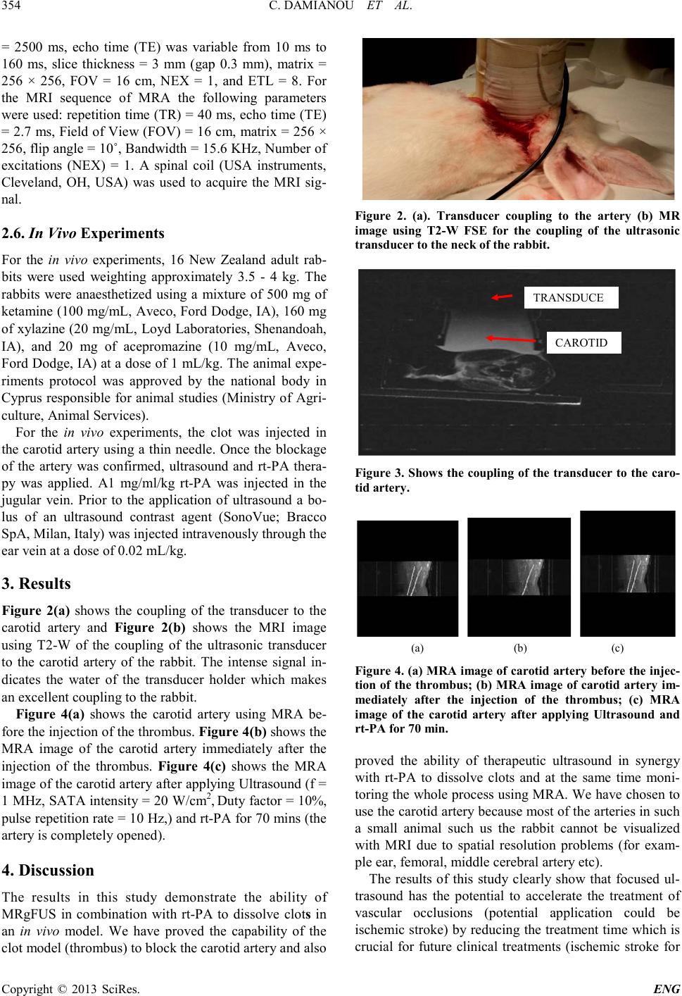 C. DAMIANOU ET AL. Copyright © 2013 SciRes. ENG 354 = 2500 ms, echo time (TE) was variable from 10 ms to 160 ms, slice thickness = 3 mm (gap 0.3 mm), matrix = 256 × 256, FOV = 16 cm, NEX = 1, and ETL = 8. For the MRI sequence of MRA the following parameters were used: rep etition time (TR) = 40 ms, echo time (TE) = 2.7 ms, Field of View (FOV) = 16 cm, matr ix = 256 × 256, flip angle = 10˚, Ban dwidth = 15.6 KHz, Number of excitations (NEX) = 1. A spinal coil (USA instruments, Cleveland, OH, USA) was used to acquire the MRI sig- nal. 2.6. In Vi v o Experiments For the in vivo experiments, 16 New Zealand adult rab- bits were used weighting approximately 3.5 - 4 kg. The rabbits were anaesthetized using a mixture of 500 mg of ketamine (100 mg/mL, Aveco, Ford Dodge, IA), 160 mg of xylazine (20 mg/mL, Loyd Laboratories, Shenandoah, IA), and 20 mg of acepromazine (10 mg/mL, Aveco, Ford Dodge, IA) at a dose of 1 mL/ kg. T he ani mal exp e- riments protocol was approved by the national body in Cyprus responsible for animal studies (Ministry of Agri- culture, Animal Services). For the in vivo experi ments, the clot was injected in the carotid artery using a thin needle. Once the blockage of the artery was confirmed, ultrasound and rt-PA thera- py was applied. A1 mg/ml/kg rt-PA was injected in the jugular vein. Prior to the application of ultrasound a bo- lus of an ultrasound contrast agent (SonoVue; Bracco SpA, Milan, Ita ly) was injected intravenously through the ear vein at a dose of 0.02 mL/kg. 3. Resul ts Figure 2(a) shows the coupling of the transducer to the carotid artery and Figure 2(b) shows the MRI image using T2-W of the coupling of the ultrasonic transducer to the carotid artery of the rabbit. The intense signal in- dicates the water of the transducer holder which makes an excellent coupling to the ra bbit. Figure 4(a) shows the carotid artery using MRA be- fore the i nj ec ti on of t he t hr ombus. Figure 4(b) shows t he MRA image of the carotid artery immediately after the injection of the thrombus. Figure 4(c) shows the MRA image of the carotid artery after app lying Ultrasound (f = 1 M Hz, SAT A intensity = 20 W/cm2, Dut y factor = 10%, pulse repetitio n rate = 10 Hz,) and rt-PA for 70 mins (the artery is completely opened). 4. Discussion The results in this study demonstrate the ability of MRgFUS in combination with rt-PA to dissolve clots in an in vivo model. We have proved the capability of the clot model ( thrombus) to blo c k the car otid artery and also Figure 2. (a). Transducer coupling to the artery (b) MR image using T2-W FSE for the coupling of the ultrasonic transducer to the neck of th e r abbit. Figure 3. Show s the coupli ng of the transducer to the car o- tid artery . (a) (b) (c) Figure 4. (a) M RA image of carotid art ery before the injec- tion of the thrombus; (b) MRA i mage of carotid artery im- mediately after the injection of the thrombus; (c) MRA image of the carotid artery after applying Ultrasound and rt-PA for 70 min. proved the ability of therapeutic ultrasound in synergy with rt-PA to dissolve clots and at the same time moni- tor ing the whole p ro cess usi ng M R A. We have cho se n to use the carotid artery because most of the arteries in such a small animal such us the rabbit cannot be visualized with MRI due to spatial resolution problems (for exam- ple ear, femoral, middle cerebral artery etc). The results of this study clearly show that focused ul- trasound has the potential to accelerate the treatment of vascular occlusions (potential application could be ischemic stro ke) b y reducing the treat ment time whic h is crucial for future clinical treatments (ischemic stroke for TRANSDUCE CAROTID 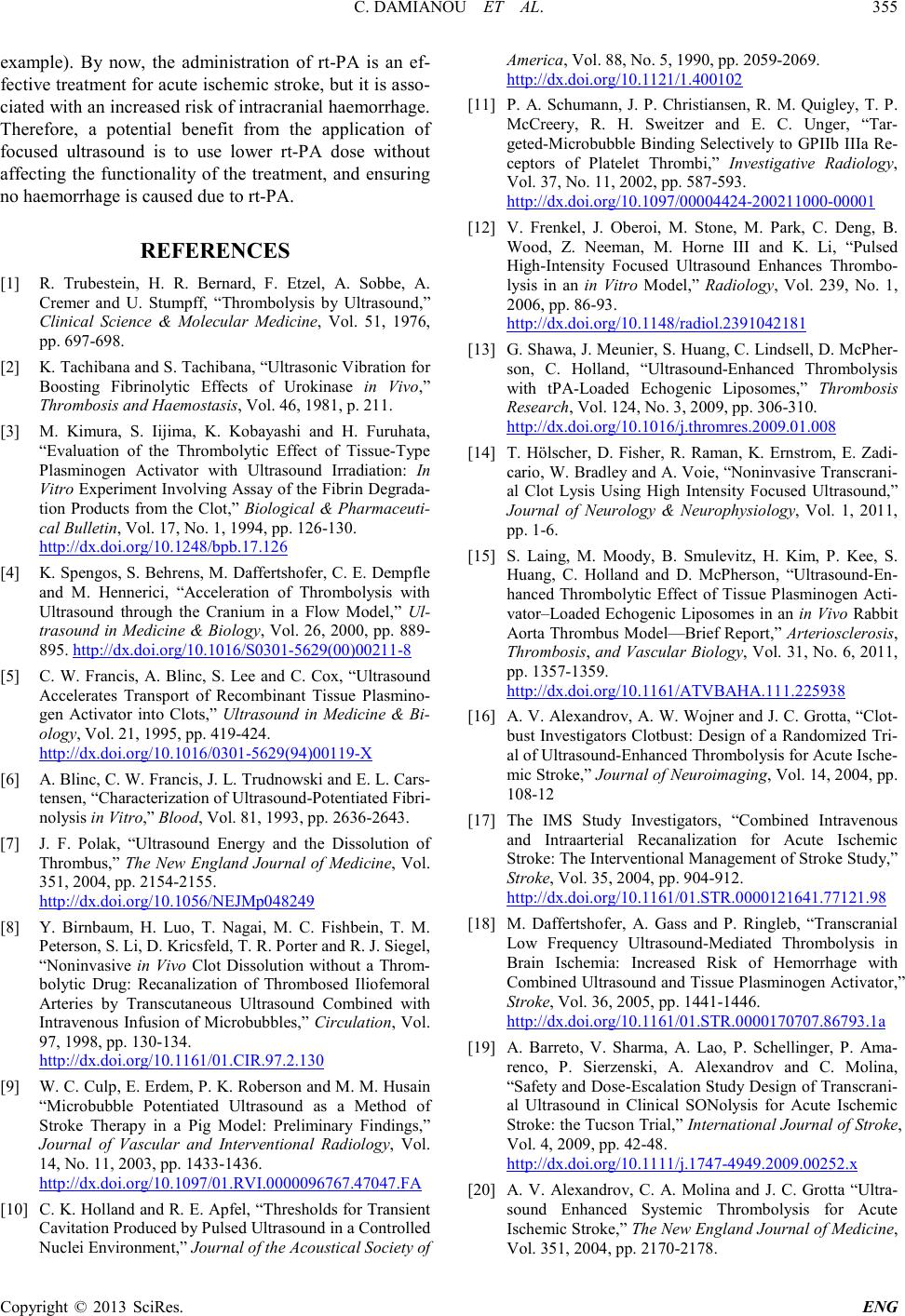 C. DAMIANOU ET AL. Copyright © 2013 SciRes. ENG 355 example). By now, the administration of rt-PA is an ef- fective treatment for acute ischemic stroke, but it i s as so- ciated with an increased risk of intracranial haemorrhage. Therefore, a potential benefit from the application of focused ultrasound is to use lower rt-PA dose without affecting the functionality of the treatment, and ensuring no haemorrhage is cause d due to rt-PA. REFERENCES [1] R. Trubestein, H. R. Bernard, F. Etzel, A. Sobbe, A. Cremer and U. Stumpff, “Thrombolysis by Ultrasound,” Clinical Science & Molecular Medicine, Vol. 51, 1976, pp. 697-698. [2] K. Tachibana and S. Tachi bana, “Ultrasonic Vibration for Boosting Fibrinolytic Effects of Urokinase in Vivo,” Thrombo si s an d Haemostasis, Vol. 46, 1981, p. 211. [3] M. Kimura, S. Iijima, K. Kobayashi and H. Furuhata, “Evaluation of the Thrombolytic Effect of Tissue-Type Plasminogen Activator with Ultrasound Irradiation: In Vitro Experiment Involving Assay of the Fibrin Degrada- tion Products from the Clot,” Biological & Pharmaceuti- cal Bulletin, Vol. 17, No. 1, 1994, pp. 126-130. http://dx.doi.org/10.1248/bpb.17.126 [4] K. Spengos, S. Behrens, M. Daffertshofer, C. E. Demp fl e and M. Hennerici, “Acceleration of Thrombolysis with Ultrasound through the Cranium in a Flow Model,” Ul- trasound in Medicine & Biology, Vol. 26, 2000, pp. 889- 895. http://dx.doi.org/10.1016/S0301-5629(00)00211-8 [5] C. W. Francis, A. Blinc, S. Lee and C. Cox, “Ultrasound Accelerates Transport of Recombinant Tissue Plasmino- gen Activator into Clots,” Ultrasound in Medicine & Bi- ology, Vol. 21, 1995, pp. 419-424. http://dx.doi.org/10.1016/0301-5629(94)00119-X [6] A. Blinc, C. W. Francis, J. L. Trudnowski and E. L. Cars- tensen , “Ch aracteri zati on o f Ultrasound-Potentiated Fibri- nolysis in Vitro,” Blood, Vol. 81, 1993, pp. 2636-2643. [7] J. F. Polak, “Ultrasound Energy and the Dissolution of Thrombus,” The New England Journal of Medicine, Vol. 351, 2004, pp. 2154-2155. http://dx.doi.org/10.1056/NEJMp048249 [8] Y. Birnbaum, H. Luo, T. Nagai, M. C. Fishbein, T. M. Peterson, S. Li, D. Kricsfeld, T. R. Porter and R. J. Siegel, “Noninvasive in Vivo Clot Dissolution without a Throm- bolytic Drug: Recanalization of Thrombosed Iliofemoral Arteries by Transcutaneous Ultrasound Combined with Intravenous Infusion of Microbubbles,” Circulation, Vol. 97, 1998, pp. 130-134. http://dx.doi.org/10.1161/01.CIR.97.2.130 [9] W. C. Culp, E. Erdem, P. K. Roberson and M. M. Husain “Microbubble Potentiated Ultrasound as a Method of Stroke Therapy in a Pig Model: Preliminary Findings,” Journal of Vascular and Interventional Radiology, Vol. 14, No. 11, 2003, pp. 1433-1436. http://dx.doi.org/10.1097/01.RVI.0000096767.47047.FA [10] C. K. Holland and R. E. Apfel, “Thresholds for Transient Cavitation Produced by Pulsed Ultrasound in a Controlled Nuclei Environment,” Journal of the Acoustical Society of America, Vol. 88, No. 5, 1990, pp. 2059-2069. http://dx.doi.org/10.1121/1.400102 [11] P. A. Schumann, J. P. Christiansen, R. M. Quigley, T. P. McCreery, R. H. Sweitzer and E. C. Unger, “Tar- geted-Microbubble Binding Selectively to GPIIb IIIa Re- ceptors of Platelet Thrombi,” Investigative Radiology, Vol. 37, No. 11, 2002, pp. 587-593. http://dx.doi.org/10.1097/00004424-200211000-00001 [12] V. Frenkel , J. Oberoi, M. Stone, M. Park, C. Deng, B. Wood, Z. Neeman, M. Horne III and K. Li, “Pulsed High-Intensity Focused Ultrasound Enhances Thrombo- lysis in an in Vitro Model,” Radiology, Vol. 239, No. 1, 2006, pp. 86-93. http://dx.doi.org/10.1148/radiol.2391042181 [13] G. Shawa, J. Meunier, S. Huang, C. Lindsell, D. M cPh e r - son, C. Holland, “Ultrasound-Enhanced Thrombolysis with tPA-Loaded Echogenic Liposomes,” Thrombosis Research, Vol. 124, No. 3, 2009, pp. 306-310. http://dx.doi.org/10.1016/j.thromres.2009.01.008 [14] T. Hölscher, D. Fisher, R. Raman, K. Ernstrom, E. Zad i- cario, W. Bradley and A. Voie, “Noninvasive Tran scrani- al Clot Lysis Using High Intensity Focused Ultrasound,” Journal of Neurology & Neurophysiology, Vol. 1, 2011, pp. 1-6. [15] S. Laing, M. Moody, B. Smulevitz, H. Kim, P. Kee, S. Huang, C. Holland and D. McPherson, “Ultrasound-En - hanced Thrombolytic Effect of Tissue Plasminogen Acti- vator–Loaded Echogenic Liposomes in an in Vivo Rabbit Aorta Thrombus Model—Brief Report,” Arteriosclerosis, Thrombo sis, and Vascular Biology, Vol. 31, N o. 6, 2011, pp. 1357-1359. http://dx.doi.org/10.1161/ATVBAHA.111.225938 [16] A. V. Alexandrov, A. W. Wojner and J. C. Grotta, “Clot- bust Investigators Clotbust: Design of a Randomized Tri- al of Ultrasound-Enhanced Thrombolysis for Acute Ische- mic Stroke,” Journal of Neuroimaging, Vol. 14, 2004, pp. 108-12 [17] The IMS Study Investigators, “Combined Intravenous and Intraarterial Recanalization for Acute Ischemic Stro ke: The In terven ti onal M an agement o f S t r o ke S tudy,” Stroke, Vol. 35, 2004, pp. 904-912. http://dx.doi.org/10.1161/01.STR.0000121641.77121.98 [18] M. Daffertshofer, A. Gass and P. Ringleb, “Transcranial Low Frequency Ultrasound-Mediated Thrombolysis in Brain Ischemia: Increased Risk of Hemorrhage with Combined Ultrasound and Tissue Plasminogen Activator,” Stroke, Vol. 36, 2005, pp. 1441-1446. http://dx.doi.org/10.1161/01.STR.0000170707.86793.1a [19] A. Barreto, V. Sharma, A. Lao, P. Schellinger, P. Ama- renco, P. Sierzenski, A. Alexandrov and C. Molina, “Safety and Dose-Escalatio n Study Design of Transcr ani- al Ultrasound in Clinical SONolysis for Acute Ischemic Stroke: the Tucson Trial,” International Journal of Stroke, Vol. 4, 2009, pp. 42-48. http://dx.doi.org/10.1111/j.1747-4949.2009.00252.x [20] A. V. Alexandrov, C. A. Molina and J. C. Grotta “Ultra- sound Enhanced Systemic Thrombolysis for Acute Ische mic Stroke,” The New England Journal of Medicine, Vol. 351, 2004, pp. 2170-2178.  C. DAMIANOU ET AL. Copyright © 2013 SciRes. ENG 356 http://dx.doi.org/10.1056/NEJMoa041175 [21] C. K. Holland, S. S Vaidya, S. Datta, C. C. Coussios and G. J. Shaw, “Ultrasound Enhanced Tissue Plasminogen Activator Thrombolysis in an in Vitro Porcine Clot Mod- el,” Thrombosis Research, Vol. 121, 2008, pp. 663-673. http://dx.doi.org/10.1016/j.thromres.2007.07.006 [22] T. Saguchi, H. Onoue, M. Urashima, T. Ishibashi, T. Abe and H. Furuhata, “Effective and Safe Conditions of Low-Frequency Transcranial Ultrasonic Thrombolysis for Acute Ischaemic Stroke: Neurologic and Histologic Eva- luation in a Rat Middle Cerebral Artery Stroke Model,” Stroke, Vol. 39, 2008, pp. 1007-1011. http://dx.doi.org/10.1161/STROKEAHA.107.496117 [23] E. Jürgen, R. K. Inke, K. Björn, H. Götz and S. Günter, “Sonothrombolysis with Transcranial Color-Coded So- nography and Recombinant Tissue-Type Plasminogen Activator in Acute Middle Cerebral Artery Main Stem Occlusion: Results from a Randomized Study,” Stroke, Vol. 39, 2008, pp. 1470-1475. http://dx.doi.org/10.1161/STROKEAHA.107.503870 |