

Paper Menu >>
Journal Menu >>
 Vol.2, No.4, 66-68 (2013) Modern Chemotherapy http://dx.doi.org/10.4236/mc.2013.24008 An autop sy case of met astatic extramammary Paget’s disease treated with multimodality treatment including anti-HER2 therapy: What is the clinical and pathological significance of trastuzumab to the patient? Noriko Yoshimura1*, Koji Arihiro2, Shunsuke Takahagi3, Michihiro Hide3 1Department of Surgery, Hiroshima General Hospital, Hiroshima, Japan; *Corresponding Author: [email protected] 2Department of Pathology, Hiroshima University Hospital, Hiroshima, Japan 3Department of Dermatology, Hiroshima University Hospital, Hiroshima, Ja pa n Received 1 October 2013; revised 21 October 2013; accepted 27 October 2013 Copyright © 2013 Noriko Yoshimura et al. This is an open access article distributed under the Creative Commons Attribution Li- cense, which permits unrestricted use, distribution, and reproduction in any medium, provided the original work is properly cited. ABSTRACT Advanced Extramammary Paget’s disease (AEMPD) shows a poor prognosis despite multi- modality therapy. In recent y ears, it is sugge sted that anti-HER2 therapy may be promising for HER2-positive AEMPD. We herein present an autop sy case of a p atient with AEMPD treated with multimodality treatment including anti-HER2 the- rapy. A 78-year-old man who diagnosed with AEMPD died after surgery and systemic chemo- therapy including anti-HER2 therapy (trastuzu- mab). The metastatic skin lesions were immno- histologically HER2-positive. While the patients were administrated trastuzumab plus taxan (do- cetaxel, and paclitaxel) regimen, the metastatic skin lesion decreased, however, brain metasta- ses were found in his brain and trastuzumab is discontinued. The skin metastasis rapidly spread over his body, leading to weakness, and he eventually died. At autopsy, the lesions of EMPD were extended distant organs including brain, although each metastasis was small and as- ymptomatic. The wide lesion of skin metastasis was exacerbated after discontinuation of tras- tuzumab, and transudate was observed due to the extensive necrosis and erosion. Our au topsy findings showed one progressive pattern of AEMPD, and indicated what is the clinical and pathological significance of anti HER2 therapy for HER2-positive AEMPD. Keywords: Extramammary Paget’s Disease; HER2; Autopsy 1. INTRODUCTION Extramammary Paget’s disease (EMPD) is a distinct type of skin carcinoma originating in the apocrine gland. Advanced EMPD (AEMPD) cases show a poor progno- sis despite multimodality therapy. In recent years, some authors reported the overexpression of human epidermal growth factor receptor 2 (HER2) [1-3] in AEMPD, thus suggesting that anti-HER2 therap y may be promising for the disease. We herein present an autopsy case of a pa- tient with AEMPD who was treated with multimodality therapy, including trastuzumab. The detailed clinical presentation of this patient has been reported previously [4]. In this letter, we highlight the autopsy findings of our patient and examine the clinical and pathological sig- nificance of trastuzumab to the patient. 2. CASE A 78-year-old male patient presented with erythema on the scrotum, which he had first noted almost five years prior. The erythema had gradually extended, these lesions were diagnosed as EMPD by a skin biopsy 28 months before his death and skin resection and dissection of the left inguinal lymph nodes were performed. After surgery, the patient was treated with systemic chemothe- rapy using Mitomycin (20 mg/m2) + 5-FU (1000 mg/m2) at first, and then weekly with Docetaxel (60 mg/m2). However, the skin recurrence worsened. At this point, a Copyright © 2013 SciRes. OPEN A CCESS 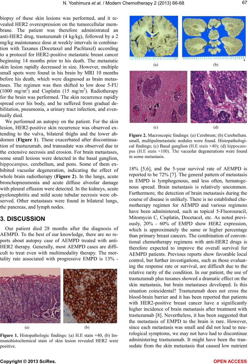 N. Yoshimura et al. / Modern Chemotherapy 2 (2013) 66-68 67 biopsy of these skin lesions was performed, and it re- vealed HER2 overexpression on the tumorcellular mem- brane. The patient was therefore administrated an anti-HER2 drug, trastuzumab (4 kg/kg), followed by a 2 mg/kg maintenance dose at weekly intervals in combina- tion with Taxanes (Docetaxel and Paclitaxel) according to a protocol for HER2-positive metastatic breast cancer beginning 14 months prior to his death. The metastatic skin lesion rapidly decreased in size. However, multiple small spots were found in his brain by MRI 10 months before his death, which were diagnosed as brain metas- tases. The regimen was then shifted to low dose 5-FU (1000 mg/m2) and Cisplatin (15 mg/m2). Radiotherapy for the brain was performed. The skin recurrence rapidly spread over his body, and he suffered from gradual de- bilitation, pneumonia, a urinary tract in fection, and ev en- tually died. We performed an autopsy on the patient. For the skin lesion, HER2-positive skin recurrence was observed ex- tending to the vulva, bilateral thighs and the lower ab- domen (Figure 1). These exacerbated after discontinua- tion of trastuzumab, and transudate was observed due to the extensive necrosis and erosion. For brain metastases, some small lesions were detected in the basal ganglion, hippocampus, cerebellum, and pons. Some of them ex- hibited vacuolar degeneration, indicating the effect of whole brain radiotherapy (Figure 2). In the lungs, acute bronchopneumonia and acute diffuse alveolar damage with pleural effusion were detected. In the kidneys, acute pyelonephritis and mild acute tubular necrosis were ob- served. Other metastases were found in bilateral lungs, the pancreas, and lymph nodes. 3. DISCUSSION Our patient died 28 months after the diagnosis of AEMPD. To the best of our knowledge, there are no re- ports about autopsy case of AEMPD treated with anti- HER2 therapy. Generally, most AEMPD cases are diffi- cult to treat even with multimodality therapy. The mor- tality rate associated with progressive EMPD is 13% - (a) (b) Figure 1. Histopathologic findings: (a) H.E stain ×40, (b) Im- munohistochemical stain of skin lesion revealed HER2 were positive. (a) (b) (c) (d) Figure 2. Macroscopic findings: (a) Cerebrum; (b) Cerebellum. small, multipulmetastatic nodules were found. Histopathologi- cal findings; (c) Basal ganglion (H.E stain ×40); (d) hippocam- pus (H.E stain ×100). The vacuolar degenerations were found in some metastasis. 18% [5,6], and the 5-year survival rate of AEMPD is reported to be 72% [7]. The general pattern of metastasis in EMPD is lymphogenous, and less often, hematoge- nous spread. Brain metastasis is relatively uncommon. Furthermore, the detection of brain metastasis during the course of disease is unlikely. There is no established che- motherapy regimen for AEMPD and various regimens have been administered, such as topical 5-Fluorouracil, Mitomycin C, Cisplatin, Docetacel, etc. As noted previ- ously, 20% - 60% of EMPD show HER2 expression, which is approximately the same or higher percentage than primary breast cancers. The combination of con ven- tional chemotherapy regimens with anti-HER2 drugs is therefore expected to improve the overall survival for AEMPD patients. Previous reports show favorable local control, but further investigations, such as those evaluat- ing the response rate or survival, are difficult due to the relative rarity of the condition. In our patient, the use of trastuzumab plus taxanes showed a dramatic effect on the skin metastasis, but brain metastases developed. Is this situation coincidental? Trastuzumab does not cross the blood-brain barrier and it has been reported that patients with HER2-positive breast cancer have a significantly higher incidence of brain metastasis after treatment with trastuzumab [8]. Nevertheless, it has been suggested that the metastasis of EMPD to the brain is rare. However, since each metastasis was small and did not lead to neu- rological symptoms, we may not have had to discontinue administering trastuzumab. It might have been the tran- sudate from the skin metastasis that caused low nutrient Copyright © 2013 SciRes. OPEN A CCESS  N. Yoshimura et al. / Modern Chemotherapy 2 (2013) 66-68 Copyright © 2013 SciRes. OPEN A CCESS 68 condition, generalized weakness, and eventually death. In many AEMPD cases, there have been no reports on lethal events such as hemorrhage due to metastases to other organs that resulted in death. Rather, patients pro- gressively weaken and die. Hence, we suggest that con- trol of skin lesions be assigned a priority instead of fo- cusing on treating small metastases in other organs. New anti-HER2 drugs have been developed recently. Lapatinib, for example, is a small molecule anti-HER2 agent used for breast cancer which is known to penetrate the blood-brain barrier and contribute to overall survival of patients with br ain metastases. Although clinical indi- cations need to be clearer and the cost might hinder posi- tive use, lapatinib could be an effective treatment strat- egy for AE MPD. 4. CONCLUSION We confirmed one progressive pattern of AEMPD treated with multimodality therapy, including anti-HER2 therapy. More amassed reports and further investigations will be necessary. REFERENCES [1] Ogawa, T., Nagash im a, Y., Wada, H., Akimoto, K., Chiba, Y. and Nagatani, T. (2005) Extramammary Paget’s: Analysis of growth signal pathway from the human epi- dermal growth factor receptor 2 protein. Human Pathol- ogy, 36, 1237-1280. [2] Plaza, J.A., Torres-Cabala, C., Ivan, D. and Prieto, V.G. (2009) HER-2/neu expression in extramammary Paget disease: A clinicopathologic and immunohistochemistry study of 47 cases with and without underlying malign- nancy. Journal of Cutaneous Pathology, 36, 729-733. h tt p:// dx. doi. org/1 0.1111 /j.1600-0560.2008.01148.x [3] Richter, C.E., Hui, P., Buza, N., Silasi, D.A., Azodi, M., Santin, A.D., et al. (2010) HER-2/NEU overexpression in vulvar Paget disease: The Yale experience. Journal of Clinical Pathology, 63, 544-547. http://dx.doi.org/10.1136/jcp.2010.077446 [4] Takahagi, S., Noda, H., Kamegashira, A., Madokoro, N., Hori, I. and Shindo, H. (2009) Metastatic extramammary Paget’s disease treated with paclitaxel and trastuzumab combination. The Journal of Dermatology, 36, 457-461. h tt p:// dx. doi. org/1 0.1111 /j.1346-8138.2009.00676.x [5] Chanda, J.J. (1985) Extramammary Paget’s disease: Prog- nosis and relationship of internal malignancy. Journal of the American Academy of Dermatology, 13, 1009-1014. http://dx.doi.org/10.1016/S0190-9622(85)70254-X [6] Hatta, N., Yamada, M., Hisano, T., Fujimoto, A. and Mo- rita, R. (2008) Extramammary Paget’s disease: Treatment, prognosis and outcome in 76 patients. British Journal of Dermatology, 158, 313-318. [7] Siesling, S., Elferink, M.A.G., van Dijck, J.A., Pierie, J.P. and Blokx, W.A. (2007) Epidemiology and treatment of extramammary Paget disease in the Netherlands. Euro- pean Journal of Surgical Oncology, 33, 951-955. http://dx.doi.org/10.1016/j.ejso.2006.11.028 [8] Musolino, A., Cinnolallo, L., Panebianco, M., Fontana, E., Zanoni, D., Bozzetti, C., et al. (2011) Multifactorial cen- tral nervous system recurrence susceptibility in patients with HER2-positive breast cancer. Cancer, 117, 1837- 1846. http://dx.doi.org/10.1002/cncr.25771 |