

Paper Menu >>
Journal Menu >>
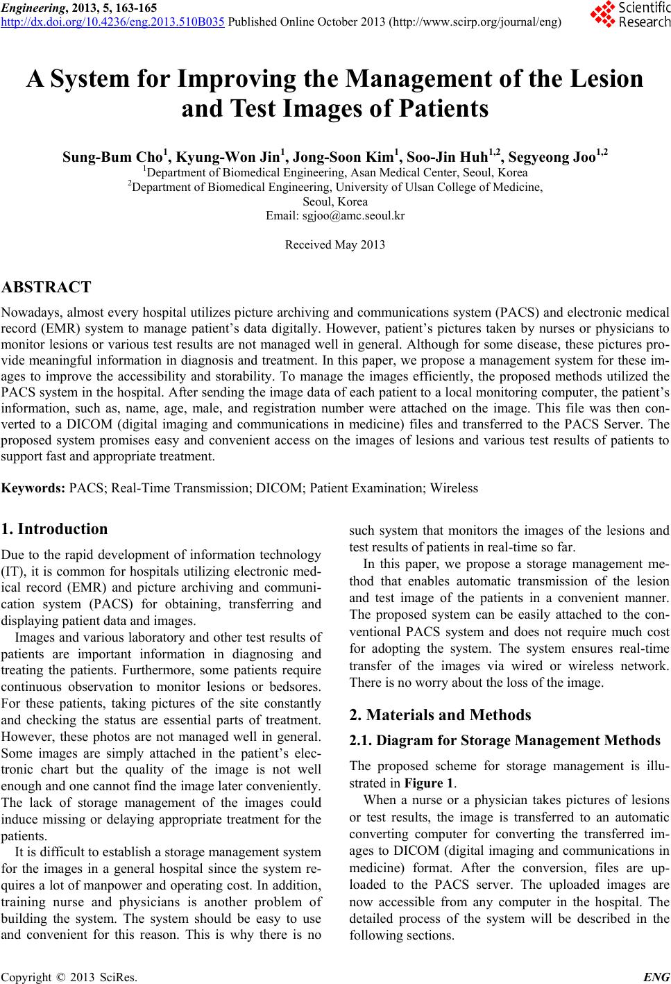 Engineering, 2013, 5, 163-165 http://dx.doi.org/10.4236/eng.2013.510B035 Published Online October 2013 (http://www.scirp.org/journal/eng) Copyright © 2013 SciRes. ENG A System for Improving the Management of the Lesion and Test Images of P atients Sung-Bum Cho1, Kyung-Won Jin1, Jong-Soon Kim1, Soo-Jin Huh1,2, Segyeong Joo1,2 1Department of Biomedical Engineering, Asan Medical Center, Seoul, Korea 2Department of Biomedical Engineering, University of Ulsan College of Medicine, Seoul, Korea Email: [email protected] Received May 2013 ABSTRACT Nowadays, almost every hospital u tilizes picture archiving and communications system (PACS) and electronic medical record (EMR) system to manage patient’s data digitally. However, patient’s pictures taken by nurses or physicians to monitor lesions or various test results are not managed well in general. Although for some disease, these pictures pro- vide meaningful information in diagnosis and treatment. In this paper, we propose a management system for these im- ages to improve the accessibility and storability. To manage the images efficiently, the proposed methods utilized the PACS system in the hospital. A fter sending the image data of each patient to a local monitoring computer, the patient’s information, such as, name, age, male, and registration number were attached on the image. This file was then con- verted to a DICOM (digital imaging and communications in medicine) files and transferred to the PACS Server. The proposed system promises easy and convenient access on the images of lesions and various test results of patients to support fast and appropriate treatment. Keywords: PACS; Real-Time Transmission; DICOM; Patient Examination; Wireless 1. Introduction Due to the rapid development of information technology (IT), it is common for hospitals utilizing electronic med- ical record (EMR) and picture archiving and communi- cation system (PACS) for obtaining, transferring and displaying patient data and images. Images and various laboratory and other test results of patients are important information in diagnosing and treating the patients. Furthermore, some patients require continuous observation to monitor lesions or bedsores. For these patients, taking pictures of the site constantly and checking the status ar e essential parts of treatment. However, these photos are not managed well in general. Some images are simply attached in the patient’s elec- tronic chart but the quality of the image is not well enough and one cannot find the image later conveniently. The lack of storage management of the images could induce missing or d elaying appropriate treatment for the patients. It is difficult to establish a storage management system for the images in a general hospital since the system re- quires a lot of manpower and operating cost. I n addition, training nurse and physicians is another problem of building the system. The system should be easy to use and convenient for this reason. This is why there is no such system that monitors the images of the lesions and test results of patients in real-time so far. In this paper, we propose a storage management me- thod that enables automatic transmission of the lesion and test image of the patients in a convenient manner. The proposed system can be easily attached to the con- ventional PACS system and does not require much cost for adopting the system. The system ensures real-time transfer of the ima ge s via wired or wireless network. There is no worry about the loss of the image. 2. Materials and Methods 2.1. Diagram for Storage Management Methods The proposed scheme for storage management is illu- strated in Figure 1. When a nurse or a physician takes pictures of lesions or test results, the image is transferred to an automatic converting computer for converting the transferred im- ages to DICOM (digital imaging and communications in medicine) format. After the conversion, files are up- loaded to the PACS server. The uploaded images are now accessible from any computer in the hospital. The detailed process of the system will be described in the following sections. 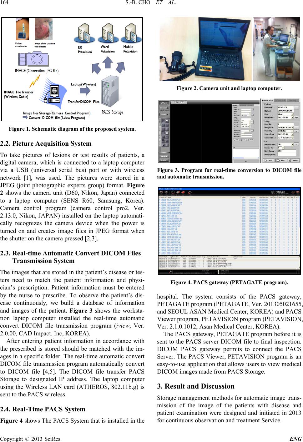 S.-B. CHO ET AL. Copyright © 2013 SciRes. ENG 164 Figure 1. Schematic diagram of the proposed system. 2.2. Picture Acquisition System To take pictures of lesions or test results of patients, a digital camera, which is connected to a laptop computer via a USB (universal serial bus) port or with wireless network [1], was used. The pictures were stored in a JPEG (joint photographic experts group) format. Figure 2 shows the camera unit (D60, Nikon, Japan) connected to a laptop computer (SENS R60, Samsu ng , Korea). Camera control program (camera control pro2, Ver. 2.13.0, Nikon, JAPAN) installed on the laptop automati- cally recognizes the camera device when the power is turned on and creates image files in JPEG format when the sh utter on the camera pressed [2,3]. 2.3. Real-time Automatic Convert DICOM Files Transmission System The images that are stored in the patient’s disease or tes- ters need to match the patient information and physi- cian’s prescription. Patient information must be entered by the nurse to prescribe. To observe the patient’s dis- ease continuously, we build a database of information and images of the patient. Figure 3 shows the worksta- tion laptop computer installed the real-time automatic convert DICOM file transmission program (iview, Ver. 2.0.00, CAD I mpact. Inc, K OR E A ). After entering patient information in accordance with the prescribed is stored should be matched with the im- ages in a specific folder. The real-time automatic convert DICOM file transmission progra m automatically convert to DICOM file [4,5]. The DICOM file transfer PACS Storage to designated IP address. The laptop computer using the Wireless LAN card (ATHEROS, 802.11b.g) is sent to the PACS wireless. 2.4. Real-Time PACS System Figure 4 shows The PACS System that is installed in the Figure 2. Camera unit and laptop computer. Figure 3. Program for real-time conversion to DICOM file and automatic transmission. Figure 4. PACS gateway (PETAGATE program). hospital. The system consists of the PACS gateway, PETAGATE program (PETAGATE, Ver. 201305021655, and SEO U L ASAN Medical Center, KO REA) and PACS Viewer program, PETAVISION program (PETAVISION, Ver. 2.1.0.1012, Asan Medical Center, KOREA). The PACS gateway, PETAGATE program before it is sent to the PACS server DICOM file to final inspection. DICOM PACS gateway permits to connect the PACS Server. The PACS Viewer, P ETAVISION prog ram is an easy-to-use application that allows users to view medical DICOM images made from PACS Storage. 3. Result and Discussion Storage management methods for automatic image trans- mission of the image of the patients with disease and patient examination were designed and initiated in 2013 for continuous observation and treatment Service. 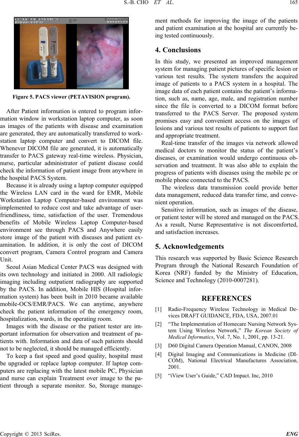 S.-B. CHO ET AL. Copyright © 2013 SciRes. ENG 165 Figure 5. PACS viewer (PETAVISION program). After Patient information is entered to program infor- mation window in workstation laptop computer, as soon as images of the patients with disease and examination are generated, they are automatically transferred to work- station laptop computer and convert to DICOM file. Whenever DICOM file are generated, it is automatically transfer to PACS gateway real-time wireless. Physician, nurse, particular administrator of patient disease could check the information of patient image from anywhere in the hospital PACS System. Because it is already using a laptop computer equipped the Wireless LAN card in the ward for EMR, Mobile Workstat ion Laptop Computer-based environment was implemented to reduce cost and take advantage of user- friendliness, time, satisfaction of the user. Tremendous benefits of Mobile Wireless Laptop Computer-based environment see through PACS and Anywhere easily store image of the patient with diseases and patient ex- amination. In addition, it is only the cost of DICOM convert program, Camera Control program and Camera Unit. Seoul Asian Medical Center PACS was designed with its own technology and initiated in 2000. All radiologic imaging including outpatient radiography are supported by the PACS. In addition, Mobile HIS (Hospital infor- mation system) has been built in 2010 became available mobile-OCS/EMR/PACS. We can anytime, anywhere check the patient information of the emergency room, hospitalization, wards, in the operating room. Images with the disease or the patient tester are im- portant information for observation and treatment of pa- tients with. Information and data of such patients should not to be neglected, it should be managed efficiently. To keep a fast speed and good quality, hospital must be upgraded or replace laptop computer. If laptop com- puters are replacing with the latest mobile PC, Physician and nurse can explain Treatment over image to the pa- tient through a separate monitor. So, Storage manage- ment methods for improving the image of the patients and patient examination at the hospital are currently be- ing tested continuously. 4. Conclusions In this study, we presented an improved management system for managing patient pictures of specific lesion or various test results. The system transfers the acquired image of patients to a PACS system in a hospital. The image data of each patient contains the patient’s informa- tion, such as, name, age, male, and registration number since the file is converted to a DICOM format before transferred to the PACS Server. The proposed system promises easy and convenient access on the images of lesions and various test results of patients to support fast and appropriate treatment. Real-time transfer of the images via network allowed medical doctors to monitor the status of the patient’s diseases, or examination would undergo continuous ob- servation and treatment. It was also able to explain the progress of patients with diseases using the mobile pc or mobile phone connected to the PACS. The wireless data transmission could provide better data management, reduced data transfer time, and conve- nient operation. Sensitive information, such as images of the disease, or patient tester will be stored and managed on the PACS. As a result, Nurse Representative is not discomforted, and satisfaction increases. 5. Acknowledgements This research was supported by Basic Science Research Program through the National Research Foundation of Korea (NRF) funded by the Ministry of Education, Science and Tech nology (2010-0007281). REFERENCES [1] Radio-Frequency Wireless Technology in Medical De- vices DRAFT GUIDANCE, FDA, USA, 2007.01 [2] “The Implementation of Homecare Nursing Network Sys- tem Using Wireless Network,” The Korean Society of Medical Informatics, Vol. 7, No. 1, 2001, pp. 13-21. [3] D60 Digital Camera Operation Manual, CANON, 2008 [4] Digital Imaging and Communications in Medicine (DI- COM), National Electrical Manufactures Association, 2001. [5] “iView User’s Guide ,” CAD Impact. Inc, 2010 |