

Paper Menu >>
Journal Menu >>
 Surgical Science, 2013, 4, 459-463 http://dx.doi.org/10.4236/ss.2013.410090 Published Online October 2013 (http://www.scirp.org/journal/ss) Transfer of the Trapezius to the Deltoid for Treatment of Shoulder Instability after Lesions of the Brachial Plexus* Antônio L. Severo1#, Philipe E. C. Maia1, Marcelo B. Lemos1, Paulo C. F. Piluski1, Osvandré L. C. Lech1, Walter Y. Fukushima2 1Department of Hand and Microsurgery, Institute of Orthopedics and Traumatology (IOT), Passo Fundo, Brazil 2Faculty of Medicine of ABC, Santo André, Brazil Email: #[email protected] Received May 20, 2013; revised June 18, 2013; accepted June 26, 2013 Copyright © 2013 Antônio L. Severo et al. This is an open access article distributed under the Creative Commons Attribution Li- cense, which permits unrestricted use, distribution, and reproduction in any medium, provided the original work is properly cited. ABSTRACT Objective: To clarify the indications and to describe the surgical technique and outcomes of surgery involving transfer of the trapezius to the deltoid for the treatment of lesions of the brachial plexus in patients with multidirectional instability in the sh oulder. Method: In 17 patients (mean age, 23 years) operated at São Vicente de Paulo Hospital and the Institute of Orthopedics and Traumatology of Passo Fundo, Brazil from 1999 to 2009, we performed trapezius transfer to the proximal humerus. In these patients, the mean interval between trauma and surgery was 8 months. Results: Functional improvement and resolution of multidirection al instability of the shou lder were observed in all the patients. No patient showed immediate postoperative complications. The mean active mobility was as follows: 95˚ flexion, 50˚ abduction, 45˚ external rotation, and internal rotation at the level of the first lumbar vertebra (L1). The trapezius muscle strength was classified as grade III, and the UCLA functional outcome was 22 points. The postoperative satisfaction was excellent, and occasional pain and weakness was reported by all the patients. Conclusions: Transfer of the trapezius muscle to the proximal humerus provides better results in patients with a more than 6-month- old lesion. This procedure also preserves passive mobility of the limb, confers shoulder stability, provides active mobility, and prevents osteo arthrosis. Keywords: Brachial Plexus Lesion; Shoulder Instability; Trapezius Transfer; Active Mobility; Shoulder Stability 1. Introduction Thus far, no consensus has been reached regarding the treatment of instability of the shoulder secondary to bra- chial plexus lesion. There are, however, a few available treatment options. Neurotization and nerve grafting are the best treatment options for acute injury [1-5]. Since the occurrence of the choice, if it is too late to undergo nerve reconstruction surgery, muscle transfer and ar- throdesis remain the only treatment options [6-8]. Muscle transfer was first performed in 1927 by Mayer for the treatment of poliomyelitis sequels [9]. In 1967, Saha [10], in his monograph, described in detail the technique for trapezius transfer, using a modification of the procedure originally described by Bateman [11]. Muscle transfer enhances the function and stability of the shoulder in paralysis of the deltoid and supraspinatus [12]. The other treatment option, arthrodesis, also re- stores stability; howev er, its indication is limited and it is less suitable as the first treatment option for treatment in cases of paralysis of the deltoid [5,13]. The aim of this study was to clarify the indicatio ns and to describe the surgical technique and outcomes of the transfer of the trapezius to the deltoid after brachial plexus lesion in patients with shoulder instability. 2. Subjects and Methods Seventeen patients (14 men and 3 women) with a bra- chial plexus lesion of traumatic origin underwent transfer of the trapezius to the proximal humerus at São Vicente de Paulo Hospital and the Institute of Orthopedics and Traumatology of Passo Fundo, Brazil between 1999 to 2009 The average age of the patients was 23 years (age *Study performed at the Institute of Orthopedics and Traumatology (IOT) and the University Hospital São Vicente de Paulo, Passo Fundo/ RS, in association with the Faculty of Medicine of ABC, Santo André/ SP-Brazil. #Corresponding author. range, 17 - 37 years), and the median follow-up period was 12 months (range, 8 - 24 months). The average interval C opyright © 2013 SciRes. SS 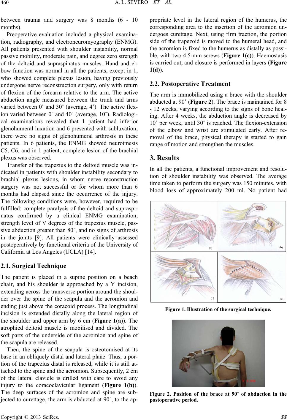 A. L. SEVERO ET AL. 460 between trauma and surgery was 8 months (6 - 10 months). Preoperative evaluation included a physical examina- tion, radiography, and electroneuromyography (ENMG). All patients presented with shoulder instability, normal passive mobility, moderate pain, and degree zero strength of the deltoid and supraspinatus muscles. Hand and el- bow function was normal in all the patients, except in 1, who showed complete plexus lesion, having previously undergone nerve reconstruction surgery, only with return of flexion of the forearm relative to the arm. The active abduction angle measured between the trunk and arms varied between 0˚ and 30˚ (average, 4˚). The active flex- ion varied between 0˚ and 40˚ (average, 10˚). Radiologi- cal examinations revealed that 1 patient had inferior glenohumeral luxation and 6 presented with subluxation; there were no signs of glenohumeral arthrosis in these patients. In 6 patients, the ENMG showed neurotmesis C5, C6, and in 1 patient, complete lesion of the brachial plexus was observed. Transfer of the trapezius to the deltoid muscle was in- dicated in patients with shoulder instability secondary to brachial plexus lesions, in whom nerve reconstruction surgery was not successful or for whom more than 6 months had elapsed since the occurrence of the injury. The following conditions were, however, required to be fulfilled: complete paralysis of the deltoid and supraspi- natus confirmed by a clinical ENMG examination, strength level of V degrees of the trapezius muscle, pas- sive abduction g reater than 80˚, and no signs of arthrosis in the joints [9]. All patients were clinically assessed postoperatively by functional criteria of the University of California at Los Angeles (UCLA) [14]. 2.1. Surgical Technique The patient is placed in a supine position on a beach chair, and his shoulder is approached by a Y incision, extending across the tran sverse portion around the shou l- der over the spine of the scapula and the acromion and ending just above the coracoid process. The longitudinal incision is extended distally along the lateral region of the shoulder and upper arm by 6 cm (Figure 1(a)). The atrophied deltoid muscle is mobilised and divided. The soft parts of the underside of the acromion and spine of the scapula are released. Then, the spine of the scapula is osteotomised at its base in an obliquely distal and lateral plane. Thus, a por- tion of the trapezius distal is released, while it is still at- tached to the spine and the acromion. Subsequently, 2 cm of the lateral clavicle is drilled with care to avoid any injury to the coracoclavicular ligament (Figure 1(b)). The deep surfaces of the acromion and spine are sub- jected to curettage, the arm is abducted at 90˚, to the ap- propriate level in the lateral region of the humerus, the corresponding area to the insertion of the acromion un- dergoes curettage. Next, using firm traction, the portion side of the trapezoid is moved to the humeral head, and the acromion is fixed to the humerus as distally as possi- ble, with two 4.5-mm screws (Figure 1(c)). Haemostasis is carried out, and closure is performed in layers (Figure 1(d)). 2.2. Postoperative Treatment The arm is immobilized using a brace with the shoulder abducted at 90˚ (Figure 2). The brace is maintained for 8 - 12 weeks, varying according to the signs of bone heal- ing. After 4 weeks, the abduction angle is decreased by 10˚ per week, until 30˚ is reached. Th e flexion-exten sion of the elbow and wrist are stimulated early. After re- moval of the brace, physical therapy is started to gain range of motion and strengthen the muscles. 3. Results In all the patients, a functional improvement and resolu- tion of shoulder instability was observed. The average time taken to perfo rm the surgery was 150 minutes, with blood loss of approximately 200 ml. No patient had (a) (b) (c) (d) Figure 1. Illustration of the surgical technique. Figure 2. Position of the brace at 90˚ of abduction in the postoperative period. Copyright © 2013 SciRes. SS 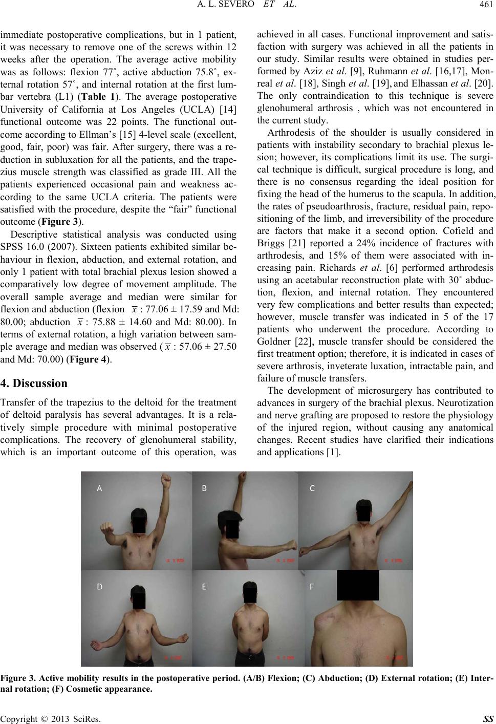 A. L. SEVERO ET AL. Copyright © 2013 SciRes. SS 461 achieved in all cases. Functional improvement and satis- faction with surgery was achieved in all the patients in our study. Similar results were obtained in studies per- formed by Aziz et al. [9], Ruhmann et al. [16,17], Mon- real et al. [18], Singh et al. [19], and Elhassan et al. [20]. The only contraindication to this technique is severe glenohumeral arthrosis , which was not encountered in the current study. immediate postoperative complications, but in 1 patient, it was necessary to remove one of the screws within 12 weeks after the operation. The average active mobility was as follows: flexion 77˚, active abduction 75.8˚, ex- ternal rotation 57˚, and internal rotation at the first lum- bar vertebra (L1) (Table 1). The average postoperative University of California at Los Angeles (UCLA) [14] functional outcome was 22 points. The functional out- come according to Ellman’s [15] 4-level scale (excellent, good, fair, poor) was fair. After surgery, there was a re- duction in subluxation for all the patients, and the trape- zius muscle strength was classified as grade III. All the patients experienced occasional pain and weakness ac- cording to the same UCLA criteria. The patients were satisfied with the procedure, despite the “fair” functional outcome (Figure 3). Arthrodesis of the shoulder is usually considered in patients with instability secondary to brachial plexus le- sion; however, its complications limit its use. The surgi- cal technique is difficult, surgical procedure is long, and there is no consensus regarding the ideal position for fixing the head of the humerus to the scapu la. In addition, the rates of pseudoarthro sis, fracture, residual pain, repo- sitioning of the limb, and irreversibility of the procedure are factors that make it a second option. Cofield and Briggs [21] reported a 24% incidence of fractures with arthrodesis, and 15% of them were associated with in- creasing pain. Richards et al. [6] performed arthrodesis using an acetabular reconstruction plate with 30˚ abduc- tion, flexion, and internal rotation. They encountered very few complications and better results than expected; however, muscle transfer was indicated in 5 of the 17 patients who underwent the procedure. According to Goldner [22], muscle transfer should be considered the first treatment option; therefore, it is indicated in cases of severe arthrosis, inveterate luxatio n, intractable pain, and failure of muscle transfers. Descriptive statistical analysis was conducted using SPSS 16.0 (2007). Sixteen patients exhibited similar be- haviour in flexion, abduction, and external rotation, and only 1 patient with total brachial plexus lesion showed a comparatively low degree of movement amplitude. The overall sample average and median were similar for flexion and abduction (flexion x : 77.06 ± 17.59 and Md: 80.00; abduction x : 75.88 ± 14.60 and Md: 80.00). In terms of external rotation, a high variation between sam- ple average and median was observed ( x : 57.06 ± 27 .50 and Md: 70.00) (Figure 4). 4. Discussion The development of microsurgery has contributed to advances in surgery of the brachial plexus. Neurotization and nerve grafting are proposed to restore the physiology of the injured region, without causing any anatomical changes. Recent studies have clarified their indications and applications [1]. Transfer of the trapezius to the deltoid for the treatment of deltoid paralysis has several advantages. It is a rela- tively simple procedure with minimal postoperative complications. The recovery of glenohumeral stability, which is an important outcome of this operation, was Figure 3. Active mobility results in the postope rative period. (A/B) Flexion; (C) Abduction; (D) External rotation; (E) Inter- al rotation; (F) Cosmetic appearance. n 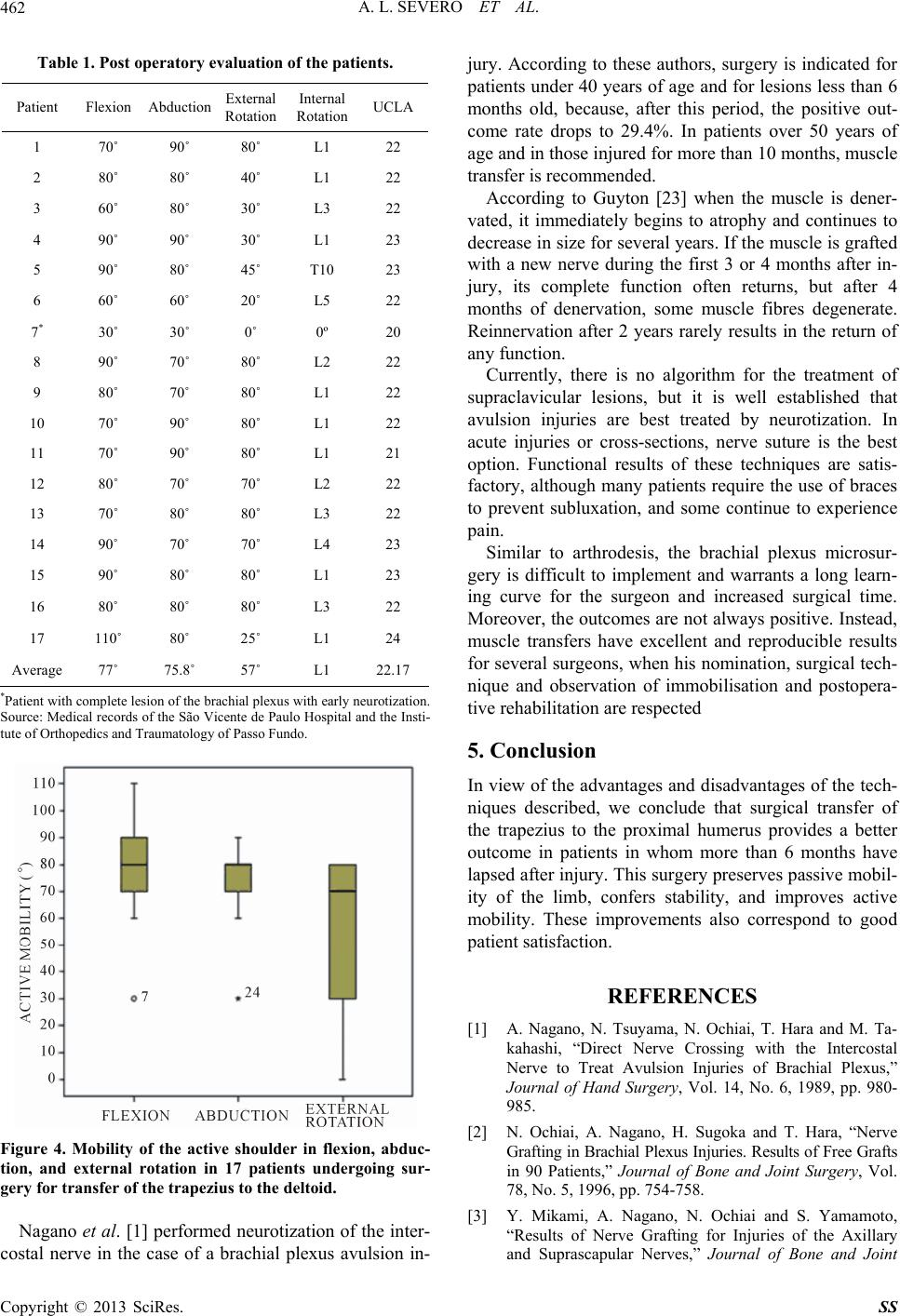 A. L. SEVERO ET AL. 462 Table 1. Post operatory evaluation of the patients. Patient Flexion Abduction External Rotation Internal Rotation UCLA 1 70˚ 90˚ 80˚ L1 22 2 80˚ 80˚ 40˚ L1 22 3 60˚ 80˚ 30˚ L3 22 4 90˚ 90˚ 30˚ L1 23 5 90˚ 80˚ 45˚ T10 23 6 60˚ 60˚ 20˚ L5 22 7* 30˚ 30˚ 0˚ 0º 20 8 90˚ 70˚ 80˚ L2 22 9 80˚ 70˚ 80˚ L1 22 10 70˚ 90˚ 80˚ L1 22 11 70˚ 90˚ 80˚ L1 21 12 80˚ 70˚ 70˚ L2 22 13 70˚ 80˚ 80˚ L3 22 14 90˚ 70˚ 70˚ L4 23 15 90˚ 80˚ 80˚ L1 23 16 80˚ 80˚ 80˚ L3 22 17 110˚ 80˚ 25˚ L1 24 Average 77˚ 75.8˚ 57˚ L1 22.17 *Patient with complete lesion of the brachial plexus with early neurotization. Source: Medical records of the São Vicente de Paulo Hospital and the Insti- tute of Orthopedics a nd Traumatology of Passo Fundo. Figure 4. Mobility of the active shoulder in flexion, abduc- tion, and external rotation in 17 patients undergoing sur- gery for transfer of the trapezius to the deltoid. Nagano et al. [1] performed neurotization of the in ter- jury. According to these authors, surgery is indicated for patients under 40 years of age and for lesions less than 6 months old, because, after this period, the positive out- come rate drops to 29.4%. In patients over 50 years of age and in those injured for more than 10 months, muscle transfer is recommended. According to Guyton [2 costal nerve in the case of a brachial plexus avulsio 3] when the muscle is dener- va here is no algorithm for the treatment of su ilar to arthrodesis, the brachial plexus microsur- ge 5. Conclusion antages and disadvantages of the tech- REFERENCES [1] A. Nagano, N.. Hara and M. Ta- hiai, A. Nagano, H. Sugoka and T. Hara, “Nerve Yamamoto, n in- “Results of Nerve Grafting for Injuries of the Axillary and Suprascapular Nerves,” Journal of Bone and Joint ted, it immediately begins to atrophy and continues to decrease in size for several years. If the muscle is grafted with a new nerve during the first 3 or 4 months after in- jury, its complete function often returns, but after 4 months of denervation, some muscle fibres degenerate. Reinnervation after 2 years rarely results in the return of any function. Currently, t praclavicular lesions, but it is well established that avulsion injuries are best treated by neurotization. In acute injuries or cross-sections, nerve suture is the best option. Functional results of these techniques are satis- factory, although many patients require the use of braces to prevent subluxation, and some continue to experience pain. Sim ry is difficult to implement and warrants a long learn- ing curve for the surgeon and increased surgical time. Moreover, the outcomes are not always positiv e. Instead, muscle transfers have excellent and reproducible results for several surgeons, when his nomination, surgical tech- nique and observation of immobilisation and postopera- tive rehabilitation are respected In view of the adv niques described, we conclude that surgical transfer of the trapezius to the proximal humerus provides a better outcome in patients in whom more than 6 months have lapsed after injury. This surg ery preserves passive mobil- ity of the limb, confers stability, and improves active mobility. These improvements also correspond to good patient satisfaction. Tsuyama, N. Ochiai, T kahashi, “Direct Nerve Crossing with the Intercostal Nerve to Treat Avulsion Injuries of Brachial Plexus,” Journal of Hand Surgery, Vol. 14, No. 6, 1989, pp. 980- 985. [2] N. Oc Grafting in Brachial Plexus Injuries. Results of Free Grafts in 90 Patients,” Journal of Bone and Joint Surgery, Vol. 78, No. 5, 1996, pp. 754-758. [3] Y. Mikami, A. Nagano, N. Ochiai and S. Copyright © 2013 SciRes. SS  A. L. SEVERO ET AL. 463 Surgery, Vol. 79, No. 4, 1997, pp. 527-531. http://dx.doi.org/10.1302/0301-620X.79B4.7481 [4] A. Severo and M. C. Costa, “Reconstrução do Membro Superior pós Trauma—A Importância das Transferências eatment of Adult rodesis Using a Pelvic-Recons- aralysis,” Bone and Joint Surgery Tendíneas,” In: O. Lech, Ed., Fundamentos em Cirurgia do Ombro: Ortopedia e Reabilitação, 2nd Edition, Revin- ter, Rio de Janeiro, 2005, pp. 485-491. [5] J. L. Giuffre, S. Kakar, A. T. Bishop, R. J. Spinner and A. Y. Shin, “Current Concepts of the Tr Brachial Plexus Injuries,” Journal of Hand Surgery, Vol. 35, No. 4, 2010, pp. 678-688. [6] R. R. Richards, J. P. Waddell and A. R. Hudson, “Shoul- der Arthrodesis for the Treatment of Brachial Plexus Palsy,” Clinical Orthopaedics and Related Research, No. 198, 1985, pp. 250-258. [7] R. R. Richard, R. M. P. Sherman, A. R. Hudson and J. P. Waddell, “Shoulder Arth truction Plate: A Report of Eleven Cases,” Journal of Bone and Joint Surgery (American Volume), Vol. 70, No. 3, 1988, pp. 416-421. [8] P. P. Kotwal, R. Mittal and R. Malhotra, “Trapezius Transfer for Deltoid P (British Volume), Vol. 80, No. 1, 1998, pp. 114-116. http://dx.doi.org/10.1302/0301-620X.80B1.8251 [9] W. Aziz, R. M. Singer and T. W. Wolff, “Transfe r of Trapezius for Flail Shoulder after Brachial Plexusthe Injur ca, Suppl. 97, 1967, pp. 5 uis, 1955. ll, Vol. 2, Manole, São Paulo, 2006, pp sfer after Brachial Plexus Palsy: Indica- y,” Journal of Bone and Joint Surgery (British Volume), Vol. 72, No. 4, 1990, pp. 701-704. [10] A. K. Saha, “Surgery of the Paralyzed and Fail Shoulder,” Acta Orthopaedica Scandinavi- 90. [11] J. E. Bateman, “The Shoulder and Environs,” CV Mosby, St Lo [12] S. T. Canale, “Paralytic Disorders,” In: Orthopaedic Surgery Campbe. 1281-1362. [13] O. Rühmann, C. J. Wirth, F. Gosse and S. Schmolke, “Trapezius Tran tions, Difficulties and Complications,” Journal of Bone and Joint Surgery (British Volume), Vol. 80, No. 1, 1998, pp. 109-113. http://dx.doi.org/10.1302/0301-620X.80B1.8195 [14] E. C. Oku, A. P. Andrade , S. P. Stadiniky, et al., “Trans- 6000400003 lation and Cultural Adaptation of the Modified-University of California at Los Angeles Shoulder Rating Scale to Portuguese Language,” Revista Brasileira de Reumatolo- gia, Vol. 46, No. 4, 2006, pp. 246-252. http://dx.doi.org/10.1590/S0482-5004200 n: 016/S0749-8063(87)80061-0 [15] H. Ellman, “Arthroscopc Subacromial Decompressio Analysis of 1-3-Year Results,” Arthroscopy, Vol. 3, No. 3, 1987, pp. 173-181. http://dx.doi.org/10.1 and C. and C. .87B2.14906 [16] F. Atlan, S. Durand, M. Fox, P. Levy, Z. Belkheyar Oberlin, “Functional Outcome of Glenohumeral Fusion in Brachial Plexus Palsy: A Report of 54 Cases,” Journal of Hand Surgery, Vol. 37, No. 4, 2012, pp. 683-687. [17] O. Rühmann, S. Schmolke, M. Bohnsack, J. Carls J. Wirth, “Trapezius Transfer in Brachial Plexus Palsy: Correlation of the Outcome with Muscle Power and Op- erative Technique,” Journal of Bone and Joint Surgery, Vol. 87, No. 2, 2005, pp. 184-190. http://dx.doi.org/10.1302/0301-620X pezius 49-7221-2-2 [18] R. Monreal, L. Paredes, H. Diaz and P. Leon, “Tra Transfer to Treat Fail Shoulder after Brachial Plexus Palsy,” Journal of Brachial Plexus and Peripheral Nerve Injury, Vol. 2, 2007, p. 2. http://dx.doi.org/10.1186/17 pezius Transfer hin, “Trapezius Transfer JS.H.00745 [19] A. K. Singh and D. Karki, “Modified Tra Technique for Restoration of Shoulder Abduction in Bra- chial Plexus Injury,” Indian Journal of Plastic Surgery, Vol. 40, No. 1, 2007, pp. 39-48. [20] B. Elhassan, A. Bishop and A. S to Restore External Rotation in a Patient with a Brachial Plexus Injury,” Journal of Bone & Joint Surgery, Vol. 91, No. 4, 2009, pp. 939-944. http://dx.doi.org/10.2106/JB meral Arthrode- ing of the Partially Paralyzed h Edition, [21] R. H. Cofield and B. T. Briggs, “Glenohu sis: Operative and Longterm Functional Results,” Journal of Bone and Joint Surgery (American Volume), Vol. 61, No. 5, 1979, pp. 668-677. [22] J. L. Goldner, “Strengthen Shoulder Girdle by Multiple Muscle-Tendon Transfers,” Hand Clinics, Vol. 4, No. 2, 1988, pp. 323-336. [23] A. Guyton, “Textbook of Medical Phy siology,” 6t Guanabara, Rio de Janeiro, 1981, pp. 103-116. Copyright © 2013 SciRes. SS |