

Paper Menu >>
Journal Menu >>
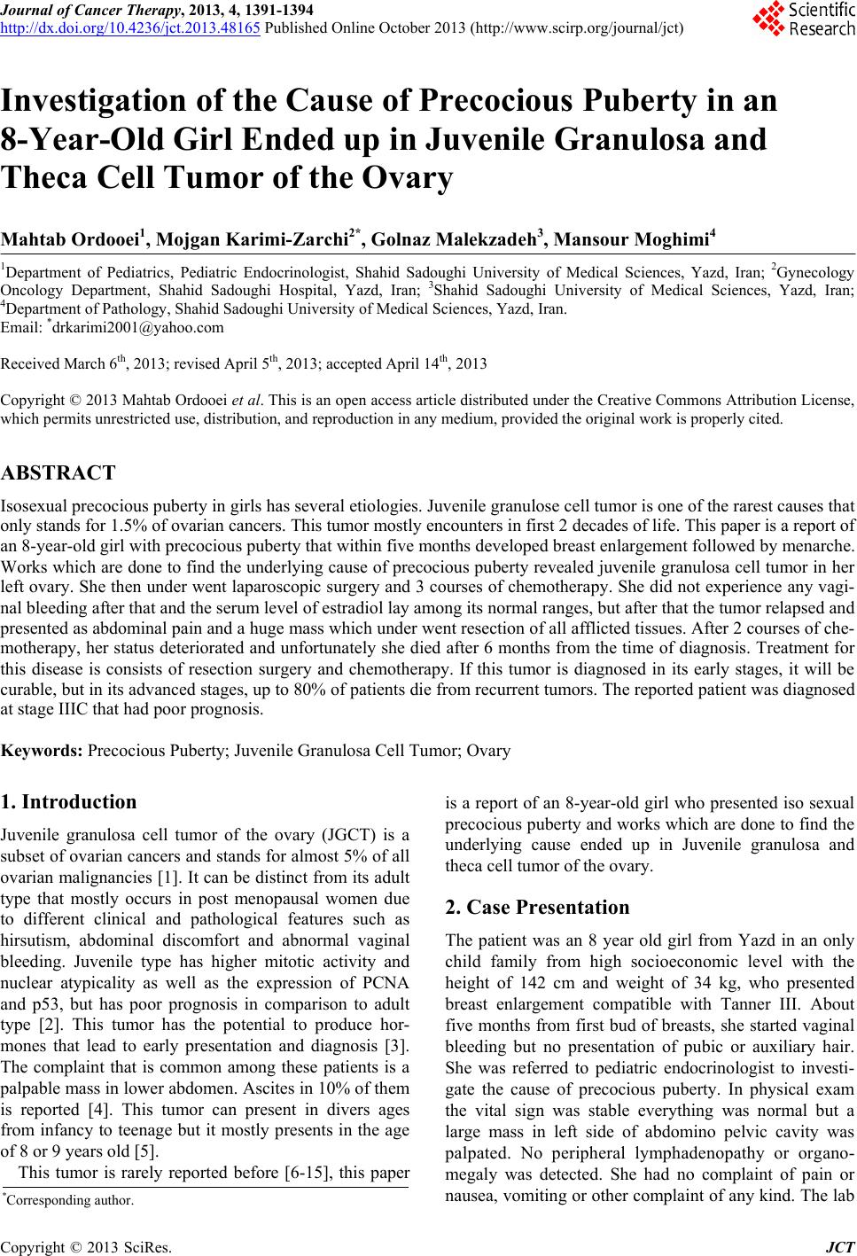 Journal of Cancer Therapy, 2013, 4, 1391-1394 http://dx.doi.org/10.4236/jct.2013.48165 Published Online October 2013 (http://www.scirp.org/journal/jct) 1391 Investigation of the Cause of Precocious Puberty in an 8-Year-Old Girl Ended up in Juvenile Granulosa and Theca Cell Tumor of the Ovary Mahtab Ordooei1, Mojgan Karimi-Zarchi2*, Golnaz Malekzadeh3, Mansour Moghimi4 1Department of Pediatrics, Pediatric Endocrinologist, Shahid Sadoughi University of Medical Sciences, Yazd, Iran; 2Gynecology Oncology Department, Shahid Sadoughi Hospital, Yazd, Iran; 3Shahid Sadoughi University of Medical Sciences, Yazd, Iran; 4Department of Pathology, Shahid Sadoughi University of Medical Sciences, Yazd, Iran. Email: *[email protected] Received March 6th, 2013; revised April 5th, 2013; accepted April 14th, 2013 Copyright © 2013 Mahtab Ordooei et al. This is an open access article distributed under the Creative Commons Attribution License, which permits unrestricted use, distribution, and reproduction in any medium, provided the original work is properly cited. ABSTRACT Isosexual precocious puberty in girls has several etiologies. Juvenile granulose cell tumor is one of the rarest causes that only stands for 1.5% of ovarian cancers. This tumor mostly encounters in first 2 decades of life. This paper is a report of an 8-year-old girl with precocious puberty that within five months developed breast enlargement followed by menarche. Works which are done to find the underlying cause of precocious puberty revealed juvenile granulosa cell tumor in her left ovary. She then under went laparoscopic surgery and 3 courses of chemotherapy. She did not experience any vagi- nal bleeding after that and the serum level of estradiol lay among its normal ranges, but after that the tumor relapsed and presented as abdominal pain and a huge mass which under went resection of all afflicted tissues. After 2 courses of che- motherapy, her status deteriorated and unfortunately she died after 6 months from the time of diagnosis. Treatment for this disease is consists of resection surgery and chemotherapy. If this tumor is diagnosed in its early stages, it will be curable, but in its advanced stages, up to 80% of patients die from recurrent tumors. The reported patient was diagnosed at stage IIIC that had poor prognosis. Keywords: Precocious Puberty; Juvenile Granulosa Cell Tumor; Ovary 1. Introduction Juvenile granulosa cell tumor of the ovary (JGCT) is a subset of ovarian cancers and stands for almost 5% of all ovarian malignancies [1]. It can be distinct from its adult type that mostly occurs in post menopausal women due to different clinical and pathological features such as hirsutism, abdominal discomfort and abnormal vaginal bleeding. Juvenile type has higher mitotic activity and nuclear atypicality as well as the expression of PCNA and p53, but has poor prognosis in comparison to adult type [2]. This tumor has the potential to produce hor- mones that lead to early presentation and diagnosis [3]. The complaint that is common among these patients is a palpable mass in lower abdomen. Ascites in 10% of them is reported [4]. This tumor can present in divers ages from infancy to teenage but it mostly presents in the age of 8 or 9 years old [5]. This tumor is rarely reported before [6-15], this paper is a report of an 8-year-old girl who presented iso sexual precocious puberty and works which are done to find the underlying cause ended up in Juvenile granulosa and theca cell tumor of the ovary. 2. Case Presentation The patient was an 8 year old girl from Yazd in an only child family from high socioeconomic level with the height of 142 cm and weight of 34 kg, who presented breast enlargement compatible with Tanner III. About five months from first bud of breasts, she started vaginal bleeding but no presentation of pubic or auxiliary hair. She was referred to pediatric endocrinologist to investi- gate the cause of precocious puberty. In physical exam the vital sign was stable everything was normal but a large mass in left side of abdomino pelvic cavity was palpated. No peripheral lymphadenopathy or organo- megaly was detected. She had no complaint of pain or nausea, vomiting or other complaint of any kind. The lab *Corresponding author. Copyright © 2013 SciRes. JCT 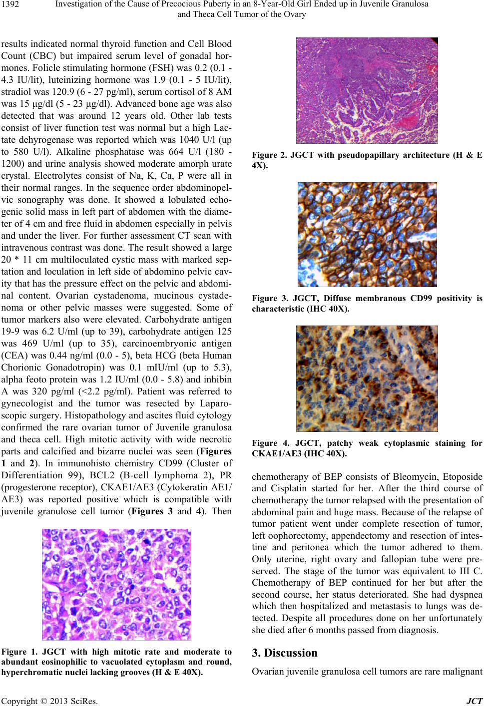 Investigation of the Cause of Precocious Puberty in an 8-Year-Old Girl Ended up in Juvenile Granulosa and Theca Cell Tumor of the Ovary 1392 results indicated normal thyroid function and Cell Blood Count (CBC) but impaired serum level of gonadal hor- mones. Folicle stimulating hormone (FSH) was 0.2 (0.1 - 4.3 IU/lit), luteinizing hormone was 1.9 (0.1 - 5 IU/lit), stradiol was 120.9 (6 - 27 pg/ml), serum cortisol of 8 AM was 15 μg/dl (5 - 23 μg/dl). Advanced bone age was also detected that was around 12 years old. Other lab tests consist of liver function test was normal but a high Lac- tate dehyrogenase was reported which was 1040 U/l (up to 580 U/l). Alkaline phosphatase was 664 U/l (180 - 1200) and urine analysis showed moderate amorph urate crystal. Electrolytes consist of Na, K, Ca, P were all in their normal ranges. In the sequence order abdominopel- vic sonography was done. It showed a lobulated echo- genic solid mass in left part of abdomen with the diame- ter of 4 cm and free fluid in abdomen especially in pelvis and under the liver. For further assessment CT scan with intravenous contrast was done. The result showed a large 20 * 11 cm multiloculated cystic mass with marked sep- tation and loculation in left side of abdomino pelvic cav- ity that has the pressure effect on the pelvic and abdomi- nal content. Ovarian cystadenoma, mucinous cystade- noma or other pelvic masses were suggested. Some of tumor markers also were elevated. Carbohydrate antigen 19-9 was 6.2 U/ml (up to 39), carbohydrate antigen 125 was 469 U/ml (up to 35), carcinoembryonic antigen (CEA) was 0.44 ng/ml (0.0 - 5), beta HCG (beta Human Chorionic Gonadotropin) was 0.1 mIU/ml (up to 5.3), alpha feoto protein was 1.2 IU/ml (0.0 - 5.8) and inhibin A was 320 pg/ml (<2.2 pg/ml). Patient was referred to gynecologist and the tumor was resected by Laparo- scopic surgery. Histopathology and ascites fluid cytology confirmed the rare ovarian tumor of Juvenile granulosa and theca cell. High mitotic activity with wide necrotic parts and calcified and bizarre nuclei was seen (Figures 1 and 2). In immunohisto chemistry CD99 (Cluster of Differentiation 99), BCL2 (B-cell lymphoma 2), PR (progesterone receptor), CKAE1/AE3 (Cytokeratin AE1/ AE3) was reported positive which is compatible with juvenile granulose cell tumor (Figures 3 and 4). Then Figure 1. JGCT with high mitotic rate and moderate to abundant eosinophilic to vacuolated cytoplasm and round, hyperchromatic nuclei lacking grooves (H & E 40X). Figure 2. JGCT with pseudopapillary architecture (H & E 4X). Figure 3. JGCT, Diffuse membranous CD99 positivity is characteristic (IHC 40X). Figure 4. JGCT, patchy weak cytoplasmic staining for CKAE1/AE3 (IHC 40X). chemotherapy of BEP consists of Bleomycin, Etoposide and Cisplatin started for her. After the third course of chemotherapy the tumor relapsed with the presentation of abdominal pain and huge mass. Because of the relapse of tumor patient went under complete resection of tumor, left oophorectomy, appendectomy and resection of intes- tine and peritonea which the tumor adhered to them. Only uterine, right ovary and fallopian tube were pre- served. The stage of the tumor was equivalent to III C. Chemotherapy of BEP continued for her but after the second course, her status deteriorated. She had dyspnea which then hospitalized and metastasis to lungs was de- tected. Despite all procedures done on her unfortunately she died after 6 months passed from diagnosis. 3. Discussion Ovarian juvenile granulosa cell tumors are rare malignant Copyright © 2013 SciRes. JCT 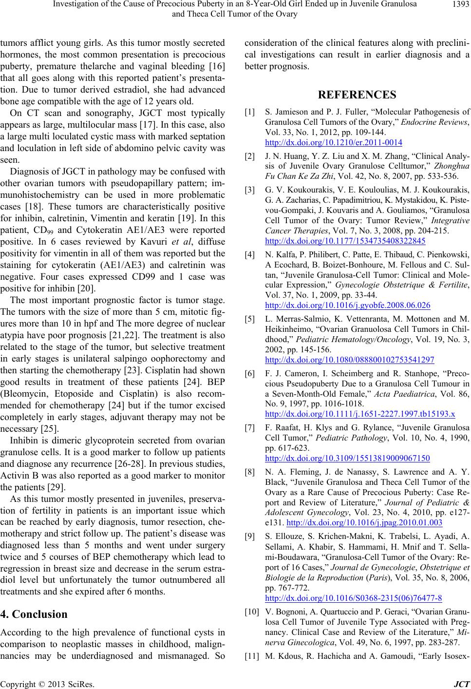 Investigation of the Cause of Precocious Puberty in an 8-Year-Old Girl Ended up in Juvenile Granulosa and Theca Cell Tumor of the Ovary 1393 tumors afflict young girls. As this tumor mostly secreted hormones, the most common presentation is precocious puberty, premature thelarche and vaginal bleeding [16] that all goes along with this reported patient’s presenta- tion. Due to tumor derived estradiol, she had advanced bone age compatible with the age of 12 years old. On CT scan and sonography, JGCT most typically appears as large, multilocular mass [17]. In this case, also a large multi loculated cystic mass with marked septation and loculation in left side of abdomino pelvic cavity was seen. Diagnosis of JGCT in pathology may be confused with other ovarian tumors with pseudopapillary pattern; im- munohistochemistry can be used in more problematic cases [18]. These tumors are characteristically positive for inhibin, calretinin, Vimentin and keratin [19]. In this patient, CD99 and Cytokeratin AE1/AE3 were reported positive. In 6 cases reviewed by Kavuri et al, diffuse positivity for vimentin in all of them was reported but the staining for cytokeratin (AE1/AE3) and calretinin was negative. Four cases expressed CD99 and 1 case was positive for inhibin [20]. The most important prognostic factor is tumor stage. The tumors with the size of more than 5 cm, mitotic fig- ures more than 10 in hpf and The more degree of nuclear atypia have poor prognosis [21,22]. The treatment is also related to the stage of the tumor, but selective treatment in early stages is unilateral salpingo oophorectomy and then starting the chemotherapy [23]. Cisplatin had shown good results in treatment of these patients [24]. BEP (Bleomycin, Etoposide and Cisplatin) is also recom- mended for chemotherapy [24] but if the tumor excised completely in early stages, adjuvant therapy may not be necessary [25]. Inhibin is dimeric glycoprotein secreted from ovarian granulose cells. It is a good marker to follow up patients and diagnose any recurrence [26-28]. In previous studies, Activin B was also reported as a good marker to monitor the patients [29]. As this tumor mostly presented in juveniles, preserva- tion of fertility in patients is an important issue which can be reached by early diagnosis, tumor resection, che- motherapy and strict follow up. The patient’s disease was diagnosed less than 5 months and went under surgery twice and 5 courses of BEP chemotherapy which lead to regression in breast size and decrease in the serum estra- diol level but unfortunately the tumor outnumbered all treatments and she expired after 6 months. 4. Conclusion According to the high prevalence of functional cysts in comparison to neoplastic masses in childhood, malign- nancies may be underdiagnosed and mismanaged. So consideration of the clinical features along with preclini- cal investigations can result in earlier diagnosis and a better prognosis. REFERENCES [1] S. Jamieson and P. J. Fuller, “Molecular Pathogenesis of Granulosa Cell Tumors of the Ovary,” Endocrine Rev i e ws, Vol. 33, No. 1, 2012, pp. 109-144. http://dx.doi.org/10.1210/er.2011-0014 [2] J. N. Huang, Y. Z. Liu and X. M. Zhang, “Clinical Analy- sis of Juvenile Ovary Granulose Celltumor,” Zhonghua Fu Chan Ke Za Zhi, Vol. 42, No. 8, 2007, pp. 533-536. [3] G. V. Koukourakis, V. E. Kouloulias, M. J. Koukourakis, G. A. Zacharias, C. Papadimitriou, K. Mystakidou, K. Piste- vou-Gompaki, J. Kouvaris and A. Gouliamos, “Granulosa Cell Tumor of the Ovary: Tumor Review,” Integrative Cancer Therapies, Vol. 7, No. 3, 2008, pp. 204-215. http://dx.doi.org/10.1177/1534735408322845 [4] N. Kalfa, P. Philibert, C. Patte, E. Thibaud, C. Pienkowski, A Ecochard, B. Boizet-Bonhoure, M. Fellous and C. Sul- tan, “Juvenile Granulosa-Cell Tumor: Clinical and Mole- cular Expression,” Gynecologie Obstetrique & Fertilite, Vol. 37, No. 1, 2009, pp. 33-44. http://dx.doi.org/10.1016/j.gyobfe.2008.06.026 [5] L. Merras-Salmio, K. Vettenranta, M. Mottonen and M. Heikinheimo, “Ovarian Granuolosa Cell Tumors in Chil- dhood,” Pediatric Hematology/Oncology, Vol. 19, No. 3, 2002, pp. 145-156. http://dx.doi.org/10.1080/088800102753541297 [6] F. J. Cameron, I. Scheimberg and R. Stanhope, “Preco- cious Pseudopuberty Due to a Granulosa Cell Tumour in a Seven-Month-Old Female,” Acta Paediatrica, Vol. 86, No. 9, 1997, pp. 1016-1018. http://dx.doi.org/10.1111/j.1651-2227.1997.tb15193.x [7] F. Raafat, H. Klys and G. Rylance, “Juvenile Granulosa Cell Tumor,” Pediatric Pathology, Vol. 10, No. 4, 1990, pp. 617-623. http://dx.doi.org/10.3109/15513819009067150 [8] N. A. Fleming, J. de Nanassy, S. Lawrence and A. Y. Black, “Juvenile Granulosa and Theca Cell Tumor of the Ovary as a Rare Cause of Precocious Puberty: Case Re- port and Review of Literature,” Journal of Pediatric & Adolescent Gynecology, Vol. 23, No. 4, 2010, pp. e127- e131. http://dx.doi.org/10.1016/j.jpag.2010.01.003 [9] S. Ellouze, S. Krichen-Makni, K. Trabelsi, L. Ayadi, A. Sellami, A. Khabir, S. Hammami, H. Mnif and T. Sella- mi-Boudawara, “Granulosa-Cell Tumor of the Ovary: Re- port of 16 Cases,” Journal de Gynecologie, Obstetrique et Bi ologie de la Reproduction (Paris), Vol. 35, No. 8, 2006, pp. 767-772. http://dx.doi.org/10.1016/S0368-2315(06)76477-8 [10] V. Bognoni, A. Quartuccio and P. Geraci, “Ovarian Granu- losa Cell Tumor of Juvenile Type Associated with Preg- nancy. Clinical Case and Review of the Literature,” Mi- nerva Ginecologica, Vol. 49, No. 6, 1997, pp. 283-287. [11] M. Kdous, R. Hachicha and A. Gamoudi, “Early Isosex- Copyright © 2013 SciRes. JCT 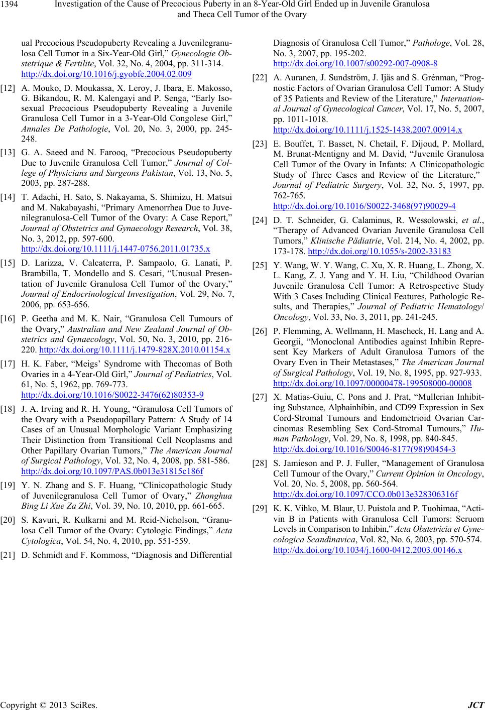 Investigation of the Cause of Precocious Puberty in an 8-Year-Old Girl Ended up in Juvenile Granulosa and Theca Cell Tumor of the Ovary Copyright © 2013 SciRes. JCT 1394 ual Precocious Pseudopuberty Revealing a Juvenilegranu- losa Cell Tumor in a Six-Year-Old Girl,” Gynecologie Ob - stetrique & Fertilite, Vol. 32, No. 4, 2004, pp. 311-314. http://dx.doi.org/10.1016/j.gyobfe.2004.02.009 [12] A. Mouko, D. Moukassa, X. Leroy, J. Ibara, E. Makosso, G. Bikandou, R. M. Kalengayi and P. Senga, “Early Iso- sexual Precocious Pseudopuberty Revealing a Juvenile Granulosa Cell Tumor in a 3-Year-Old Congolese Girl,” Annales De Pathologie, Vol. 20, No. 3, 2000, pp. 245- 248. [13] G. A. Saeed and N. Farooq, “Precocious Pseudopuberty Due to Juvenile Granulosa Cell Tumor,” Journal of Col- lege of Physicians and Surgeons Pakistan, Vol. 13, No. 5, 2003, pp. 287-288. [14] T. Adachi, H. Sato, S. Nakayama, S. Shimizu, H. Matsui and M. Nakabayashi, “Primary Amenorrhea Due to Juve- nilegranulosa-Cell Tumor of the Ovary: A Case Report,” Jou rn al of Ob st etrics an d Gynaecology Research, Vol. 38, No. 3, 2012, pp. 597-600. http://dx.doi.org/10.1111/j.1447-0756.2011.01735.x [15] D. Larizza, V. Calcaterra, P. Sampaolo, G. Lanati, P. Brambilla, T. Mondello and S. Cesari, “Unusual Presen- tation of Juvenile Granulosa Cell Tumor of the Ovary,” Journal of Endocrinological Investigation, Vol. 29, No. 7, 2006, pp. 653-656. [16] P. Geetha and M. K. Nair, “Granulosa Cell Tumours of the Ovary,” Australian and New Zealand Journal of Ob- stetrics and Gynaecology, Vol. 50, No. 3, 2010, pp. 216- 220. http://dx.doi.org/10.1111/j.1479-828X.2010.01154.x [17] H. K. Faber, “Meigs’ Syndrome with Thecomas of Both Ovaries in a 4-Year-Old Girl,” Journal of Pediatrics, Vol. 61, No. 5, 1962, pp. 769-773. http://dx.doi.org/10.1016/S0022-3476(62)80353-9 [18] J. A. Irving and R. H. Young, “Granulosa Cell Tumors of the Ovary with a Pseudopapillary Pattern: A Study of 14 Cases of an Unusual Morphologic Variant Emphasizing Their Distinction from Transitional Cell Neoplasms and Other Papillary Ovarian Tumors,” The American Journal of Surgical Pathology, Vol. 32, No. 4, 2008, pp. 581-586. http://dx.doi.org/10.1097/PAS.0b013e31815c186f [19] Y. N. Zhang and S. F. Huang, “Clinicopathologic Study of Juvenilegranulosa Cell Tumor of Ovary,” Zhonghua Bing Li Xue Za Zhi, Vol. 39, No. 10, 2010, pp. 661-665. [20] S. Kavuri, R. Kulkarni and M. Reid-Nicholson, “Granu- losa Cell Tumor of the Ovary: Cytologic Findings,” Acta Cytologica, Vol. 54, No. 4, 2010, pp. 551-559. [21] D. Schmidt and F. Kommoss, “Diagnosis and Differential Diagnosis of Granulosa Cell Tumor,” Pathologe, Vol. 28, No. 3, 2007, pp. 195-202. http://dx.doi.org/10.1007/s00292-007-0908-8 [22] A. Auranen, J. Sundström, J. Ijäs and S. Grénman, “Prog- nostic Factors of Ovarian Granulosa Cell Tumor: A Study of 35 Patients and Review of the Literature,” Internation- al Jo urnal of Gynecological C ancer, Vol. 17, No. 5, 2007, pp. 1011-1018. http://dx.doi.org/10.1111/j.1525-1438.2007.00914.x [23] E. Bouffet, T. Basset, N. Chetail, F. Dijoud, P. Mollard, M. Brunat-Mentigny and M. David, “Juvenile Granulosa Cell Tumor of the Ovary in Infants: A Clinicopathologic Study of Three Cases and Review of the Literature,” Journal of Pediatric Surgery, Vol. 32, No. 5, 1997, pp. 762-765. http://dx.doi.org/10.1016/S0022-3468(97)90029-4 [24] D. T. Schneider, G. Calaminus, R. Wessolowski, et al., “Therapy of Advanced Ovarian Juvenile Granulosa Cell Tumors,” Klinische Pädiatrie, Vol. 214, No. 4, 2002, pp. 173-178. http://dx.doi.org/10.1055/s-2002-33183 [25] Y. Wang, W. Y. Wang, C. Xu, X. R. Huang, L. Zhong, X. L. Kang, Z. J. Yang and Y. H. Liu, “Childhood Ovarian Juvenile Granulosa Cell Tumor: A Retrospective Study With 3 Cases Including Clinical Features, Pathologic Re- sults, and Therapies,” Journal of Pediatric Hematology/ Oncology, Vol. 33, No. 3, 2011, pp. 241-245. [26] P. Flemming, A. Wellmann, H. Mascheck, H. Lang and A. Georgii, “Monoclonal Antibodies against Inhibin Repre- sent Key Markers of Adult Granulosa Tumors of the Ovary Even in Their Metastases,” The American Journal of Surgical Pathology, Vol. 19, No. 8, 1995, pp. 927-933. http://dx.doi.org/10.1097/00000478-199508000-00008 [27] X. Matias-Guiu, C. Pons and J. Prat, “Mullerian Inhibit- ing Substance, Alphainhibin, and CD99 Expression in Sex Cord-Stromal Tumours and Endometrioid Ovarian Car- cinomas Resembling Sex Cord-Stromal Tumours,” Hu- man Pathology, Vol. 29, No. 8, 1998, pp. 840-845. http://dx.doi.org/10.1016/S0046-8177(98)90454-3 [28] S. Jamieson and P. J. Fuller, “Management of Granulosa Cell Tumour of the Ovary,” Current Opinion in Oncology, Vol. 20, No. 5, 2008, pp. 560-564. http://dx.doi.org/10.1097/CCO.0b013e328306316f [29] K. K. Vihko, M. Blaur, U. Puistola and P. Tuohimaa, “Acti- vin B in Patients with Granulosa Cell Tumors: Seruom Levels in Comparison to Inhibin,” Acta Obstetricia et Gyne- cologica Scandinavica, Vol. 82, No. 6, 2003, pp. 570-574. http://dx.doi.org/10.1034/j.1600-0412.2003.00146.x |