

Paper Menu >>
Journal Menu >>
 Open Journal of Gastroenterology, 2013, 3, 298-302 OJGas http://dx.doi.org/10.4236/ojgas.2013.36051 Published Online October 2013 (http://www.scirp.org/journal/ojgas/) Rectal bleeding in adults over 20 y ears: Endoscopic investigations and results in current hospital practices in Yaoundé, Cameroon Firmin Ankouane Andoulo1, Dominique Noah Noah2*, Roger Djapa1, Michele Tagni Sartre3, Elie Claude Ndjitoyap Ndam4, Kathleen Ngu Blacket1 1CHU Yaounde, Department of Internal Medicine and Specialties, Faculty of Medicine and Biomedical Sciences, University of Yaounde I, Yaounde, Cameroon 2Yaounde Central Hospital, Department of Internal Medicine, Faculty of Medicine and Pharmaceuticals Sciences, University of Douala, Douala, Cameroon 3La Cathédrale Medical Centre, Yaounde, Cameroon 4Yaounde General Hospital, Department of Internal Medicine and Specialties, Faculty of Medicine and Biomedical Sciences, Uni- versity of Yaounde I, Yaounde, Cameroon Email: *[email protected] Received 25 July 2013; revised 29 August 2013; accepted 12 September 2013 Copyright © 2013 Firmin Ankouane Andoulo et al. This is an open access article distributed under the Creative Commons Attribu- tion License, which permits unrestricted use, distribution, and reproduction in any medium, provided the original work is properly cited. ABSTRACT Introduction: In order to identify the modes of inves- tigation and the results of the assessment of rectal bleeding in the Cameroonian adult, we retrospective- ly analysed the records of 287 patients aged above 20 years diagnosed with rectal bleeding with the aim to know the prescription patterns according to age, the diagnostic performance of tests and the results. Me- thodology: Between the 1st of January 2009 and the 30th of June 2010, we examined patients at the Uni- versity Teaching Hospital and the “La Cathédrale” Medical Centre in Yaounde. Age, sex, endoscopic tests and results were evaluated. Results: 287 proto- cols met our selection criteria, sex ratio (M/F) 2.4/1, median age 46 years interquartile range [36 , 55 ]. N o r- mal tests were 57 (19.2%). Main lesions: haemorrhoids (42.4% CI95 36.7 - 48.3), colorectal cancer (10.8% CI95 7.5 - 14.9), anal fissure (8.8% CI95 5.8 - 12.6) and colorectal polyps (8.4% CI95 5.5 - 12.2). The pre- valence of significant lesions (polyps and cancer) re- corded 7% for those under age 40. 20.5% in those were between 40 - 50 years, and 28.9% for those over 50 years. For anoscopies, 44.4% were under 40 years, 39.3% of cases of sigmoidoscopy affected those be- tween 40 - 50 years and colonoscopy affected 54% of those over 50 years. For the diagnostic yield, 26.2% had a significant lesion for flexible sigmoidoscopy and only 16.7% and 1.6% for colonoscopy and anoscopy respectively. Conclusion: The study shows that ano- scopy and sigmoidoscopy are the main initial tests conducted in Cameroon in the assessment of rectal bleeding in adults of less than 50 years and they are quite sufficient. Haemorrhoids and colorectal cancer are the main pathologies. Keywords: Rectal Bleeding; Colon Polyps; Colorectal Cancer; Sigmoidoscopy; Colonoscopy; Cameroon Cancer 1. INTRODUCTION Rectal bleeding accounts for 20% of gastrointestinal bleeding. It is equal to the most common detection mode for lower gastrointestinal bleeding especially in the eld- erly [1]. Several studies have been conducted in order to know what mode of investigation is appropriate depend- ing on the age of the patients, the risk of colorectal can- cer and the clinical presentation mode [2-4]. Even though colonoscopy is unanimously recommended for patients aged 50 and above in the assessment of rectal bleeding [5], there seem to be no consensus guidelines on how to deal with rectal bleeding in young patients under the age of 50 [1,3,6,7]. In sub-Saharan Africa and more specifically Camer- oon, few studies have focused on the topic and there is no consensus [1,8,9]. We sought to know, through a retrospective study, which investigation modes existed in our context and the *Corresponding author. OPEN ACCESS 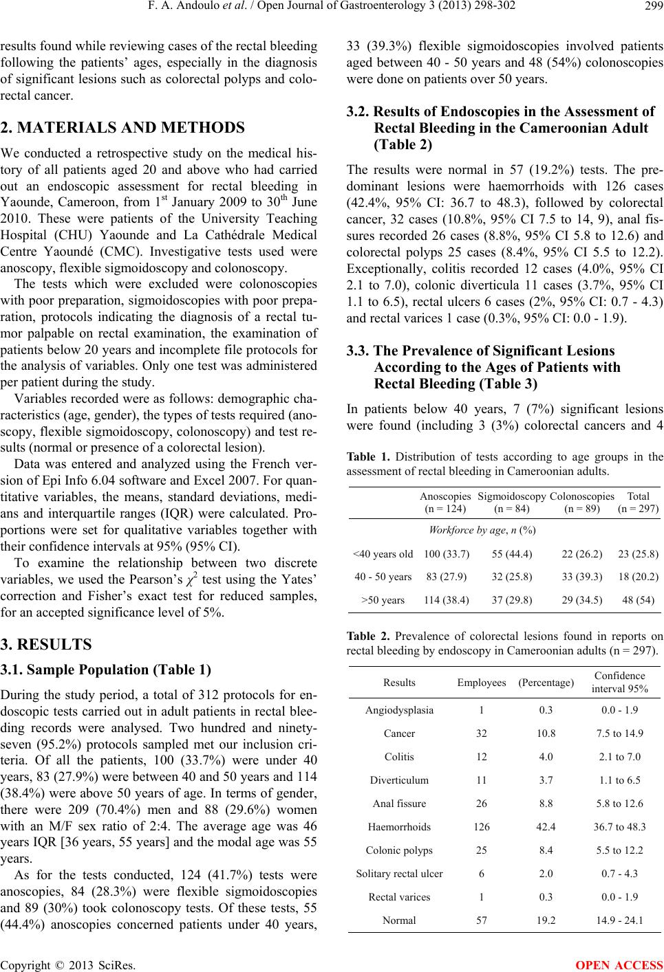 F. A. Andoulo et al. / Open Journal of Gastroenterology 3 (2013) 298-302 299 results found while reviewing cases of the rectal bleeding following the patients’ ages, especially in the diagnosis of significant lesions such as colorectal polyps and colo- rectal cancer. 2. MATERIALS AND METHODS We conducted a retrospective study on the medical his- tory of all patients aged 20 and above who had carried out an endoscopic assessment for rectal bleeding in Yaounde, Cameroon, from 1st January 2009 to 30th June 2010. These were patients of the University Teaching Hospital (CHU) Yaounde and La Cathédrale Medical Centre Yaoundé (CMC). Investigative tests used were anoscopy, flexible sigmoidoscopy and colonoscopy. The tests which were excluded were colonoscopies with poor preparation, sigmoidoscopies with poor prepa- ration, protocols indicating the diagnosis of a rectal tu- mor palpable on rectal examination, the examination of patients below 20 years and incomplete file protocols for the analysis of variables. Only one test was administered per patient during the study. Variables recorded were as follows: demographic cha- racteristics (age, gender), the types of tests required (ano- scopy, flexible sigmoidoscopy, colonoscopy) and test re- sults (normal or presence of a colorectal lesion). Data was entered and analyzed using the French ver- sion of Epi Info 6.04 software and Excel 2007. For quan- titative variables, the means, standard deviations, medi- ans and interquartile ranges (IQR) were calculated. Pro- portions were set for qualitative variables together with their confidence intervals at 95% (95% CI). To examine the relationship between two discrete variables, we used the Pearson’s χ2 test using the Yates’ correction and Fisher’s exact test for reduced samples, for an accepted significance level of 5%. 3. RESULTS 3.1. Sample Population (Table 1) During the study period, a total of 312 protocols for en- doscopic tests carried out in adult patients in rectal blee- ding records were analysed. Two hundred and ninety- seven (95.2%) protocols sampled met our inclusion cri- teria. Of all the patients, 100 (33.7%) were under 40 years, 83 (27.9%) were between 40 and 50 years and 114 (38.4%) were above 50 years of age. In terms of gender, there were 209 (70.4%) men and 88 (29.6%) women with an M/F sex ratio of 2:4. The average age was 46 years IQR [36 years, 55 years] and the modal age was 55 years. As for the tests conducted, 124 (41.7%) tests were anoscopies, 84 (28.3%) were flexible sigmoidoscopies and 89 (30%) took colonoscopy tests. Of these tests, 55 (44.4%) anoscopies concerned patients under 40 years, 33 (39.3%) flexible sigmoidoscopies involved patients aged between 40 - 50 years and 48 (54%) colonoscopies were done on patients over 50 years. 3.2. Results of Endoscopies in the Assessment of Rectal Bleeding in the Cameroonian Adult (Table 2) The results were normal in 57 (19.2%) tests. The pre- dominant lesions were haemorrhoids with 126 cases (42.4%, 95% CI: 36.7 to 48.3), followed by colorectal cancer, 32 cases (10.8%, 95% CI 7.5 to 14, 9), anal fis- sures recorded 26 cases (8.8%, 95% CI 5.8 to 12.6) and colorectal polyps 25 cases (8.4%, 95% CI 5.5 to 12.2). Exceptionally, colitis recorded 12 cases (4.0%, 95% CI 2.1 to 7.0), colonic diverticula 11 cases (3.7%, 95% CI 1.1 to 6.5), rectal ulcers 6 cases (2%, 95% CI: 0.7 - 4.3) and rectal varices 1 case (0.3%, 95% CI: 0.0 - 1.9). 3.3. The Prevalence of Significant Lesions According to the Ages of Patients with Rectal Bleeding (Table 3) In patients below 40 years, 7 (7%) significant lesions were found (including 3 (3%) colorectal cancers and 4 Table 1. Distribution of tests according to age groups in the assessment of rectal bleeding in Cameroonian adults. Anoscopies (n = 124) Sigmoidoscopy (n = 84) Colonoscopies (n = 89) Total (n = 297) Workforce by age, n (%) ˂40 years old100 (33.7)55 (44.4) 22 (26.2) 23 (25.8) 40 - 50 years83 (27.9)32 (25.8) 33 (39.3) 18 (20.2) >50 years 114 (38.4)37 (29.8) 29 (34.5) 48 (54) Table 2. Prevalence of colorectal lesions found in reports on rectal bleeding by endoscopy in Cameroonian adults (n = 297). Results Employees (Percentage) Confidence interval 95% Angiodysplasia 1 0.3 0.0 - 1.9 Cancer 32 10.8 7.5 to 14.9 Colitis 12 4.0 2.1 to 7.0 Diverticulum 11 3.7 1.1 to 6.5 Anal fissure 26 8.8 5.8 to 12.6 Haemorrhoids 126 42.4 36.7 to 48.3 Colonic polyps 25 8.4 5.5 to 12.2 Solitary rectal ulcer6 2.0 0.7 - 4.3 Rectal varices 1 0.3 0.0 - 1.9 Normal 57 19.2 14.9 - 24.1 Copyright © 2013 SciRes. OPEN ACCESS 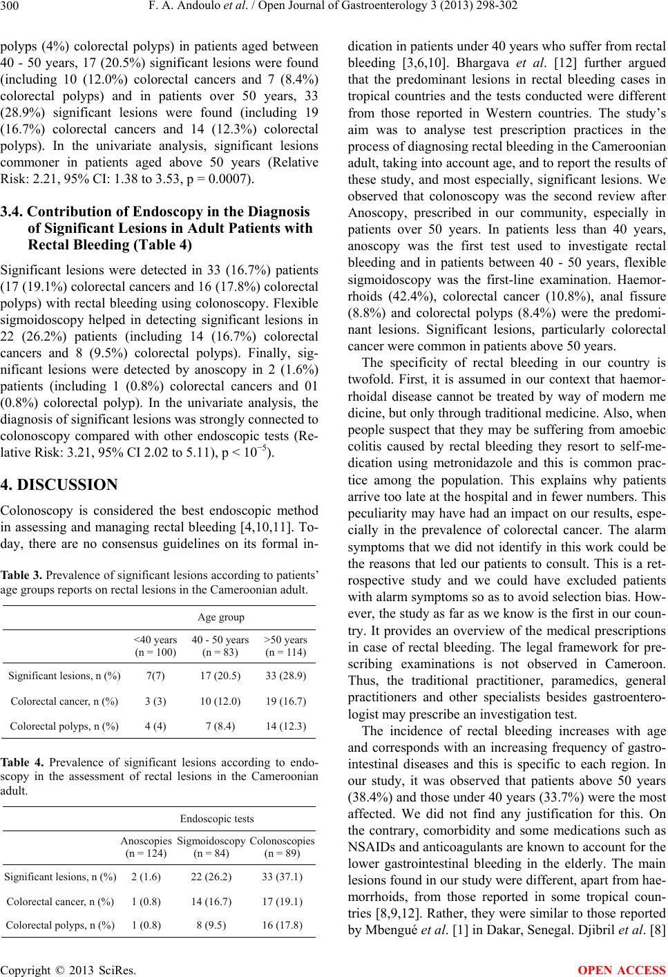 F. A. Andoulo et al. / Open Journal of Gastroenterology 3 (2013) 298-302 300 polyps (4%) colorectal polyps) in patients aged between 40 - 50 years, 17 (20.5%) significant lesions were found (including 10 (12.0%) colorectal cancers and 7 (8.4%) colorectal polyps) and in patients over 50 years, 33 (28.9%) significant lesions were found (including 19 (16.7%) colorectal cancers and 14 (12.3%) colorectal polyps). In the univariate analysis, significant lesions commoner in patients aged above 50 years (Relative Risk: 2.21, 95% CI: 1.38 to 3.53, p = 0.0007). 3.4. Contribution of Endoscopy in the Diagnosis of Significant Lesions in Adult Patients with Rectal Bleeding (Table 4) Significant lesions were detected in 33 (16.7%) patients (17 (19.1%) colorectal cancers and 16 (17.8%) colorectal polyps) with rectal bleeding using colonoscopy. Flexible sigmoidoscopy helped in detecting significant lesions in 22 (26.2%) patients (including 14 (16.7%) colorectal cancers and 8 (9.5%) colorectal polyps). Finally, sig- nificant lesions were detected by anoscopy in 2 (1.6%) patients (including 1 (0.8%) colorectal cancers and 01 (0.8%) colorectal polyp). In the univariate analysis, the diagnosis of significant lesions was strongly connected to colonoscopy compared with other endoscopic tests (Re- lative Risk: 3.21, 95% CI 2.02 to 5.11), p < 10−5). 4. DISCUSSION Colonoscopy is considered the best endoscopic method in assessing and managing rectal bleeding [4,10,11]. To- day, there are no consensus guidelines on its formal in- Table 3. Prevalence of significant lesions according to patients’ age groups reports on rectal lesions in the Cameroonian adult. Age group <40 years (n = 100) 40 - 50 years (n = 83) >50 years (n = 114) Significant lesions, n (%) 7(7) 17 (20.5) 33 (28.9) Colorectal cancer, n (%) 3 (3) 10 (12.0) 19 (16.7) Colorectal polyps, n (%) 4 (4) 7 (8.4) 14 (12.3) Table 4. Prevalence of significant lesions according to endo- scopy in the assessment of rectal lesions in the Cameroonian adult. Endoscopic tests Anoscopies (n = 124) Sigmoidoscopy (n = 84) Colonoscopies (n = 89) Significant lesions, n (%) 2 (1.6) 22 (26.2) 33 (37.1) Colorectal cancer, n (%) 1 (0.8) 14 (16.7) 17 (19.1) Colorectal polyps, n (%) 1 (0.8) 8 (9.5) 16 (17.8) dication in patients under 40 years who suffer from rectal bleeding [3,6,10]. Bhargava et al. [12] further argued that the predominant lesions in rectal bleeding cases in tropical countries and the tests conducted were different from those reported in Western countries. The study’s aim was to analyse test prescription practices in the process of diagnosing rectal bleeding in the Cameroonian adult, taking into account age, and to report the results of these study, and most especially, significant lesions. We observed that colonoscopy was the second review after Anoscopy, prescribed in our community, especially in patients over 50 years. In patients less than 40 years, anoscopy was the first test used to investigate rectal bleeding and in patients between 40 - 50 years, flexible sigmoidoscopy was the first-line examination. Haemor- rhoids (42.4%), colorectal cancer (10.8%), anal fissure (8.8%) and colorectal polyps (8.4%) were the predomi- nant lesions. Significant lesions, particularly colorectal cancer were common in patients above 50 years. The specificity of rectal bleeding in our country is twofold. First, it is assumed in our context that haemor- rhoidal disease cannot be treated by way of modern me dicine, but only through traditional medicine. Also, when people suspect that they may be suffering from amoebic colitis caused by rectal bleeding they resort to self-me- dication using metronidazole and this is common prac- tice among the population. This explains why patients arrive too late at the hospital and in fewer numbers. This peculiarity may have had an impact on our results, espe- cially in the prevalence of colorectal cancer. The alarm symptoms that we did not identify in this work could be the reasons that led our patients to consult. This is a ret- rospective study and we could have excluded patients with alarm symptoms so as to avoid selection bias. How- ever, the study as far as we know is the first in our coun- try. It provides an overview of the medical prescriptions in case of rectal bleeding. The legal framework for pre- scribing examinations is not observed in Cameroon. Thus, the traditional practitioner, paramedics, general practitioners and other specialists besides gastroentero- logist may prescribe an investigation test. The incidence of rectal bleeding increases with age and corresponds with an increasing frequency of gastro- intestinal diseases and this is specific to each region. In our study, it was observed that patients above 50 years (38.4%) and those under 40 years (33.7%) were the most affected. We did not find any justification for this. On the contrary, comorbidity and some medications such as NSAIDs and anticoagulants are known to account for the lower gastrointestinal bleeding in the elderly. The main lesions found in our study were different, apart from hae- morrhoids, from those reported in some tropical coun- tries [8,9,12]. Rather, they were similar to those reported by Mbengué et al. [1] in Dakar, Senegal. Djibril et al. [8] Copyright © 2013 SciRes. OPEN ACCESS  F. A. Andoulo et al. / Open Journal of Gastroenterology 3 (2013) 298-302 301 in Lome, Togo limited their study to subjects aged be- tween 24 - 45 years; individuals aged 50 and above were excluded. These subjects are known to be most affected by colorectal cancer [13] cancer which could explain the low prevalence (5.88%) of colorectal cancer in this series compared to ours. Also, Assi et al. [9] in Abidjan, Côte d’Ivoire, reported a prevalence of colorectal cancer of 5.53% among adult subjects with rectal bleeding using colonoscopy as a diagnostic test. The difference com- pared to our series would be based on the type of diag- nostic tests. It is important to note the high prevalence of colorectal cancer in our study and in that of Mbengué et al. [1] of 10.8% and 11.9% respectively. Changes in our eating habits have been observed in recent years, initially predominantly vegetarian and meat-rich today may partly explain these results. The prevalence of significant lesions in patients 40 - 50 years and in patients over 50 years was high, whereas in patients below 40 years, it was low. These results are consistent with those contained in the literature and es- pecially those in the work of Lewis et al. [6] who re- ported similar results in a series of patients under 50 years with rectal bleeding. During the study and that of Lewis et al. [6] the warning signs which are changing bowel habits, iron deficiency anaemia and weight loss, these signs are known to have a high positive predictive value [14-16] in diagnosing colorectal cancer in a patient with signs of rectal bleeding which were not mentioned. The diverticular bleeding is a common cause of rectal bleeding in the West [17]. In sub-Saharan Africa, diver- ticular disease is a rare disease [18] and diverticular bleeding is rarely described [19]. The 3.7% prevalence rate is therefore in accordance with these arguments. Bleeding from rectal ulcer seemed relatively frequent compared to the literature [20,21]. Lin et al. [21] have identified comorbid conditions associated with this dis- ease including diabetes, bed-rest, renal failure, hypogly- caemia albumin, cerebrovascular accidents, respiratory failure and atherosclerosis. None of these factors was re- ported in our series. Their cause is not mechanical. The diagnostic value for significant lesions, particu- larly colorectal cancer was higher with flexible sigmoi- doscopy (26.2%) for lesions of the distal segment of the colon. This is not a new observation [11,22,23]. We noted that 65.5% of flexible sigmoidoscopy was per- formed in patients less than 50 years. This test is consid- ered sufficient in the assessment of rectal bleeding with- out alarming symptoms in patients less than 50 years by some authors which is due to the preferential localization of significant lesions in the distal colon in this age group on the one hand [2-4,11,24], but also due to a low preva- lence of significant lesions, particularly colorectal cancer in this age group [6,16,25,26]. Marderstein et al. [10] in a study of simple rectal bleeding emphasised that the diagnostic yield of colonoscopy was low in indicating this. The authors added that flexible sigmoidoscopy was sufficient to exclude significant injury if other formal indicators for colonoscopy were not associated. We drew three arguments from the findings in relation to our study: 1) that flexible sigmoidoscopy was sufficient in the as- sessment of our patients under 50 years, 2) that the per- formance of this test was high and 3) that our patients probably had warning signs associated with rectal bleed- ing to explain the high frequency of significant lesions in the age group of those below 50 years (27.5%). Late ar- rival to the hospital as already mentioned is a contribut- ing factor. Colonoscopy in the assessment of rectal bleeding without warning signs has a low yield according to some authors [10,14,23] and is costly for a patient who has no health insurance. In Cameroon, the price of a colono- scopy is three times that of sigmoidoscopy. Colonoscopy was the second examination prescribed in our study (30%), especially in patients over 50 years. It is recom- mended by most published guidelines and EPAGE II criteria (available at http://www.epage.ch) in screening for colorectal cancer in patients at high risk of colorectal cancer [27] and in subjects aged above 50 years. Risk factors for colorectal cancer and alarm symptoms were not identified in this study. However, the contribution of colonoscopy was instrumental in the diagnosis of sig- nificant lesions. Late arrivals at hospitals explain this result. 5. CONCLUSION The absence of a legal framework and a consensus on the follow-up of rectal bleeding on the one hand and the tra- ditional methods of individuals in the face of rectal blee- ding on the other hand are major obstacles in assisting the patient. However, the requirements of investigative tests do not really deviate from what is recommended in several publications in the existing literature. Test results need to be reported taking into account the characteris- tics of rectal bleeding and its associated warning signs. The high cost of testing and the lack of health coverage oblige one to adopt a systematic approach focusing on anoscopy and sigmoidoscopy such as initial tests for young patients with no warning signs and colonoscopy for eld- erly patients or those at the risk of colorectal cancer. REFERENCES [1] Mbengue, M., Dia, D., Diouf, M.L., Bassène, M.L., Fall, S., Diallo, S., et al. (2009) Apport de la coloscopie dans le diagnostic des rectorragies à Dakar (Sénégal). Mé- decine Tropicale, 69, 286-288. [2] Nikpour, S. and Ali Asgari, A. (2008) Colonoscopic eval- uation of minimal rectal bleeding in average-risk patients Copyright © 2013 SciRes. OPEN ACCESS  F. A. Andoulo et al. / Open Journal of Gastroenterology 3 (2013) 298-302 Copyright © 2013 SciRes. 302 OPEN ACCESS for colorectal cancer. World Journal of Gastroenterology, 14, 6536-6540. http://dx.doi.org/10.3748/wjg.14.6536 [3] Khalid, A.B., Majid, S., Salih, M. and Jafri, W. (2011) Is full colonoscopic examination necessary in young pa- tients with fresh bleeding per rectum? Endoscopy, 43, 692-696. http://dx.doi.org/10.1055/s-0030-1256356 [4] Wong, R.F., Khosla, R., Moore, J.H. and Kuwada, S.K. (2004) Consider colonoscopy for young patients with hematochezia. Journal of Family Practice, 53, 879-884. [5] Peytremann-Bridevaux, I., Arditi, C., Froehlich, F., O’Mal- ley, J., Fairclough, P., Le Moine, O., et al. (2009) Appro- priateness of colonoscopy in Europe (EPAGE II). Iron- deficiency anaemia and hematochezia. Endoscopy, 41, 227-233. http://dx.doi.org/10.1055/s-0028-1119644 [6] Lewis, J.D., Shih, C.E. and Blecker, D. (2001) Endo- scopy for hematochezia in patients under 50 years of age. Digestive Diseases and Sciences, 12, 2660-2665. http://dx.doi.org/10.1023/A:1012715110440 [7] Burling, D., East, J.E. and Taylor, S.A. (2007) Investi- gating rectal bleeding. BMJ, 335, 1260-1262. http://dx.doi.org/10.1136/bmj.39379.598090.AD [8] Djibril, A.M., M’ba, K.B., Bagny, A., Kaaga, L., Redah, D. and Agbetra, A. (2009) Etiologies des rectorragies chez l’adulte en milieu Africain: A propos de 85 cas col- ligés en 12 ans au Chu-Campus de Lomé. Mali Médical, 24, 40-42. [9] Assi, C., Lohoues-Kouacou, M.J., Toth’o, A., Traoré, F., Allah-Kouadio, E. and Camara, B.M. (2006) Quelques aspects épidémiologiques des rectorragies chez l’adulte noir Africain. Médecine d’Afrique Noire, 5, 315-319. [10] Marderstein, E.L. and Church, J.M. (2008) Classic outlet rectal bleeding does not require full colonoscopy to ex- clude significant pathology. Diseases of the Colon & Rec- tum, 2, 202-206. http://dx.doi.org/10.1007/s10350-007-9123-1 [11] Choi, H.K., Law, W.L. and Chu, K.W. (2003) The value of flexible sigmoidoscopy for patients with bright red rectal bleeding. Hong Kong Medical Journal, 9, 171-174. [12] Bhargava, D.K., Rai, R.R. and Chopra, P. (1990) Colo- noscopy for investigation of unexplained rectal bleeding in tropical country. Journal of Gastroenterology, 6, 781- 785. [13] Karsten, B., Kim, J., King, J. and Kumar, R.R. (2008) Characteristics of colorectal cancer in young patients at an urban county hospital. American Surgeon, 74, 973- 976. [14] Adelstein, B.A., Macaskill, P., Chan, S.F., Katelaris, P.H. and Irwig, L. (2011) Most bowel cancer symptoms do not indicate colorectal cancer and polyps: A systematic re- view. BMC Gastroenterology, 30, 65. http://dx.doi.org/10.1186/1471-230X-11-65 [15] Astin, M., Griffin, T., Neal, R.D., Rose, P. and Hamilton, W. (2011) The diagnostic value of symptoms for colo- rectal cancer in primary care: A systematic review. Brit- ish Journal of General Practice, 61, 231-243. http://dx.doi.org/10.3399/bjgp11X572427 [16] Robertson, R., Campbell, C., Weller, D.P., Elton, R., Mant, D., Primrose, J., et al. (2006) Predicting colorectal cancer risk in patients with rectal bleeding. British Jour- nal of General Practice, 56, 763-767. [17] Wilkins, T., Baird, C., Pearson, A.N. and Schade, R.R. (2009) Diverticular bleeding. American Family Physician, 80, 977-983. [18] Mbengué, M., Dia, D. and Diouf, M.I. (2009) La diverti- culose colique à Dakar: Aspects épidémiologiques, clini- ques et endoscopiques chez 34 patients. Journal Africain d’Hépato-Gastroentérologie, 3, 112-114. [19] Ankouane Andoulo, F., Tagni-Sartre, M., Dang Babagna, I. and Ndjitoyap Ndam, E.C. (2012) Hémorragie diverti- culaire colique rare chez un noir africain: A propos d’un cas. Journal Africain d’Hépato-Gastroentérologie, 6, 68- 71. [20] Motomura, Y., Akahoshi, K., Matsui, N., Kubokawa, M., Higuchi, N., Oda, M., et al. (2010) Clinical and endo- scopic characteristics of acute hemorrhagic rectal ulcer, and endoscopic hemostatic treatment: A retrospective study of 95 patients. Colorectal Disease, 12, e320-e325. http://dx.doi.org/10.1111/j.1463-1318.2009.02091.x [21] Lin, C.K., Liang, C.C., Chang, H.T., Hung, F.M. and Lee, T.H. (2011) Acute hemorrhagic rectal ulcer: An impor- tant cause of lower gastrointestinal bleeding in the criti- cally ill patients. Digestive Diseases and Sciences, 56, 3631-3637. http://dx.doi.org/10.1007/s10620-011-1759-y [22] Hafeez Bhatt, A.B. and Quraishy, M.S. (2011) Flexible sigmoidoscopic findings in patients with fresh rectal bleeding. Journal of College of Physicians and Surgeons Pakistan, 21, 577-578. [23] Thompson, M.R., Flashman, K.G., Wooldrage, K., Rogers, P.A., Senapati, A., O’Leary, D.P., et al. (2008) Flexible sigmoidoscopy and whole colonic imaging in the diagno- sis of cancer in patients with colorectal symptoms. British Journal of Surgery, 95, 1140-1146. http://dx.doi.org/10.1002/bjs.6234 [24] Kumar, R.R., King, J., Holt, A., Huynh, R., Mittal, R., Deen, R., et al. (2007) Prevalence of left-sided colorectal cancer and benefit of flexible sigmoidoscopy: A county hospital experience. American Surgeon, 73, 994-997. [25] Zbuk, K., Sidebotham, E.L., Bleyer, A. and La Quaglia, M.P. (2009) Colorectal cancer in young adults. Seminars in Oncology, 36, 439-450. http://dx.doi.org/10.1053/j.seminoncol.2009.07.008 [26] Mathew, J., Shankar, P. and Aldean, I.M. (2004) Audit on flexible sigmoidoscopy for rectal bleeding in a district general hospital: Are we over-loading the resources? Postgraduate Medical Journal, 80, 38-40. http://dx.doi.org/10.1136/pmj.2003.008284 [27] Arditi, C., Peytremann-Bridevaux, I., Burnand, B., Ec- kardt, V.F., Bytzer, P., Agréus, L., et al. (2009) Appro- priateness of colonoscopy in Europe (EPAGE II). Scr- eening for colorectal cancer. Endoscopy, 41, 200-208. http://dx.doi.org/10.1055/s-0028-1119626 |