

Paper Menu >>
Journal Menu >>
 International Journal of Medical Physics, Clinical Engineering and Radiation Oncology, 2013, 2, 111-116 Published Online November 2013 (http://www.scirp.org/journal/ijmpcero) http://dx.doi.org/10.4236/ijmpcero.2013.24015 Open Access IJMPCERO Dosimetric Impact of Inter-Fraction Variation in Interstitial HDR Brachytherapy Saravanan Kandasam y1*, K. S. Reddy1, Vivekanandan Nagarajan2, Parthasarathy Vedasoundaram1, Gunaseelan Karunanidhi1 1Department of Radiotherapy, Regional Cancer Centre, Jawaharlal Institute of Postgraduate Medical Education and Research (JIPMER), Puducherry, India 2Medical Physics Department, Cancer Institute (WIA), Chennai, India Email: *[email protected] Received August 9, 2013; revised September 10, 2013; accepted October 1, 2013 Copyright © 2013 Saravanan Kandasamy et al. This is an open access article distributed under the Creative Commons Attribution License, which permits unrestricted use, distribution, and reproduction in any medium, provided the original work is properly cited. ABSTRACT Background: Patient setup errors in External Beam Radiotherapy (EBRT) are minimized to a great extent, due to recent technological developments, but in contrary brachytherapy received less attention in inter-fraction catheter movement and its impact in dose delivery. This article deals with inter-fraction interstitial catheter movement and its impact in dose delivery to the target. An attempt is made to study the dosimetric impact of this variation. Objectives: The objec- tive of the study is to evaluate the inter-fraction variation in the position of implanted interstitial applicators and to as- sess the dosimetric impact in interstitial High Dose Rate (HDR) brachytherapy. Materials and Methods: 55 patients treated for carcinoma tongue, breast, buccal mucosa, cervix, floor of the mouth and soft tissue sarcoma over a period of 2 years (December 2011-May 2013) were considered. All the patients underwent CT scan the next day of the implant- ing and 3D planning was done either by Eclipse or Oncentra Master Plan Treatment Planning System (TPS). Patients were treated by HDR brachytherapy remote after-loading units, either by Gamma Med iX plus or Microselectron. At the end of the last fraction, CT scan was repeated and re-planning done. The variation in the position of the implanted ap- plicators/catheters and its impact on dosimetric parameters was evaluated and analyzed. Results: The range of posi- tional displacement of the interstitial catheters ranges from 4.5 mm to 6.8 mm. The maximum variation in prescribed dose to D90 of Clinical Target Volume was 10.88%. Conclusions: If the total duration of the interstitial implant of HDR brachytherapy extends for more than a week from the day of imaging, it is recommended to do CT imaging and re-plan again. It is mandatory to suture the buttons of the implanting to the skin. Edema and movement of organs (e.g., tongue) are the main cause for the positional variation of the catheters. Keywords: HDR; Brachytherapy; Interstitial Implant; Inter-Fraction Variation; Dosimetric Variation 1. Introduction The worldwide incidence of squamous cell carcinoma of head and neck is more than 500,000 cases per year and the management of patients with head and neck cancer is complex [1]. The choice of treatment modality depends on the stage and site of the disease. Brachytherapy plays an integral role in the management of head and neck can- cers and has been described as the first form of confor- mal radiation [2]. Precise source placement enables very high doses within the tumor and sufficient dose at the margin between the tumor and normal tissue ensuring high tumor control. At the same time, only small vol- umes of normal tissue are irradiated thus decreasing the normal tissue complications. Brachytherapy involves the implantation of plastic catheters (applicators) into tumors. In 1904, Wickham and Derais used sharpened goose quills to perform intra- tumoral implantations [3]. Abbe and Morton have re- ported anecdotal reports of cure for cancers in the head and neck with brachytherapy [4,5]. In 1909, Minet de- scribed the first use of a radium tube placed in a catheter to treat prostate cancer [6]. Traditionally treatment plan- ning of brachytherapy was mainly based on radiographs and point dosimetry [6]. The dose distribution was re- lated to the geometry of the catheters. With the newer three dimensional treatment planning together with CT *Corresponding author. 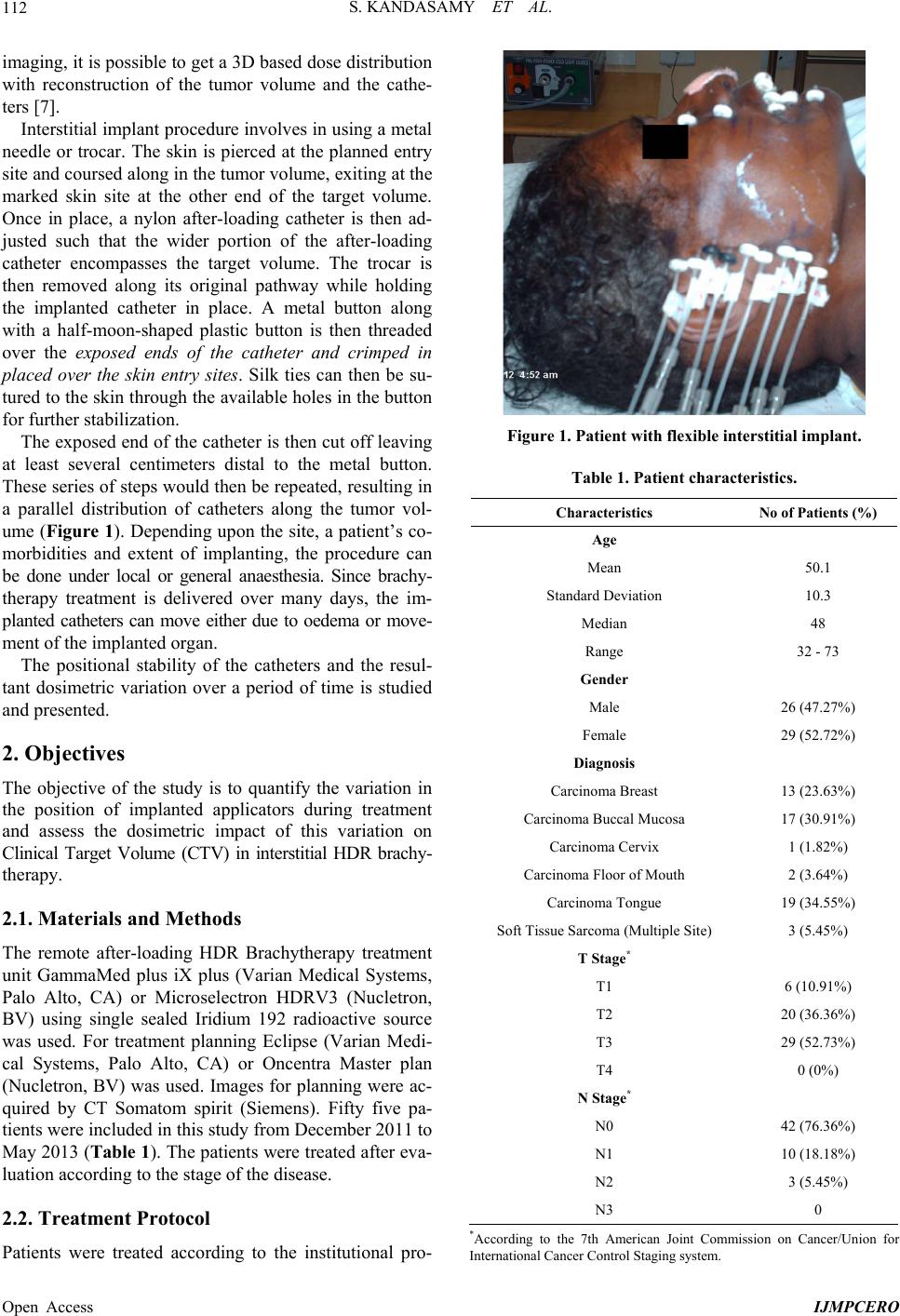 S. KANDASAMY ET AL. Open Access IJMPCERO 112 imaging, it is possible to get a 3D based dose distribution with reconstruction of the tumor volume and the cathe- ters [7]. Interstitial implant procedure involves in using a metal needle or trocar. The skin is pierced at the planned entry site and coursed along in the tumor volume, exiting at the marked skin site at the other end of the target volume. Once in place, a nylon after-loading catheter is then ad- justed such that the wider portion of the after-loading catheter encompasses the target volume. The trocar is then removed along its original pathway while holding the implanted catheter in place. A metal button along with a half-moon-shaped plastic button is then threaded over the exposed ends of the catheter and crimped in placed over the skin entry sites. Silk ties can then be su- tured to the skin through the available holes in the button for further stabilization. The exposed end of the catheter is then cut off leaving at least several centimeters distal to the metal button. These series of steps would then be repeated, resulting in a parallel distribution of catheters along the tumor vol- ume (Figure 1). Depending upon the site, a patient’s co- morbidities and extent of implanting, the procedure can be done under local or general anaesthesia. Since brachy- therapy treatment is delivered over many days, the im- planted catheters can move either due to oedema or move- ment of the implanted organ. The positional stability of the catheters and the resul- tant dosimetric variation over a period of time is studied and presented. 2. Objectives The objective of the study is to quantify the variation in the position of implanted applicators during treatment and assess the dosimetric impact of this variation on Clinical Target Volume (CTV) in interstitial HDR brachy- therapy. 2.1. Materials and Methods The remote after-loading HDR Brachytherapy treatment unit GammaMed plus iX plus (Varian Medical Systems, Palo Alto, CA) or Microselectron HDRV3 (Nucletron, BV) using single sealed Iridium 192 radioactive source was used. For treatment planning Eclipse (Varian Medi- cal Systems, Palo Alto, CA) or Oncentra Master plan (Nucletron, BV) was used. Images for planning were ac- quired by CT Somatom spirit (Siemens). Fifty five pa- tients were included in this study from December 2011 to May 2013 (Tab le 1 ). The patients were treated after eva- luation according to the stage of the disease. 2.2. Treatment Protocol Patients were treated according to the institutional pro- Figure 1. Patient with flexible interstitial implant. Table 1. Patient characteristics. Characteristics No of Patients (%) Age Mean 50.1 Standard Deviation 10.3 Median 48 Range 32 - 73 Gender Male 26 (47.27%) Female 29 (52.72%) Diagnosis Carcinoma Breast 13 (23.63%) Carcinoma Buccal Mucosa 17 (30.91%) Carcinoma Cervix 1 (1.82%) Carcinoma Floor of Mouth 2 (3.64%) Carcinoma Tongue 19 (34.55%) Soft Tissue Sarcoma (Multiple Site) 3 (5.45%) T Stage* T1 6 (10.91%) T2 20 (36.36%) T3 29 (52.73%) T4 0 (0%) N Stage* N0 42 (76.36%) N1 10 (18.18%) N2 3 (5.45%) N3 0 *According to the 7th American Joint Commission on Cancer/Union for International Cancer Control Staging system. 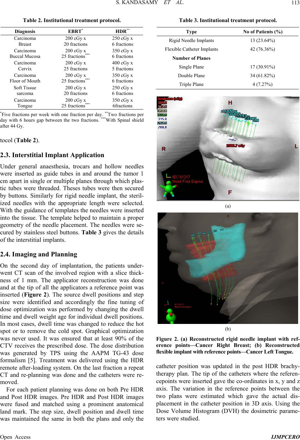 S. KANDASAMY ET AL. Open Access IJMPCERO 113 Table 2. Institutional treatment protocol. Diagnosis EBRT* HDR** Carcinoma Breast 200 cGy x 20 fractions 250 cGy x 6 fractions Carcinoma Buccal Mucosa 200 cGy x 25 fractions*** 350 cGy x 6 fractions Carcinoma Cervix 200 cGy x 25 fractions 400 cGy x 5 fractions Carcinoma Floor of Mouth 200 cGy x 25 fractions*** 350 cGy x 6 fractions Soft Tissue sarcoma 200 cGy x 20 fractions 250 cGy x 6 fractions Carcinoma Tongue 200 cGy x 25 fractions*** 350 cGy x 6fractions *Five fractions per week with one fraction per day. **Two fractions per day with 6 hours gap between the two fractions. ***With Spinal shield after 44 Gy. tocol (Table 2). 2.3. Interstitial Implant Application Under general anaesthesia, trocars and hollow needles were inserted as guide tubes in and around the tumor 1 cm apart in single or multiple planes through which plas- tic tubes were threaded. Theses tubes were then secured by buttons. Similarly for rigid needle implant, the steril- ized needles with the appropriate length were selected. With the guidance of templates the needles were inserted into the tissue. The template helped to maintain a proper geometry of the needle placement. The needles were se- cured by stainless steel buttons. Table 3 gives the details of the interstitial implants. 2.4. Imaging and Planning On the second day of implantation, the patients under- went CT scan of the involved region with a slice thick- ness of 1 mm. The applicator reconstruction was done and at the tip of all the applicators a reference point was inserted (Figure 2). The source dwell positions and step size were identified and accordingly the fine tuning of dose optimization was performed by changing the dwell time and dwell weight age for individual dwell positions. In most cases, dwell time was changed to reduce the hot spot or to remove the cold spot. Graphical optimization was never used. It was ensured that at least 90% of the CTV receives the prescribed dose. The dose distribution was generated by TPS using the AAPM TG-43 dose formalism [5]. Treatment was delivered using the HDR remote after-loading system. On the last fraction a repeat CT and re-planning was done and the catheters were re- moved. For each patient planning was done on both Pre HDR and Post HDR images. Pre HDR and Post HDR images were fused and matched using a prominent anatomical land mark. The step size, dwell position and dwell time was maintained the same in both the plans and only the Table 3. Institutional treatment protocol. Type No of Patients (%) Rigid Needle Implants 13 (23.64%) Flexible Catheter Implants 42 (76.36%) Number of Planes Single Plane 17 (30.91%) Double Plane 34 (61.82%) Triple Plane 4 (7.27%) (a) (b) Figure 2. (a) Reconstructed rigid needle implant with ref- erence points—Cancer Right Breast; (b) Reconstructed flexible implant with reference points—Cancer Left Tongue. catheter position was updated in the post HDR brachy- therapy plan. The tip of the catheters where the referen- cepoints were inserted gave the co-ordinates in x, y and z axis. The variation in the reference points between the two plans were estimated which gave the actual dis- placement in the catheter position in 3D axis. Using the Dose Volume Histogram (DVH) the dosimetric parame- ters were studied. 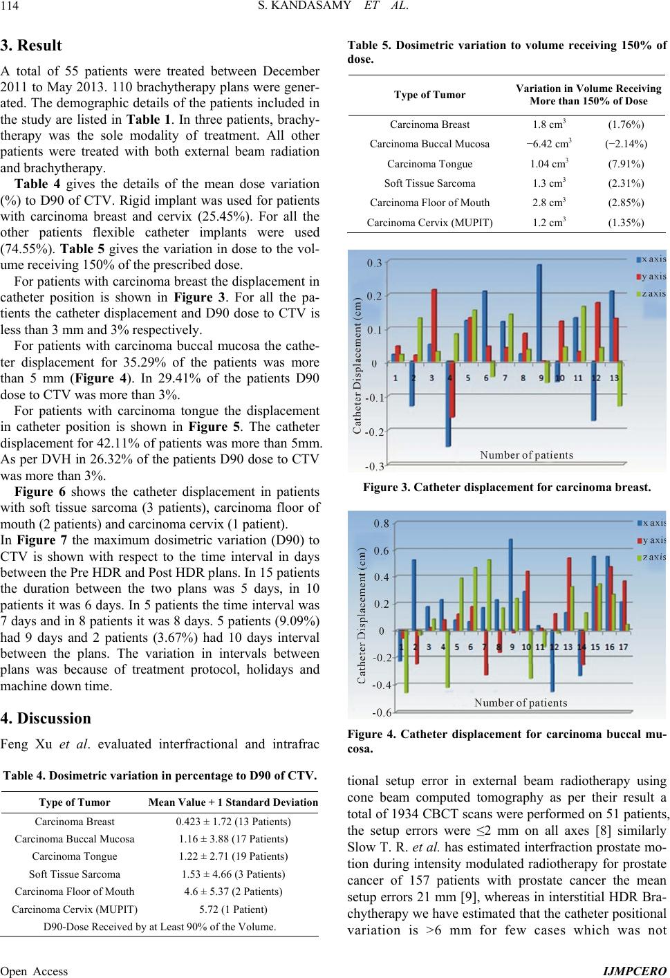 S. KANDASAMY ET AL. Open Access IJMPCERO 114 3. Result A total of 55 patients were treated between December 2011 to May 2013. 110 brachytherapy plans were gener- ated. The demographic details of the patients included in the study are listed in Table 1. In three patients, brachy- therapy was the sole modality of treatment. All other patients were treated with both external beam radiation and brachytherapy. Table 4 gives the details of the mean dose variation (%) to D90 of CTV. Rigid implant was used for patients with carcinoma breast and cervix (25.45%). For all the other patients flexible catheter implants were used (74.55%). Table 5 gives the variation in dose to the vol- ume receiving 150% of the prescribed dose. For patients with carcinoma breast the displacement in catheter position is shown in Figure 3. For all the pa- tients the catheter displacement and D90 dose to CTV is less than 3 mm and 3% respectively. For patients with carcinoma buccal mucosa the cathe- ter displacement for 35.29% of the patients was more than 5 mm (Figure 4). In 29.41% of the patients D90 dose to CTV was more than 3%. For patients with carcinoma tongue the displacement in catheter position is shown in Figure 5. The catheter displacement for 42.11% of patients was more than 5mm. As per DVH in 26.32% of the patients D90 dose to CTV was more than 3%. Figure 6 shows the catheter displacement in patients with soft tissue sarcoma (3 patients), carcinoma floor of mouth (2 patients) and carcinoma cervix (1 patient). In Figure 7 the maximum dosimetric variation (D90) to CTV is shown with respect to the time interval in days between the Pre HDR and Post HDR plans. In 15 patients the duration between the two plans was 5 days, in 10 patients it was 6 days. In 5 patients the time interval was 7 days and in 8 patients it was 8 days. 5 patients (9.09%) had 9 days and 2 patients (3.67%) had 10 days interval between the plans. The variation in intervals between plans was because of treatment protocol, holidays and machine down time. 4. Discussion Feng Xu et al. evaluated interfractional and intrafrac Table 4. Dosimetric variation in percentage to D90 of CTV. Type of Tumor Mean Value + 1 Stand ard Deviation Carcinoma Breast 0.423 ± 1.72 (13 Patients) Carcinoma Buccal Mucosa 1.16 ± 3.88 (17 Patients) Carcinoma Tongue 1.22 ± 2.71 (19 Patients) Soft Tissue Sarcoma 1.53 ± 4.66 (3 Patients) Carcinoma Floor of Mouth 4.6 ± 5.37 (2 Patients) Carcinoma Cervix (MUPIT) 5.72 (1 Patient) D90-Dose Received by at Least 90% of the Volume. Table 5. Dosimetric variation to volume receiving 150% of dose. Type of Tumor Variation in Volume Receiving More than 150% of Dose Carcinoma Breast 1.8 cm3 (1.76%) Carcinoma Buccal Mucosa −6.42 cm3 (−2.14%) Carcinoma Tongue 1.04 cm3 (7.91%) Soft Tissue Sarcoma 1.3 cm3 (2.31%) Carcinoma Floor of Mouth 2.8 cm3 (2.85%) Carcinoma Cervix (MUPIT) 1.2 cm3 (1.35%) Figure 3. Catheter displacement for carcinoma breast. Figure 4. Catheter displacement for carcinoma buccal mu- cosa. tional setup error in external beam radiotherapy using cone beam computed tomography as per their result a total of 1934 CBCT scans were performed on 51 patients, the setup errors were ≤2 mm on all axes [8] similarly Slow T. R. et al. has estimated interfraction prostate mo- tion during intensity modulated radiotherapy for prostate cancer of 157 patients with prostate cancer the mean setup errors 21 mm [9], whereas in interstitial HDR Bra- chytherapy we have estimated that the catheter positional variation is >6 mm for few cases which was not 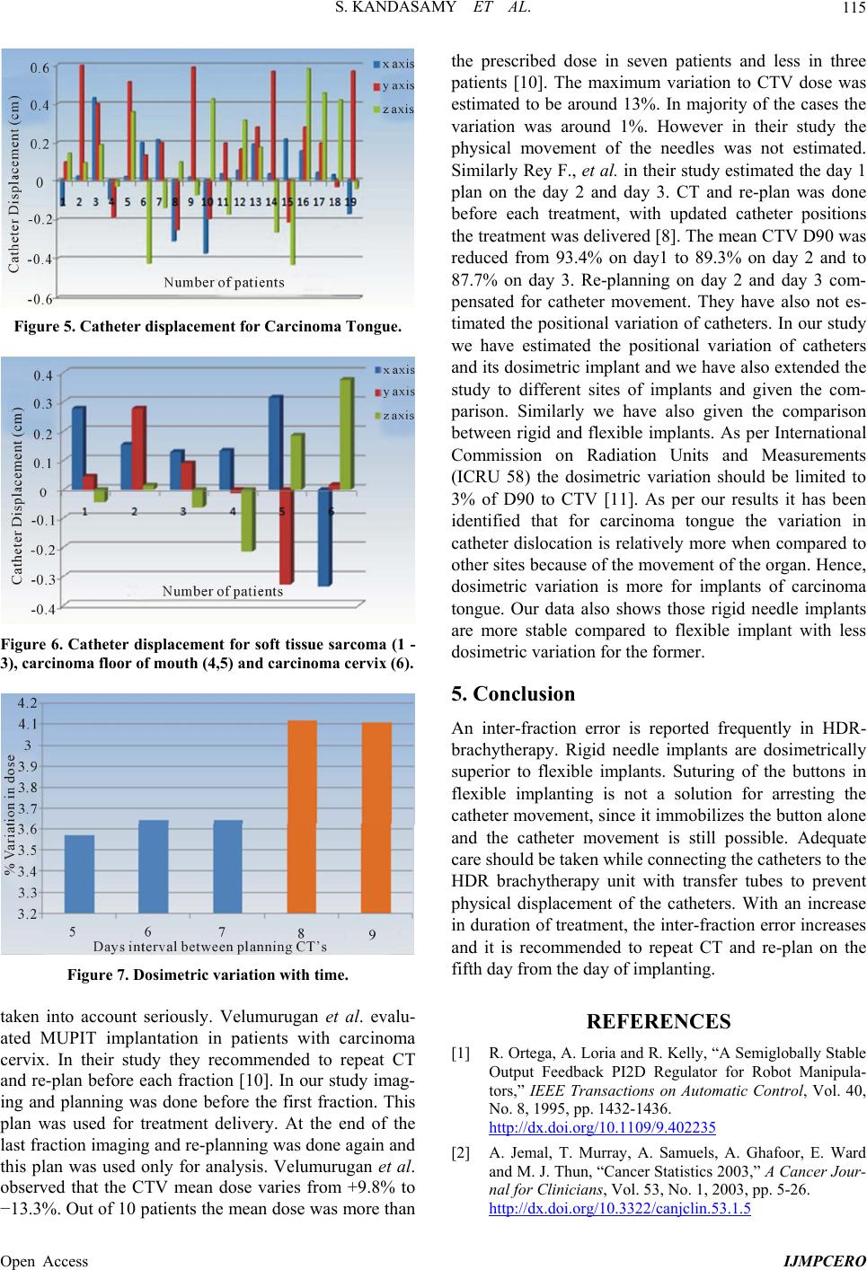 S. KANDASAMY ET AL. Open Access IJMPCERO 115 Figure 5. Catheter displacement for Carcinoma Tongue. Figure 6. Catheter displacement for soft tissue sarcoma (1 - 3), carcinoma floor of mouth (4,5) and carcinoma cervix (6). Figure 7. Dosimetric variation with time. taken into account seriously. Velumurugan et al. evalu- ated MUPIT implantation in patients with carcinoma cervix. In their study they recommended to repeat CT and re-plan before each fraction [10]. In our study imag- ing and planning was done before the first fraction. This plan was used for treatment delivery. At the end of the last fraction imaging and re-planning was done again and this plan was used only for analysis. Velumurugan et al. observed that the CTV mean dose varies from +9.8% to −13.3%. Out of 10 patients the mean dose was more than the prescribed dose in seven patients and less in three patients [10]. The maximum variation to CTV dose was estimated to be around 13%. In majority of the cases the variation was around 1%. However in their study the physical movement of the needles was not estimated. Similarly Rey F., et al. in their study estimated the day 1 plan on the day 2 and day 3. CT and re-plan was done before each treatment, with updated catheter positions the treatment was delivered [8]. The mean CTV D90 was reduced from 93.4% on day1 to 89.3% on day 2 and to 87.7% on day 3. Re-planning on day 2 and day 3 com- pensated for catheter movement. They have also not es- timated the positional variation of catheters. In our study we have estimated the positional variation of catheters and its dosimetric implant and we have also extended the study to different sites of implants and given the com- parison. Similarly we have also given the comparison between rigid and flexible implants. As per International Commission on Radiation Units and Measurements (ICRU 58) the dosimetric variation should be limited to 3% of D90 to CTV [11]. As per our results it has been identified that for carcinoma tongue the variation in catheter dislocation is relatively more when compared to other sites because of the movement of the organ. Hence, dosimetric variation is more for implants of carcinoma tongue. Our data also shows those rigid needle implants are more stable compared to flexible implant with less dosimetric variation for the former. 5. Conclusion An inter-fraction error is reported frequently in HDR- brachytherapy. Rigid needle implants are dosimetrically superior to flexible implants. Suturing of the buttons in flexible implanting is not a solution for arresting the catheter movement, since it immobilizes the button alone and the catheter movement is still possible. Adequate care should be taken while connecting the catheters to the HDR brachytherapy unit with transfer tubes to prevent physical displacement of the catheters. With an increase in duration of treatment, the inter-fraction error increases and it is recommended to repeat CT and re-plan on the fifth day from the day of implanting. REFERENCES [1] R. Ortega, A. Loria and R. Kelly, “A Semiglobally Stable Output Feedback PI2D Regulator for Robot Manipula- tors,” IEEE Transactions on Automatic Control, Vol. 40, No. 8, 1995, pp. 1432-1436. http://dx.doi.org/10.1109/9.402235 [2] A. Jemal, T. Murray, A. Samuels, A. Ghafoor, E. Ward and M. J. Thun, “Cancer Statistics 2003,” A Cancer Jour- nal for Clinicians, Vol. 53, No. 1, 2003, pp. 5-26. http://dx.doi.org/10.3322/canjclin.53.1.5  S. KANDASAMY ET AL. Open Access IJMPCERO 116 [3] R. G. Dale and B. Jones, “The Clinical Radiobiology of Brachytherapy,” British Journal of Radiology, Vol. 71, No. 845, 1998, pp. 465-483. [4] C. A. Joslin, A. Flynn and E. J. Hall, “Principles and Practice of Brachytherapy Using Afterloading Systems,” Arnold, London, 2001. [5] E. K Salminen, K. Kiel, G. S. Ibbott, M. C. Joiner, E. Rosenblatt, E. Zubizarreta, et al., International Confer- ence on Advances in Radiation Oncology (ICARO): Out- comes of an IAEA Meeting, Vienna, 27-29 April 2009. http://dx.doi.org/10.1186/1748-717X-6-11 [6] R. Nath, L. L. Anderson, G. Luxton, K. A. Weaver, J. F. Williamson and A. S. Meigooni, “Dosimetry of Intersti- tial Brachytherapy Sources: Recommendations of the AAPM Radiation Therapy Committee Task Group No. 43. American Association of Physicists in Medicine,” Medi- cal Physics, Vol. 22, No. 2, 1995, pp. 209-234. http://dx.doi.org/10.1118/1.597458 [7] F. Rey, C. Chang, C. Mesina, N. Dixit, B. K. Kevin Teo and L. L. Lin, “Dosimetric Impact of Interfraction Cathe- ter Movement and Organ Motion on MRI/CT Guided HDR Interstitial Brachytheratpy for Gynecologic Can- cer,” Radiotherapy & Oncology, Vol. 107, No. 1, 2013, pp. 112-116. http://dx.doi.org/10.1016/j.radonc.2012.12.013 [8] F. Xu, J. Wang, S. Bai, Q.-F. Xu, Y.-L. Shen and R.-M. Zhong, “Interfractional and Intrafractional Setup Errors in Radiotherapy for Tumors Analyzed by Cone-Beam Com- puted Tomography,” Chinese Journal of Cancer, Vol. 27, No. 10, 2008, pp. 373-376. [9] T. R. Slow, C. L. F. Ngol and W. K. T. Tan, “Inter-Frac- tion Prostate Motion during Internsity-Modulcated Ra- diotherapy for Prostate Cancer,” Singapore Medical Jour- nal, Vol. 52, No. 6, 2011, p. 405. [10] T. Velmurugan, P. Sukumar, C. Krishnappan and R. Boopathy, “Study of Dosimetric Variation Due to Inter- fraction Organ Movement in High Dose Rate Interstitial (MUPIT) Brachytherapy for Gynecologic Malignancies,” Polish Journal of Medical Physics and Engineering, Vol. 16, No. 2, 2010, pp. 85-95. [11] International Commission on Radiation Units and Meas- urements (ICRU), “Report 58 on Dose Volume Specifica- tion for Reporting Interstitial Therapy,” ICRU, Bethesda, 1997, pp. 1-45. |