

Paper Menu >>
Journal Menu >>
 Journal of Cancer Therapy, 2013, 4, 1335-1340 http://dx.doi.org/10.4236/jct.2013.48158 Published Online October 2013 (http://www.scirp.org/journal/jct) 1335 Primary Cervical Choriocarcinoma: Case Report and the Review of Literatures N. Behtash, M. Karimi Zarchi*, A. A. Shamshirsaz, A. A. Shamshirsaz, Ashrafosadt Miratashi-Yazd, N. Mehrdad Gynecological Oncology Fellowship, Shahid Sadoughi University of Medical Science, Yazd, Iran. Email: *[email protected] Received March 6th, 2013; revised April 6th, 2013; accepted April 14th, 2013 Copyright © 2013 N. Behtash et al. This is an open access article distributed under the Creative Commons Attribution License, which permits unrestricted use, distribution, and reproduction in any medium, provided the original work is properly cited. ABSTRACT Background: Primary choriocarcinoma of the cervix is a very rare en tity. Case: A 35-year-old patient had been admit- ted to our hospital with vaginal bleeding for 7 months. A cervical mass measured 4 × 4 cm was palpated on the anterior surface of the cervix. Biopsy of the lesion demonstrated extensive necrosis and was in favor of squamous cell carci- noma. Patient was accepted as FIGO stage Ib1-2 and underwent type II hysterectomy with bilateral salpingoopherectomy and bilateral pelvic lymph node dissection was carried out. Post operative pathologic evaluation of the surgical speci- men evinced that the case was primary cervical choriocarcinoma. Conclusion: Primary choriocarcinoma of the cervix should be considered in patients in their reproductive years with cervical lesion and negative cervical cytology. Beta-hCG assay and transvaginal color Doppler sonogra ph y are usefu l for earl y diagnosi s. Keywords: Choriocarcinoma; Cervix; Primary 1. Introduction Choriocarcinoma was categorized into two classes: ges- tational and non gestational. Non gestational choriocarci- noma is a rare germ cell tumor of the ovary that only few cases are reported in literature. Gestational choriocarci- noma belongs to the group of the gestational tropho- blastic disease (GTD) and usually arises in the uterine cavity. Extrauterine chorio carcinoma is rarely seen. Saito et al. defined following diagnostic criteria for extrau- terine choriocarcinoma in 1965: 1) absence of disease in the uterine cavity, 2) exclusion of coexistence of normal intrauterine pregnancy, 3) exclusion of molar pregnancy and 4) pathologic confirmation of disease [1]. Most of the extrauterine choriocarcinoma reported in literature, originates in uterine cervix [2-7]. However, there are also reports in the literature that choriocarcinoma located in other regions outside the uterus in genital tract such as ovary [8], tube [9], vulva [10] and vagina [11]. Our ob- jective was to describe the clinical and pathologic find- ings of one case of primary cervical choriocarcinoma in a 35-year-old woman and reviews of the literatures. 2. Materials and Methods From January 1950 to December 2008, a PUBMED/ MEDLINE search performed using the key words chori- ocarcinoma and cervix and primary. Totally 29 publica- tions including 78 patients existed (Table 1), which 18 are in the PUBMED/MEDLINE. In our review, we ex- cluded all papers other than English language. We got information regarding age, gravida and parity, symptoms and duration, preceding pregnancy, latent period from preceding pregnancy, location, lesion size and hCG level (Table 2). Also type of therapy and the outcomes were attained and mentio ned in separate table (Table 3). 3. Case Presentation A 35-year old woman, Gravid 3, Para 2 and Abortion 1, had been experienced irregular vaginal bleeding for 7 months when she was referred to our clinic on June 2008. Her first and second pregnancy ended in a full-term de- livery by caesarean section in 37 and 38 weeks of gesta- tion in 2002 and 2004, respectively. Her third pregnancy ended in a missed abortion at 6 week’s gestation in 2005. She mentioned a regular menstrual cycle with 7 days mean duration. She complained of bleeding 4 days before her menses, and in the last month, she mentioned con- *Corresponding a uthor. Copyright © 2013 SciRes. JCT 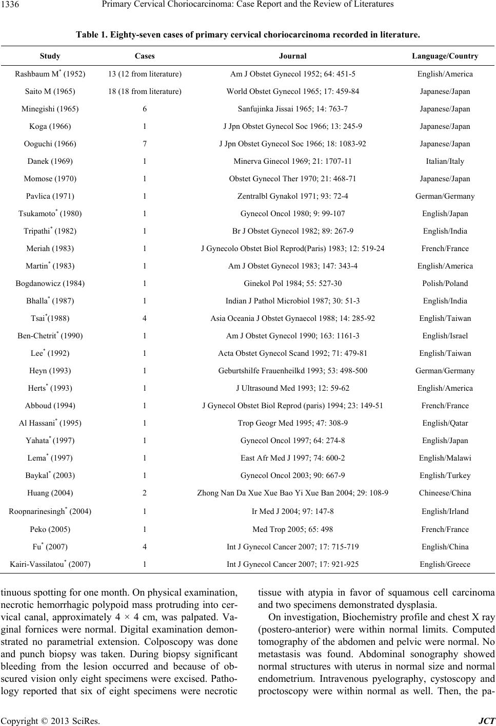 Primary Cervical Choriocarcinoma: Case Report and the Review of Literatures 1336 Table 1. Eighty-seven cases of primary cervical choriocarcinoma recorded in literature. Study Cases Journal Language/Country Rashbaum M* (1952) 13 (12 from l it erature) Am J Obstet Gynecol 1952; 6 4: 451-5 English/Amer i ca Saito M (1965) 18 (18 from literature) World Obstet Gynecol 1965; 17: 459-84 Japanese/Japan Minegishi (1965) 6 Sanfujinka Jissai 1965; 14: 763 -7 Japanese/Japan Koga (1966) 1 J Jpn Obstet Gynecol Soc 1966; 13: 245-9 Japanese/Japan Ooguchi (1966) 7 J Jpn Obstet Gynecol Soc 1966; 18: 1083-92 Japanese/Japan Danek (1969) 1 Minerva Ginecol 1969; 21: 1707-11 Italian/Italy Momose (1970) 1 Obstet Gynecol Ther 1970; 21: 468-71 Japanese/Japan Pavlica (1971) 1 Zentralbl Gynakol 1971; 93: 72- 4 German/Germany Tsukamoto* (19 80) 1 Gynecol Oncol 1980; 9: 99-107 English/Japan Tripathi* (1982) 1 Br J Obstet Gynecol 1982; 89: 267-9 English/India Meriah (1983) 1 J Gynecolo Obstet Biol Reprod(Paris) 1983; 12: 51 9-24 French/France Martin* (1983) 1 Am J Obstet Gynecol 1983; 147: 343-4 English/Americ a Bogdanowicz (19 84) 1 Ginekol Pol 1984; 55: 527-30 Polish/Poland Bhalla* (1987) 1 Indian J Pathol Microbiol 1987; 3 0: 51-3 English/India Tsai*(1988) 4 Asia Oceania J Obstet Gynaecol 1988; 14: 285-9 2 English/Taiwan Ben-Chetrit* (1990) 1 Am J Obstet Gynecol 19 90 ; 1 63: 1161-3 English/Israel Lee* (1992) 1 Acta Obstet Gynecol Scand 1992; 71: 4 79-81 English/ Taiwan Heyn (1993) 1 Geburtshilfe Frauenheilkd 1993; 53: 498-500 German/Germany Herts* (1993) 1 J Ultrasound Med 1993; 12: 59-62 English/America Abboud (1994) 1 J Gynecol Obstet Biol Reprod (paris) 1994; 23: 149- 51 French/France Al Hassani* (1995) 1 Trop Geogr Med 1995; 47: 308-9 English/Qatar Yahata* (1997) 1 Gynecol Oncol 1997; 64: 274-8 English/Japan Lema* (1997) 1 East Afr Med J 1997; 74: 600-2 English / Malawi Baykal* (2003) 1 Gynecol Oncol 2003; 90: 667-9 English/Turkey Huang (2004) 2 Zhong Nan Da Xue Xue B ao Yi Xue Ban 2004; 29: 108-9 Chineese/China Roopnarinesingh* (2004) 1 Ir Med J 2004; 97: 14 7-8 English/Irland Peko (2005) 1 Med Trop 2005; 65: 498 French/France Fu* (2007) 4 Int J Gynecol Cancer 20 07; 17: 715-719 English/China Kairi-Vassilatou* (2007) 1 Int J Gynecol Cancer 2007; 17: 921-925 English/Greece tinuous spotting for one month. On physical examination, necrotic hemorrhagic polypoid mass protruding into cer- vical canal, approximately 4 × 4 cm, was palpated. Va- ginal fornices were normal. Digital examination demon- strated no parametrial extension. Colposcopy was done and punch biopsy was taken. During biopsy significant bleeding from the lesion occurred and because of ob- scured vision only eight specimens were excised. Patho- logy reported that six of eight specimens were necrotic tissue with atypia in favor of squamous cell carcinoma and two specimens demonstrated dysplasia. On investigation, Biochemistry profile and chest X ray (postero-anterior) were within normal limits. Computed tomography of the abdomen and pelvic were normal. No metastasis was found. Abdominal sonography showed normal structures with uterus in normal size and normal endometrium. Intravenous pyelography, cystoscopy and proctoscopy were within normal as well. Then, the pa- Copyright © 2013 SciRes. JCT 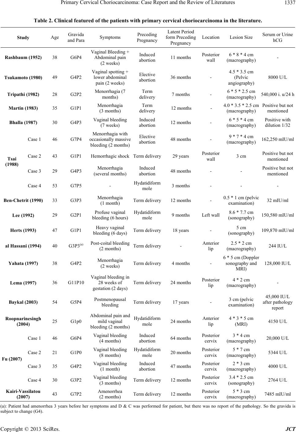 Primary Cervical Choriocarcinoma: Case Report and the Review of Literatures 1337 Table 2. Clinical featured of the patients with primary cervical choriocarcinoma in the literature. Study Age Gravida and Para Symptoms Preceding Pregnancy Latent Period form Preceding Pregnancy LocationLesion Size Serum or Urine hCG Rashbaum (1952) 38 G6P4 Vaginal Bleeding + Abdominal pain (2 weeks) Induced abortion 11 months Posterior wall 6 * 8 * 4 cm (macrography) - Tsukamoto (198 0) 49 G4P2 Vaginal spotting + lower abdominal pain (2 weeks) Elective abortion 36 months - 4.5 * 3.5 cm (Pelvic angiography) 8000 U/L Tripathi (1982) 28 G2P2 Menorrhagia (7 months) Term delivery 7 months - 6 * 5 * 2.5 cm (macrography) 540,000 i. u/24 h Martin (1983) 35 G1P1 Menorrhagia (3 months) Term delivery 12 months - 4.0 * 3.5 * 2.5 cm (macrography) Positive but not mentioned Bhalla (1987) 30 G4P3 Vaginal bleeding (7 weeks) Induced abortion 12 months - 6 * 5 * 4 cm (macrography) Positive with dilution 1/32 Case 1 46 G7P4 Menorrhagia with occasionally massive bleeding (2 months) Elective abortion 48 months - 9 * 7 * 4 cm (macrography) 162,250 mIU/ml Case 2 43 G1P1 Hemorrhagic shockTerm delivery29 years Posterior wall 3 cm Positive but not mentioned Case 3 29 G4P3 Menorrhagia (several months) Induced abortion 48 months - - Positive but not mentioned Tsai (1988) Case 4 53 G7P5 - Hydatidiform mole 3 months - - - Ben-Chetrit (199 0) 33 G3P3 Menorrhagia (1 month) Term delivery12 months - 0.5 * 1 cm (pelvic examination) 3 2 mIU/ml Lee (1992) 29 G2P1 Profuse vaginal bleeding (6 hours)Hydatidiform mole 9 months Left wall8.6 * 7.7 cm (sonography) 150,580 mIU/ml Herts (1993) 47 G1P1 Heavy vaginal bleeding (6 days) Term delivery18 years - 5 cm (sonography) 109,870 mIU/ml al Hassani (1994) 40 G3P3(a) Post-coital bleeding (2 months) Term delivery- Anterior lip 2.5 * 2 cm (macrography) 244 IU/L Yahata (1997) 38 G4P2 Menorrhagia (2 weeks) Term delivery4 months - 6 * 5 cm (Doppler sonography an d MRI) 128,000 IU/L Lema (1997) 36 G11P10 Vaginal bleeding in 28 weeks of gestation (2 days)Term delivery24 months Posterior lip 4 * 2 cm (macrography) - Baykal (2003) 54 G5P4 Postmenopausal bleeding Term delivery17 years - 3 cm (pelvic examination) 45,000 IU/L after pathology report Roopnarinesingh (2004) 25 G1p0 Abdominal pain and mild vaginal bleeding (2 months) Hydatidiform mole 24 months Anterior lip 4 * 3 * 5 cm (MRI) 4150 U/L Case 1 46 G6P4 Vaginal bleeding (4 months) Induced abortion 64 months Posterior cervix 3 * 4 cm (macrography) 20,000 U/L Case 2 21 G1P0 Vaginal bleeding (8 months) Hydatidiform mole 20 months Posterior cervix 5 * 7 cm (macrography) 5344 U/L Case 3 35 G4P2 Vaginal bleeding (1 month) Induced abortion 47 months Posterior cervix 2 * 3 cm (macrography) 4000 U/L Fu (2007) Case 4 30 G3P2 Vaginal bleeding (3 months) Term delivery12 months Posterior cervix 3.4 * 2.5 cm (sonography) 2764 U/L Kairi-Vassilatou (2007) 43 G7P2 Amenorrhea (2 months) Term delivery12 months Posterior cervix 5 * 3 cm (macrography) 7485 mIU/ml (a): Patient had amenorrhea 3 years before her symptoms and D & C was performed for patient, but there was no report of the pathology. So the gravida is subject to change (G4). Copyright © 2013 SciRes. JCT 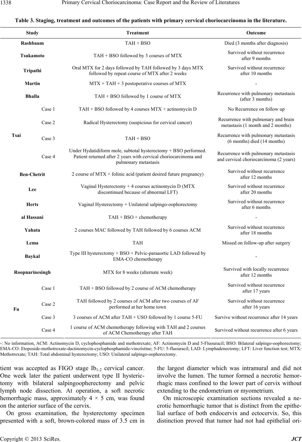 Primary Cervical Choriocarcinoma: Case Report and the Review of Literatures 1338 Table 3. Staging, treatment and outcomes of the patients with primary cervical choriocarcinoma in the literature. Study Treatment Outcome Rashbaum TAH + BSO Died (3 months after diagnosis) Tsukamoto TAH + BSO followed by 3 courses of MTX Survived without recurrence after 9 months Tripathi Oral MTX for 2 days followed by TAH followed by 3 days MTX followed by repe at course of MTX after 2 weeks Survived without recurre nce after 10 months Martin MTX + TAH + 3 postoperative courses of MTX - Bhalla TAH + BSO followed by 1 course of MTX Recurrence with pulmonary metastasis (after 3 months) Case 1 TAH + BSO followed by 4 courses MTX + actinomycin D No Recurrence on follow up Case 2 Radical Hysterectomy (suspicious for cervical cancer) Re c u rrence with pulmonary and brain metastasis (1 month and 2 months) Case 3 TAH + BSO Recurrence with pulmonary metastasis (6 months) died (14 months) Tsai Case 4 Under Hydatidiform mole, subtotal hysterectomy + BSO performed. Patient returned after 2 years with cervical choriocarcinoma and pulmonary metastasis Recurrence with pulmonary metastasis and cervical choriocarcinoma (2 years) Ben-Chetrit 2 course of MTX + folinic acid (patient desired future pregnan cy) Survived without recurrence after 12 months Lee Vaginal Hysterectomy + 4 courses actinomycin D (MTX discontinued because of abnormal LFT) Survived without recurrence after 20 months Herts Vaginal Hysterectomy + Unilateral salpingo-oophorectomy Survived without recurrence after 6 months al Hassani TAH + BSO + chemotherapy - Yahata 2 courses MAC followed by TAH followed by 6 cours es ACM Survived without recurrence after 18 months Lema TAH Missed on follow-up after surgery Baykal Type III hysterect omy + BSO + Pelvic-paraaortic LAD followed by EMA-CO chemotherapy - Roopnarinesingh MTX for 8 weeks (alternate week) Survived with locally recurrence after 12 months Case 1 TAH + BSO followed by 2 course of ACM chemotherapy Survived without recurrence after 17 years Case 2 TAH followed by 2 courses of ACM after two courses of AF performed at h e r h ome town Survived without recurrence after 16 years Case 3 3 courses of ACM after TAH + USO foll owed by 1 course 5-FU Survive without recurrence after 14 years Fu Case 4 1 course of ACM chemoth er apy following with TAH and 2 courses of ACM Chemo t h erapy after TAH Survived without recurrence after 6 years -: No information, ACM: Actinomycin D, cyclophosphamide and methotrexate; AF: Actinomycin D and 5-Fluouracil; BSO: Bilateral salpingo-oophorectomy; EMA-CO: Etoposide-methotrexate-dactinomycin-cyclophosphamide-vincristine; 5-FU: 5-fluouracil; LAD: Lymphadenectomy; LFT: Liver function test; MTX: Methotrexate; TAH: Total a bdominal hysterectomy; USO: Unilateral salpingo-oophorectomy. tient was accepted as FIGO stage Ib1-2 cervical cancer. One week later the patient underwent type II hysteric- tomy with bilateral salpingoopherectomy and pelvic lymph node dissection. At operation, a soft necrotic hemorrhagic mass, approximately 4 × 5 cm, was found on the anterior surface of the cervix. On gross examination, the hysterectomy specimen presented with a soft, brown-colored mass of 3.5 cm in the largest diameter which was intramural and did not involve the lumen. The tumor formed a necrotic hemor- rhagic mass confined to the lower part of cervix without extending to the endometrium or myometrium. On microscopic examination sections revealed a ne- crotic hemorrhagic tumor that is distinct from the epithe- lial surface of both endocervix and ectocervix. So, this distinction proved that tumor had not had epithelial ori- Copyright © 2013 SciRes. JCT  Primary Cervical Choriocarcinoma: Case Report and the Review of Literatures 1339 gin and we could exclude Squamous cell carcinoma or adenocarcinoma. On magnification Malignant tropho- blastic tumor composed of atypic bizarre polygonal tro- phoblasts with nucleolated nuclei, High nuclear to cyto- plasm ratio (N/C ratio) and atypic mitotic figures which are strongly positive for B-HCG and pancytokeratin. Human placental lactogen (HPL) was weakly positive in few cells. There was also hemorrhage (Blood Lake) in tissue and for this reason, the pathologist performed Im- munohistochemistry of CD31 and VIII Factor to exclude tumor with endothelial origin like angiosarcoma. Based on these findings, choriocarcinoma was diagnosed for the patient. An extensive sectioning of the endometrium, myometrium, parametrial tissues and lymph nodes con- firmed that these areas were free of neoplastic cells or any other site of trophoblastic cells. Patient discharged with good condition four days later and postoperative B-hCG was 6 mIU/ml after 2 weeks of operation. According to the FIGO staging system of GTD, the patient was classified as stage I, but because there was no preoperative B-hCG level we could not de- termine the risk score of the patient [12]. The patient is under follow-up and has had serial B-hCG measurement, which has been declining through 4 weeks. She is doing well without any complain so far. 4. Discussion Gestational intrauterine choriocarcinoma has higher inci- dence rate in Asia, Africa and Latin America in com- parison to United States and Europe, may be due to so- cioeconomic status [13]. However, the overall incidence of choriocarcinoma in recent years has been decreased parallel to improving socioeconomic conditions [13]. Pri- mary choriocarcinoma of the cervix is an extremely rare disease; however, most of the case reports in literatures demonstrate that extrauterine choriocarcinoma mostly originates in cervix rather than other sites in genital tract [2 -7] . O ur ca se fulfilled the criteria defin ed by Saito et a l. [1]. So, it is an ectopic choriocarcinoma originates from the cervix. The median age was 37 with the range of 21 - 54 year s old (two patients were postmenopausal). 9 patients were less than 35 years old. The latent period from preceding pregnancy had a range of 3 - 348 months with 62% less than or equal to 24 months. Long latency of choriocarci- noma after the preceding pregnancy is not common [14]. Choriocarcinoma is heterochronic if the latent period is more than two years [15]. Choriocarcinoma occurred in our patients 3 years after the preceding pregnancy, which was terminated by induced abortion. There was limited data regarding the location of tumor with mostly origi- nated from posterior cervix. Gestational choriocarcinoma may follow any type of pregnancy, which most preceded by molar pregnancy (50%), 25% developed after abortion and 25% follow an apparently normal pregnancy. The more common gesta- tional variety is associated with a coincident and antece- dent pregnancy. In this review primary cervical chorio- carcinoma preceded by 50% normal pregnancy, 32% abortion and 18% de vel o ped after hydatidi f orm mole. There are some theories expressed for developing of cervical choriocarcinoma [16,17]. It is possible that it can be a metastatic tumor from a vanishing intrauterine cho- riocarcinoma or it could be a transformation of a cervical pregnancy. The pathologic examination ruled out these possibilities, as we demonstrated that no trophoblastic cell was observed in endothelium and myometriu m of the uterus. Another theory expresses the role of therapeutic abortion in the implantation of trophoblasts in the cervix and lower genital tract. Trauma to the cervix during D & C may be a predisposing factor [6]. Our case was pre- ceded by an abortion and it supports the later theory. There was no pathological examination of the curetted material as this was performed in another city and we did not have access to the pathology. The clinical diagnosis of primary choriocarcinoma is very difficult. In this review all patients except one, complained of vaginal bleeding with different period of time ranged from 6 hours to 8 months, with different severity that one patient referred with hypovolemic shock. Only one patient complained of amenorrhea. The predo- minant symptom is abnormal vaginal bleeding which is due to angiogenesis and neovasculirization of this tumor. There are few differential diagnoses which include uter- ine cervical pregnancy, threatened abortion, a cervical polyp and malignancy [4]. In the present case, choriocar- cinoma was presented with vaginal bleeding and misdi- agnosed as malignancy. For this reason, we should con- sider choriocarcinoma as differential diagnosis in any patient in their reproductive years with a cervical lesion with high vascul ari t y and pr o fuse bleeding. How can we suspect and diagnose cervical choriocar- cinoma? As we mentioned above, in cervical lesions in women of child-bearing age with profuse bleeding, measurement of serum and urinary B-hCG is useful for diagnosis. Color Doppler transvaginal ultrasonography and MRI with con trast detect the abundan t blood flow in the tumor [4]. Transvaginal Doppler sonography allows detection of tumoral vessels, which manifests high dia- stolic blood flow and low impedance resulting in ne- ovasculirization [4]. Contrast-enhanced MRI can demon- strate tumoral vascularity and in cases of necrosis hy- pointensity of the necrotic areas are perceptible [4]. Diagnosis of such a kind of cervical tumor is very dif- ficult for pathologist both in biopsy and surgical resec- tion. In our case, the first pathologist misdiagnosed choriocarcinoma with squamous cell carcinoma as Ben- Copyright © 2013 SciRes. JCT  Primary Cervical Choriocarcinoma: Case Report and the Review of Literatures Copyright © 2013 SciRes. JCT 1340 Chetrit [18]. Necrosis is a predominant feature of these neoplasms, often to such an extent that the viable tissue is obscured. So, we should consider immunohistochemi- cal staining in extend necrotic tissue obtained from cer- vix for more information, because there is the chance for more limited surgery and more successful chemotherapy for choriocarcinoma if it is diagnosed in the first step. In our case, massive necrosis of the tumor and arising of tumor from non-epithelial origin motivate the pathologist to perform immunohistochemical staining. As we show- ed the specimen was markedly positive fo r B-hCG. The treatment of choice as in our patient was total ab- dominal hysterectomy and bilateral salpingoopherectomy with or without chemotherapy. In terestingly, Ben-Chetrit and his co-worke rs in one report and Roopnarinesingh et al. in other report, documented conservative management of primary cervical choriocarcinoma [4,18]. 5. Conclusion In conclusion, cervical choriocarcinoma should be con- sidered in the differential diagnosis of cervical lesions in their child-bearing years, especially lesions with promi- nent vascularity. A beta-hCG assay, transvaginal color Doppler sonography or contrast-enhanced MRI may form part of pre-operative investigation in such a patient. A detailed correlation between histologic findings and patient’s clinical history, lab studies and radiologic data is always required to preclude diagnostic pitfalls and de- signates the appropriate treatment. REFERENCES [1] M. Saito, T. Azuma and K. Nakamura, “On Ectopic Choriocarcinoma,” World Journal of Obstetrics and Gy- necology, Vol. 17, 1965, pp. 459-484. [2] C. Baykal, G. Tulunay, D. Bulbul, N. Boran and M. F. Kose, “Primary Choriocarcinoma of the Uterine Cervix in a Postmenopausal Patient: A Case Report,” Gynecologic Oncology, Vol. 90, No. 3, 2003, pp. 667-669. http://dx.doi.org/10.1016/S0090-8258(03)00369-X [3] Y. Fu, W. Lu, C. Zhou and X. Xie, “Primary Cervical Choriocarcinoma: Report of Four Cases and Literature Review,” International Journal of Gynecological Cancer, Vol. 17, No. 3, 2007, pp. 715-719. http://dx.doi.org/10.1111/j.1525-1438.2007.00819.x [4] T. Yahata, S. Kodama, H. Kase, N. Sekizuka, T. Kuraba- yashi, Y. Aoki and K. Tanaka, “Primary Choriocarcinoma of the Uterine Cervix: Clinical, MRI, and Color Doppler Ultrasonographic Study,” Gynecologic Oncol ogy , Vol. 64, No. 2, 1997, pp. 274-278. http://dx.doi.org/10.1006/gyno.1996.4541 [5] R. Roopnarinesingh, S. Igoe and J. E. Gillan, “Choriocar- cinoma-Presenting as a Primary Lesion of the Cervix,” Irish Medical Journal, Vol. 97, 2004, pp. 147-148. [6] S. S. Al Hassani and G. C. Ejeckam, “Primary Cervical Choriocarcinoma,” Tropical and Geographical Medicine, Vol. 47, 1995, pp. 308-309. [7] B. R. Herts, J. M. Yee a nd R. F. Porges, “Primary Cervi- cal Choriocarcinoma: Case Report and Review of Litera- ture,” Journal of Ultrasound in Medicine, Vol. 12, 1993, pp. 59-62. [8] N. P. Veridiano, D. Gal, I. Delke, Y. Rosen and M. L. Tancer, “Gestational Choriocarcinoma of the Ovary,” Gy- necologic Oncology, Vol. 10, No. 2, 1980, pp. 235-240. http://dx.doi.org/10.1016/0090-8258(80)90087-6 [9] M. G. Muto, J. M. Lage, R. S. Berkowitz, D. P. Goldste in and M. R. Bernstein, “Gestational Choriocarcinoma Dis- ease of the Fallopian Tube,” Journal of Reproductive Me- dicine, Vol. 36, 1991, pp. 57-60. [10] S. Weiss, A. Amit, M. R. Schwartz and A. L. Kaplan, “Primary Choriocarcinoma of the Vulva,” International Journal of Gynecological Cancer, Vol. 11, No. 3, 2001, pp. 251-254. http://dx.doi.org/10.1046/j.1525-1438.2001.01005.x [11] H. Sonobe, K. Taguchi, K. Ogawa and T. Yoshioka, “La- tent Vaginal Choriocarcinoma in a Postmenopausal Wo- man,” Acta Pathologica Japonica, Vol. 26, 1976, pp. 611-618. [12] FIGO Oncology Committee Report, “FIGO Staging for Gestational Trophoblastic Neoplasia 2000,” International Journal of Gynecology & Obstetrics, Vol. 77, No. 3, 2002, pp. 285-287. http://dx.doi.org/10.1016/S0020-7292(02)00063-2 [13] P. Y. Wel snd P. C. Ouyang, “Trophoblastic Disease in Taiwan: A Review of 157 Cases on a 10-Year Period,” American Journal of Obstetrics & Gynecology, Vol. 85, 1963, pp. 844-849. [14] P. C. Dyke and L. M. Fink, “Latent Choriocarcinoma,” Cancer, Vol. 20, No. 1, 1967, pp. 150-154. http://dx.doi.org/10.1002/1097-0142(1967)20:1<150::AI D-CNCR2820200121>3.0.CO;2-9 [15] W. Park, “Choriocarcinoma : A Study of Its Pathology,” F. A. Davis, Philadelphia, 1971. [16] N. T su k a mo to, M. Nakamura, M. Kashimura and T. Saito, “Primary Cervical Choriocarcinoma,” Gynecologic On- cology, Vol. 9, No. 1, 1980, pp. 99-107. http://dx.doi.org/10.1016/0090-8258(80)90014-1 [17] E. Kairi-Vassilatou, K. Papakonstantinou, D. Grapsa, A. Kondi-Paphiti and D. Hasiakos “Primary Gestational Choriocarcinoma of the Uterine Cervix,” International Journal of Gynecological Cancer, Vol. 17, No. 4, 2007, pp. 921-925. http://dx.doi.org/10.1111/j.1525-1438.2006.00852.x [18] A. Ben-Chetrit, S. Yagel, I. Ariel, D. Zacut, S. Shimono- vitz and D. Ceinikier-Hochner, “Successful Conservative Management of Primary Nonmetastatic Cervical Chorio- carcinoma,” American Journal of Obstetrics & Gynecol- ogy, Vol. 163, No. 4, 1990, pp. 1161-1163. http://dx.doi.org/10.1016/0002-9378(90)90679-2 |