

Paper Menu >>
Journal Menu >>
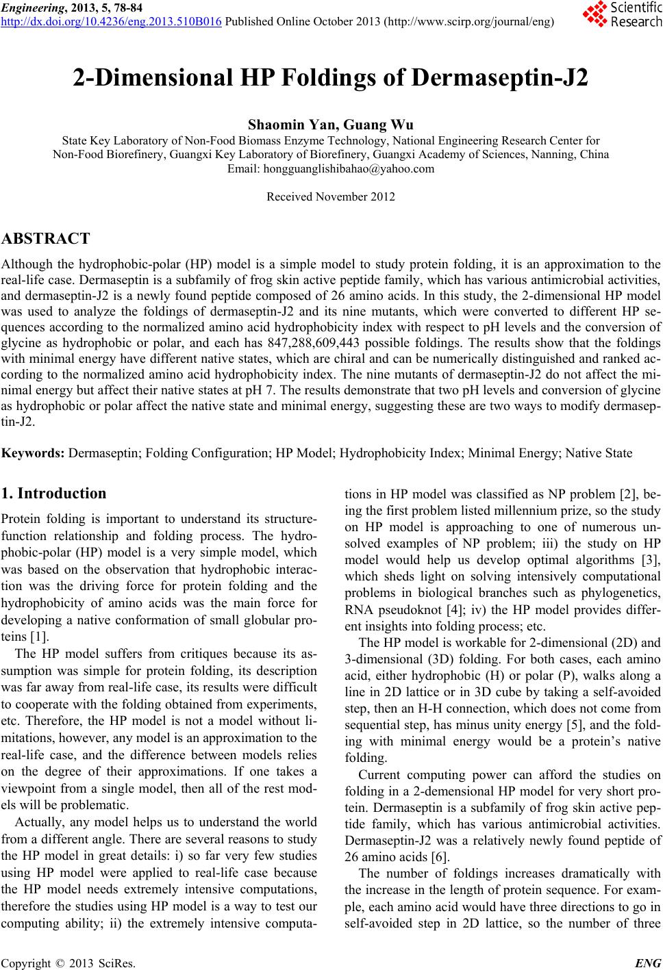 Engineering, 2013, 5, 78-84 http://dx.doi.org/10.4236/eng.2013.510B016 Published Online October 2013 (http://www.scirp.org/journal/eng) Copyright © 2013 SciRes. ENG 2-Dimensional HP Foldings of Dermaseptin-J2 Shaomin Yan, Guang Wu State Key Laboratory of Non-Food Biomass Enzyme Technology, National Engineering Research Center for Non-Food Biorefinery, Guangxi Key Laboratory of Biorefinery, Guangxi Academy of Sciences, Nanning, China Email: [email protected] Received November 2012 ABSTRACT Although the hydrophobic-polar (HP) model is a simple model to study protein folding, it is an approximation to the real-life case. Dermaseptin is a subfamily of frog skin active peptide family, which has various antimicrobial activities, and dermaseptin-J2 is a newly found peptide composed of 26 amino acids. In this study, the 2-dimensional HP model was used to analyze the foldings of dermaseptin-J2 and its nine mutants, which were converted to different HP se- quences according to the normalized amino acid hydrophobicity index with respect to pH levels and the conversion of glycine as hydrophobic or polar, and each has 847,288,609,443 possible foldings. The results show that the foldings with minimal energy have different native states, which are chiral and can be numerically distinguished and ranked ac- cording to the normalized amino acid hydrophobicity index. The nine mutants of dermaseptin-J2 do not affect the mi- nimal energy but affect their native states at pH 7. The results demonstrate that two pH levels and conversion of glycine as hydrophobic or polar affect the native state and minimal energy, suggesting these are two ways to modify dermasep- tin-J2. Keywords: Dermaseptin; Folding Configuration; HP Model; Hydrophobicity Ind ex; Minimal Energy; Native State 1. Introduction Protein folding is important to understand its structure- function relationship and folding process. The hydro- phobic-polar (HP) model is a very simple model, which was based on the observation that hydrophobic interac- tion was the driving force for protein folding and the hydrophobicity of amino acids was the main force for developing a native conformation of small globular pro- teins [1]. The HP model suffers from critiques because its as- sumption was simple for protein folding, its description was far away from real-life case, its results were difficult to cooperate with the folding obtained from experiments, etc. Therefore, the HP model is not a model without li- mitations, however, any model is an approximation to the real-life case, and the difference between models relies on the degree of their approximations. If one takes a viewpoint from a single model, then all of the rest mod- els will be problematic. Actually, any model helps us to understand the world from a different angle. There are several reasons to study the HP model in great details: i) so far very few studies using HP model were applied to real-life case because the HP model needs extremely intensive computations, therefore the studies using HP model is a way to test our computing ability; ii) the extremely intensive computa- tions in HP model was classified as NP problem [2], be- ing the first problem listed millennium prize, so the study on HP model is approaching to one of numerous un- solved examples of NP problem; iii) the study on HP model would help us develop optimal algorithms [3], which sheds light on solving intensively computational problems in biological branches such as phylogenetics, RNA pseudoknot [4]; iv) the HP model provides differ- ent insights into f olding pr oc ess; etc . The HP model is workable for 2-dimensional (2D) and 3-dimensional (3D) folding. For both cases, each amino acid, either hydrophobic (H) or polar (P), walks along a line in 2D lattice or in 3D cube by taking a self-avoided step, then an H-H connection, which does not come from sequential step, has minus unity energy [5], and the fold- ing with minimal energy would be a protein’s native folding. Current computing power can afford the studies on folding in a 2-demensional HP model for very short pro- tein. Dermaseptin is a subfamily of frog skin active pep- tide family, which has various antimicrobial activities. Dermaseptin-J2 was a relatively newly found peptide of 26 amino acids [6]. The number of foldings increases dramatically with the increase in the length of protein sequence. For exam- ple, each amino acid would have three directions to go in self-avoided step in 2D lattice, so the number of three 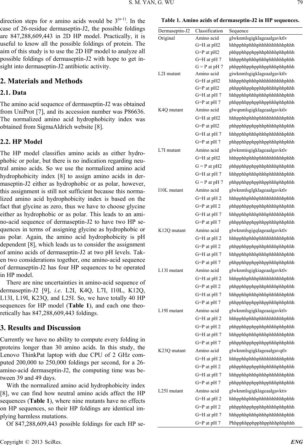 S. M. YAN, G. WU Copyright © 2013 SciRes. ENG 79 direction steps for n amino acids would be 3(n-1). In the case of 26-residue dermaseptin-J2, the possible foldings are 847,288,609,443 in 2D HP model. Practically, it is useful to know all the possible foldings of protein. The aim of this study is to use the 2D HP model to analyze all possible foldings of dermaseptin-J2 with hope to get in- sight int o de rmasept in-J2 antibiotic activity. 2. Materials and Methods 2.1. Data The amino acid sequence of dermaseptin-J2 was obtained from UniProt [7], and its accession number w as P86636. The normalized amino acid hydrophobicity index was obtaine d from SigmaAldri c h we bs i te [8]. 2.2. HP Model The HP model classifies amino acids as either hydro- phobic or polar, but there is n o indication regarding neu- tral amino acids. So we use the normalized amino acid hydrophobicity index [8] to assign amino acids in der- maseptin-J2 either as hydrophobic or as polar, however, this assignment is still not sufficient because this norma- lized amino acid hydrophobicity index is based on the fact that glycine as zero, thus we have to choose glycine either as hydrophobic or as polar. This leads to an ami- no-acid sequence of dermaseptin-J2 to have two HP se- quences in terms of assigning glycine as hydrophobic or as polar. Again, the amino acid hydrophobicity is pH dependent [8], which leads us to consider the assignment of amino acids of dermaseptin-J2 at two pH levels. Tak- en two considerations together, one amino-acid s equence of dermaseptin-J2 has four HP sequences to be operated in HP model. There are nine uncertainties in amino-acid sequence of dermaseptin-J2 [9], i.e. L2I, K4Q, L7I, I10L, K12Q, L13I, L19I, K23Q, and L25I. So, we have totally 40 HP sequences for HP model (Table 1), and each one theo- retically has 847,288,609,443 foldings. 3. Results and Discussion Currently we have no ability to compute every folding in proteins longer than 30 amino acids. In this study, the Lenovo ThinkPat laptop with due CPU of 2 GHz com- puted 200,000 to 250,000 foldings per second, for a 26- amino-acid dermaseptin-J2, the computing time was be- tween 39 a nd 4 9 da y s . With the normalized amino acid hydrophobicity index [8], we can find how neutral amino acids affect the HP sequences (Tab le 1), wh ere nine mutants have no effects on HP sequences, so their HP foldings are identical im- plying harmless mutations. Of 847,288,609,443 possible foldings for each HP se- Table 1. Amino acids of dermaseptin-J2 in HP sequence s. Dermaseptin-J2 Classification Sequence Original Amino acid glwknmlsgigklageaalgavktlv G=H at pH2 hhhpphhphhhphhhhhhhhhhphhh G=P at pH2 phhpphhpphpphhphhhhphhphhh G=H at pH 7 hhhpphhphhhphhhphhhhhhphhh G = P at pH 7 phhpphhpphpphhpphhhphhphhh L2I mutant Amino acid giwknmlsgigklageaalgavktlv G=H at pH2 hhhpphhphhhphhhhhhhhhhphhh G=P at pH2 phhpphhpphpphhphhhhphhphhh G=H at pH 7 hhhpphhphhhphhhphhhhhhphhh G=P at pH 7 phhpphhpphpphhpphhhphhphhh K4Q mutant Amino acid glwqnmlsgigklageaalgavktlv G=H at pH2 hhhpphhphhhphhhhhhhhhhphhh G=P at pH2 phhpphhpphpphhphhhhphhphhh G=H at pH 7 hhhpphhphhhphhhphhhhhhphhh G=P at pH 7 phhpphhpphpphhpphhhphhphhh L7I mutant Amino acid glwknmisgigklageaalgavktlv G=H at pH2 hhhpphhphhhphhhhhhhhhhphhh G = P at pH2 phhpphhpphpphhphhhhphhphhh G=H at pH 7 hhhpphhphhhphhhphhhhhhphhh G = P at pH 7 phhpphhpphpphhpphhhphhphhh I10L mutant Amino acid glwknmlsglgklageaalgavktlv G=H at pH 2 hhhpphhphhhphhhhhhhhhhphhh G=P at pH 2 phhpphhpphpphhphhhhphhphhh G=H at pH 7 hhhpphhphhhphhhphhhhhhphhh G=P at pH 7 phhpphhpphpphhpphhhphhphhh K12Q mutant Amino acid glwknmlsgigqlageaalgavktlv G=H at pH 2 hhhpphhphhhphhhhhhhhhhphhh G=P at pH 2 phhpphhpphpphhphhhhphhphhh G=H at pH 7 hhhpphhphhhphhhphhhhhhphhh G=P at pH 7 phhpphhpphpphhpphhhphhphhh L13I mutant Amino acid glwknmlsgigkiageaalgavktlv G=H at pH 2 hhhpphhphhhphhhhhhhhhhphhh G=P at pH 2 phhpphhpphpphhphhhhphhphhh G=H at pH 7 hhhpphhphhhphhhphhhhhhphhh G=P at pH 7 phhpphhpphpphhpphhhphhphhh L19I mutant Amino acid glwknmlsgigklageaaigavktlv G=H at pH 2 hhhpphhphhhphhhhhhhhhhphhh G=P at pH 2 phhpphhpphpphhphhhhphhphhh G=H at pH 7 hhhpphhphhhphhhphhhhhhphhh G=P at pH 7 phhpphhpphpphhpphhhphhphhh K23Q mutant Amino acid glwknmlsgigklageaalgavqtlv G=H at pH 2 hhhpphhphhhphhhhhhhhhhphhh G=P at pH 2 phhpphhpphpphhphhhhphhphhh G=H at pH 7 hhhpphhphhhphhhphhhhhhphhh G=P at pH 7 phhpphhpphpphhpphhhphhphhh L25I mutant Amino acid glwknmlsgigklageaalgavktiv G=H at pH 2 hhhpphhphhhphhhhhhhhhhphhh G=P at pH 2 phhpphhpphpphhphhhhphhphhh G=H at pH 7 hhhpphhphhhphhhphhhhhhphhh G=P at pH 7 Phhpphhpphpphhpphhhphhphhh 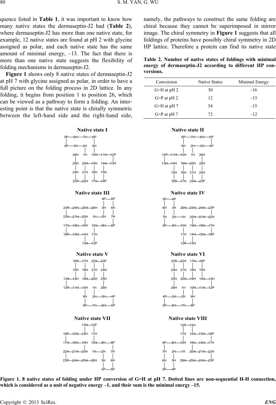 S. M. YAN, G. WU Copyright © 2013 SciRes. ENG 80 quence listed in Table 1, it was important to know how many native states the dermaseptin-J2 had (Table 2), where dermaseptin-J2 has more than one native state, for example, 12 native states are found at pH 2 with glycine assigned as polar, and each native state has the same amount of minimal energy, –13. The fact that there is more than one native state suggests the flexibility of folding mechanisms in dermaseptin-J2. Figure 1 shows only 8 native states of dermaseptin -J2 at pH 7 with glycine assigned as polar, in order to have a full picture on the folding process in 2D lattice. In any folding, it begins from position 1 to position 26, which can be viewed as a pathway to form a folding. An inter- esting point is that the native state is chirally symmetric between the left-hand side and the right-hand side, namely, the pathways to construct the same folding are chiral because they cannot be superimposed in mirror image. The chiral symmetry in Figure 1 suggests that all foldings of proteins hav e possibly chiral symmetry in 2D HP lattice. Therefore a protein can find its native state Table 2. Number of native states of foldings with minimal energy of dermaseptin-J2 according to different HP con- versions. Conversion Native States Minimal Energy G=H at pH 2 30 –16 G=P at pH 2 12 –13 G=H at pH 7 54 –15 G=P at pH 7 72 –12 Figure 1. 8 native states of folding under HP conversion of G=H at pH 7. Dotted lines are non-sequential H-H connection, which is considered as a unit o f nega t iv e energy –1, and their sum is th e m inimal energy –15. 26H 25H 24H 23P 22H 21H 20H 19H 18H 17H 16P 15H 14H 13H 12P11H10H 9H 8P7H6H5P 4P 3H 2H 1H 26H 25H 24H 23P22H 21H 20H19H 18H 17H16P 15H 14H13H 12P 11H 10H 9H 8P 7H6H 5P 4P3H2H 1H 26H25H24H23P 22H 21H 20H 19H18H17H 16P 15H 14H 13H 12P 11H 10H 9H8P 7H 6H 5P4P 3H 2H1H 26H 25H 24H 23P 22H21H20H 19H 18H 17H 16P15H14H 13H12P 11H 10H9H8P 7H 6H 5P 4P 3H 2H 1H 26H 25H 24H 23P22H 21H 20H19H 18H 17H16P 15H 14H13H 12P 11H 10H 9H 8P 7H6H 5P 4P3H2H 1H 26H 25H 24H 23P 22H 21H 20H 19H 18H 17H 16P 15H 14H 13H 12P11H10H 9H 8P7H6H5P 4P 3H 2H 1H Native state III Native state INative state II Native state IV Native state VINative state V 26H25H24H23P 22H 21H 20H 19H18H17H 16P 15H 14H 13H 12P 11H 10H 9H8P 7H 6H 5P4P 3H 2H1H 26H 25H 24H 23P 22H21H20H 19H 18H17H 16P15H14 H 13H12P 11H 10H9H8P 7H 6H 5P 4P 3H 2H 1H Native state VIIINative state VII 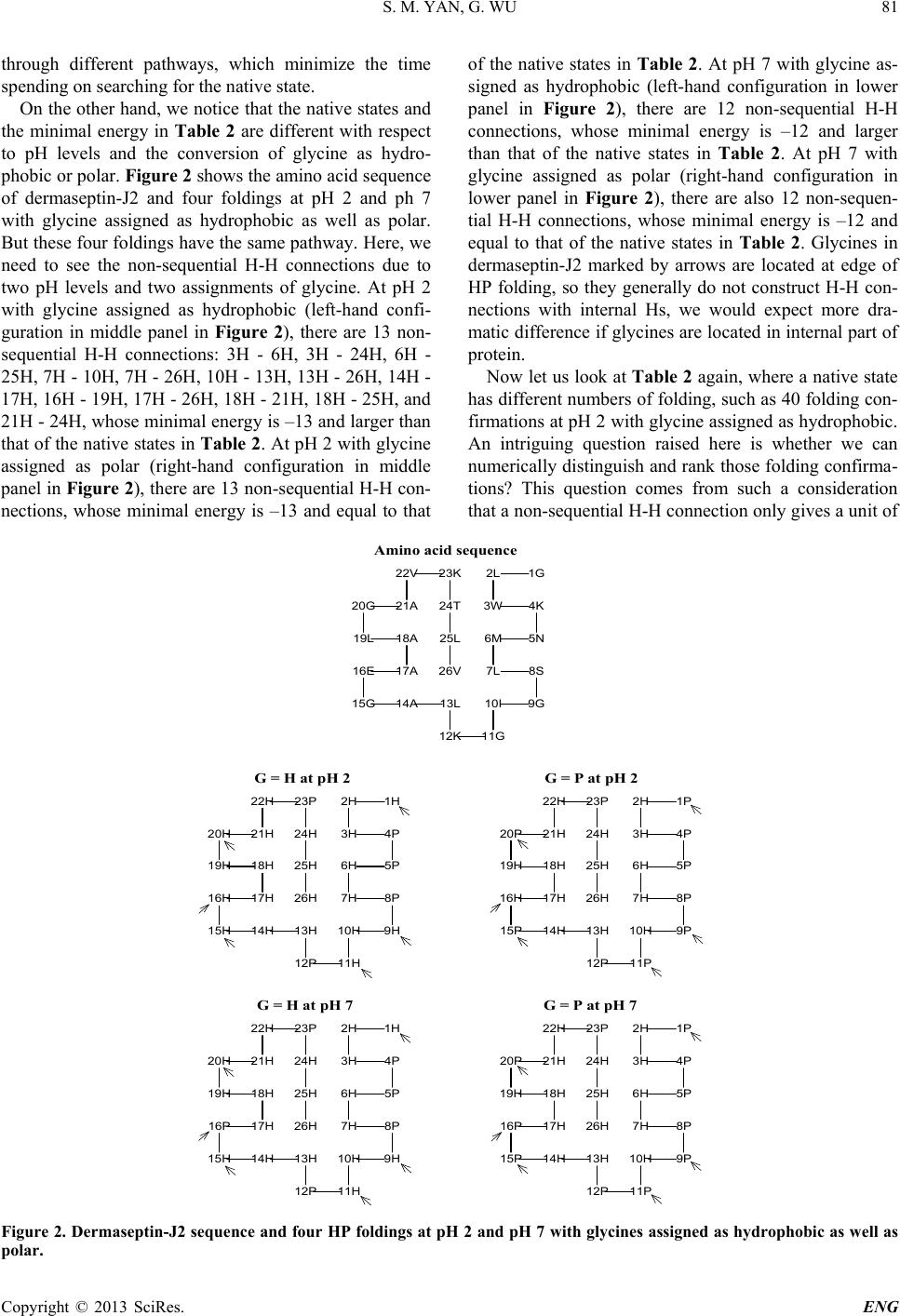 S. M. YAN, G. WU Copyright © 2013 SciRes. ENG 81 through different pathways, which minimize the time spending on searching for the native state. On the other hand, we notice that the native states and the minimal energy in Table 2 are different with respect to pH levels and the conversion of glycine as hydro- phobic or polar. Figure 2 shows the amino acid sequence of dermaseptin-J2 and four foldings at pH 2 and ph 7 with glycine assigned as hydrophobic as well as polar. But these four foldings have the same pathway. Here, we need to see the non-sequential H-H connections due to two pH levels and two assignments of glycine. At pH 2 with glycine assigned as hydrophobic (left-hand confi- guration in middle panel in Figure 2), there are 13 non- sequential H-H connections: 3H - 6H, 3H - 24H, 6H - 25H, 7H - 10H, 7H - 26H, 10H - 13H, 13H - 26 H, 14H - 17H, 16H - 19H, 17H - 26H, 18H - 21H, 18H - 25H, and 21H - 24H, whose minimal energy is –13 and larger than that of the native states in Tab le 2. At pH 2 with glycine assigned as polar (right-hand configuration in middle panel in Figu re 2), th ere are 13 non-sequential H-H con- nections, whose minimal energy is –13 and equal to that of the native states in Table 2. At pH 7 with glycine as- signed as hydrophobic (left-hand configuration in lower panel in Figure 2), there are 12 non-sequen tial H-H connections, whose minimal energy is –12 and larger than that of the native states in Table 2. At pH 7 with glycine assigned as polar (right-hand configuration in lower panel in Figure 2), there are also 12 non-seq uen- tial H-H connections, whose minimal energy is –12 and equal to that of the native states in Table 2. Glycines in dermaseptin-J2 marked by arrows are located at edge of HP folding, so they generally do not construct H-H con- nections with internal Hs, we would expect more dra- matic difference if glycines are located in internal part of protein. Now let us look at Table 2 again, where a native state has different numbers of folding, such as 40 folding con- firmations at pH 2 with glycine assigned as hydrophobic. An intriguing question raised here is whether we can numerically distinguish and rank those folding confirma- tions? This question comes from such a consideration that a non-sequential H-H connection only gives a unit of Figure 2. Dermaseptin-J2 sequence and four HP foldings at pH 2 and pH 7 with glycines assigned as hydrophobic as well as polar. 26V 25L 24T 23K22V 21A20G 19L 18A 17A16E 15G 14A13L 12K 11G 10I 9G 8S7L 6M 5N 4K3W 2L 1G 26H 25H 24H 23P22H 21H20H 19H 18H 17H16H 15H 14H 13H 12P 11H 10H 9H 8P7H 6H 5P 4P3H 2H 1H 26H 25H 24H 23P22H 21H20P 19H 18H 17H16H 15P 14H 13H 12P 11P 10H 9P 8P7H 6H 5P 4P3H 2H 1P 26H 25H 24H 23P22H 21H20H 19H 18H 17H16P 15H 14H 13H 12P 11H 10H 9H 8P7H 6H 5P 4P3H 2H 1H 26H 25H 24H 23P22H 21H20P 19H 18H 17H16P 15P 14H 13H 12P 11P 10H 9P 8P7H 6H 5P 4P3H 2H 1P G = H at pH 2 Amino acid s equence G = P at pH 2 G = P at pH 7G = H at pH 7 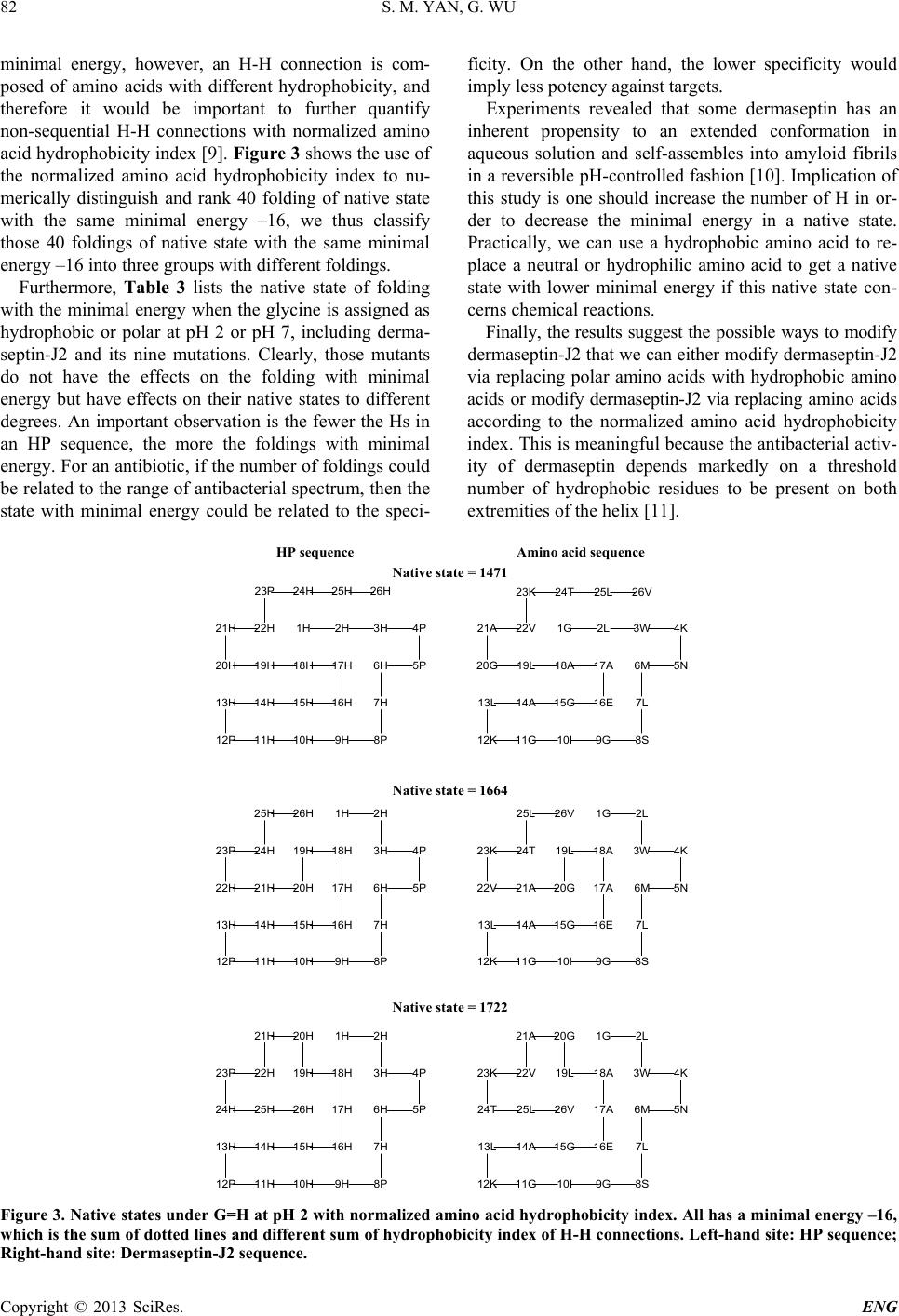 S. M. YAN, G. WU Copyright © 2013 SciRes. ENG 82 minimal energy, however, an H-H connection is com- posed of amino acids with different hydrophobicity, and therefore it would be important to further quantify non-sequential H-H connections with normalized amino acid hydrophobicity index [9]. Figure 3 shows the use of the normalized amino acid hydrophobicity index to nu- merically distinguish and rank 40 folding of native state with the same minimal energy –16, we thus classify those 40 foldings of native state with the same minimal energy –16 into three groups wi th different foldi n gs. Furthermore, Table 3 lists the native state of folding with the minimal energy when the glycine is assigned as hydrophobic or polar at pH 2 or pH 7, including derma- septin-J2 and its nine mutations. Clearly, those mutants do not have the effects on the folding with minimal energy but have effects on their native states to different degrees. An important observation is the fewer the Hs in an HP sequence, the more the foldings with minimal energy. For an antibiotic, if the number of foldings could be related to the range of antibacterial spectrum, then the state with minimal energy could be related to the speci- ficity. On the other hand, the lower specificity would imply less potency against targets. Experiments revealed that some dermaseptin has an inherent propensity to an extended conformation in aqueous solution and self-assembles into amyloid fibrils in a reversible pH-controlled fashion [10]. Implication of this study is one should increase the number of H in or- der to decrease the minimal energy in a native state. Practically, we can use a hydrophobic amino acid to re- place a neutral or hydrophilic amino acid to get a native state with lower minimal energy if this native state con- cerns chemical reactions. Finally, the results suggest the possible ways to modify dermaseptin-J2 that we can either modify dermaseptin-J2 via replacing polar amino acids with hydrophobic amino acids or modify dermaseptin-J2 via replacing amino acids according to the normalized amino acid hydrophobicity index. This is meaningful because the antibacterial activ- ity of dermaseptin depends markedly on a threshold number of hydrophobic residues to be present on both extremities of t he he l ix [11]. Figure 3. Native states under G=H at pH 2 with normalized amino acid hydrophobici ty index. All has a minimal ene rgy –16, which is the sum of dotted lines and different sum of hydrophobicity index of H-H connections. Left-hand site: HP sequence; Right-hand site: Dermaseptin-J2 sequence. 26H25H24H23P 22H21H 20H 19H18H 17H 16H15H14H13H 12P 11H10H9H8P 7H 6H 5P 4P3H2H1 H 26H25H 24H23P 22H 21H20H 19H 18H 17H 16H15H14H13H 12P 11H10H9H8P 7H 6H 5P 4P3H 2H1H 26V25L 24T23K 22V21A 20G 19L 18A 17A 16E15G14A13L 12K 11G10I9G8S 7L 6M 5N 4K3W 2L1G 26H25H24H 23P 22H 21H 20H 19H 18H 17H 16H15H14H13H 12P 11H10H9H8P 7H 6H 5P 4P3H 2H1H 26V25L24T 23K 22V 21A 20G 19L 18A 17A 16E15G14A13L 12K 11G10I9G8S 7L 6M 5N 4K3W 2L1G Native state = 1471 HP sequence 26V25L24T23K 22V21A 20G 19L 18A 17A 16E15G14A13L 12K 11G10I9G8S 7L 6M 5N 4K3W2L1G Amino acid sequence Native state = 1664 Native state = 1722 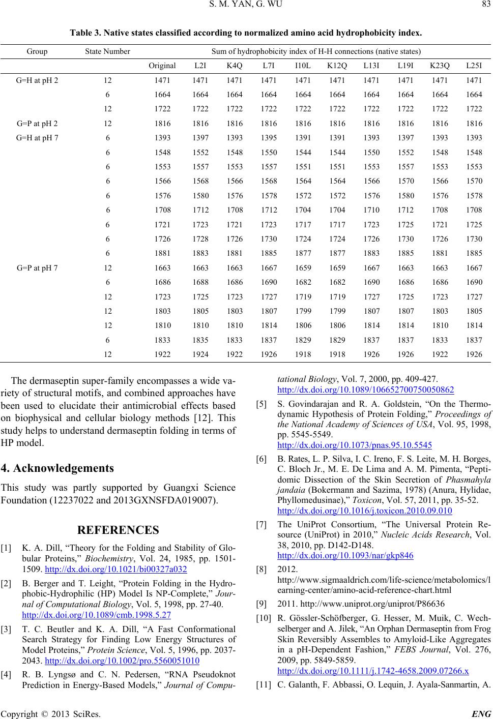 S. M. YAN, G. WU Copyright © 2013 SciRes. ENG 83 Table 3. Native states classified according to norm alized amino acid hydrophobi city index. Group State Nu mb e r Sum of hydrophobicity index of H-H connections (native states) Original L2I K4Q L7I I10L K12Q L13I L19I K23Q L25I G=H at pH 2 12 1471 1471 1471 1471 1471 1471 1471 1471 1471 1471 6 1664 1664 1664 1664 1664 1664 1664 1664 1664 1664 12 1722 1722 1722 1722 1722 1722 1722 1722 1722 1722 G=P at pH 2 12 1816 1816 1816 1816 1816 1816 1816 1816 1816 1816 G=H at pH 7 6 1393 1397 1393 1395 1391 1391 1393 1397 1393 1393 6 1548 1552 1548 1550 1544 1544 1550 1552 1548 1548 6 1553 1557 1553 1557 1551 1551 1553 1557 1553 1553 6 1566 1568 1566 1568 1564 1564 1566 1570 1566 1570 6 1576 1580 1576 1578 1572 1572 1576 1580 1576 1578 6 1708 1712 1708 1712 1704 1704 1710 1712 1708 1708 6 1721 1723 1721 1723 1717 1717 1723 1725 1721 1725 6 1726 1728 1726 1730 1724 1724 1726 1730 1726 1730 6 1881 1883 1881 1885 1877 1877 1883 1885 1881 1885 G=P at pH 7 12 1663 1663 1663 1667 1659 1659 1667 1663 1663 1667 6 1686 1688 1686 1690 1682 1682 1690 1686 1686 1690 12 1723 1725 1723 1727 1719 1719 1727 1725 1723 1727 12 1803 1805 1803 1807 1799 1799 1807 1807 1803 1805 12 1810 1810 1810 1814 1806 1806 1814 1814 1810 1814 6 1833 1835 1833 1837 1829 1829 1837 1837 1833 1837 12 1922 1924 1922 1926 1918 1918 1926 1926 1922 1926 The dermaseptin super-family encompasses a wide va- riety of structural motifs, and combined approaches have been used to elucidate their antimicrobial effects based on biophysical and cellular biology methods [12]. This study helps to understand dermaseptin folding in terms of HP model. 4. Acknowledgements This study was partly supported by Guangxi Science Foundation (12237022 and 2013GXNSFDA019007). REFERENCES [1] K. A. Dill, “Theory for the Folding and Stability of Glo- bular Proteins,” Biochemistry, Vol. 24, 1985, pp. 1501- 1509. http://dx.doi.org/10.1021/bi00327a032 [2] B. Berger and T. Leight, “Protein Folding in the Hydro- phobic-Hydrophilic (HP) Model Is NP-Complete,” Jour- nal of Computational Biology, Vol. 5, 1998, pp. 27-40. http://dx.doi.org/10.1089/cmb.1998.5.27 [3] T. C. Beutler and K. A. Dill, “A Fast Conformational Search Strategy for Finding Low Energy Structures of Model Proteins,” Protein Science, Vol. 5, 1996, pp. 2037- 2043. http://dx.doi.org/10.1002/pro.5560051010 [4] R. B. Lyngsø and C. N. Pedersen, “RNA Pseudoknot Prediction in Energy-Based Models,” Journal of Compu- tational Biology, Vol. 7, 2000, pp. 409-427. http://dx.doi.org/10.1089/106652700750050862 [5] S. Govindarajan and R. A. Goldstein, “On the Thermo- dynamic Hypothesis of Protein Folding,” Proceedings of the National Academy of Sciences of USA, Vol. 95, 1998, pp. 5545-5549. http://dx.doi.org/10.1073/pnas.95.10.5545 [6] B. Rates, L. P. Silva, I. C. Ireno, F. S. Leite, M. H. Borges, C. Bloch Jr., M. E. De Lima and A. M. Pimenta, “Pepti- domic Dissection of the Skin Secretion of Phasmahyla jandaia (Bokermann and Sazima, 1978) (Anura, Hylidae, Phyllomedusinae),” Toxicon, Vol. 57, 2011, pp. 35-52. http://dx.doi.org/10.1016/j.toxicon.2010.09.010 [7] The UniProt Consortium, “The Universal Protein Re- source (UniProt) in 2010,” Nucleic Acids Research, Vol. 38, 2010, pp. D142-D148. http://dx.doi.org/10.1093/nar/gkp846 [8] 2012. http://www.sigmaaldrich.com/life-science/metabolomics/l earning-center /ami no-acid-reference-chart.html [9] 2011. http://www.uniprot.org/uniprot/P86636 [10] R. Gössler-Schöfberger, G. Hesser, M. Muik, C. Wech- selberger and A. Jilek, “An Orphan Dermaseptin from Frog Skin Reversibly Assembles to Amyloid-Like Aggregates in a pH-Dependent Fashion,” FEBS Journal, Vol. 276, 2009, pp. 5849-5859. http://dx.doi.org/10.1111/j.1742-4658.2009.07266.x [11] C. Galanth, F. Abbassi, O. Lequin, J. Ayala-Sanmartin, A.  S. M. YAN, G. WU Copyright © 2013 SciRes. ENG 84 Ladram, P. Nicolas and M. Amiche, “Mechanism of An- tibacterial Action of Dermaseptin B2: Interplay between Helix-Hinge-Helix Structure and Membrane Curvature Strain,” Biochemistry, Vol. 48, 2009, pp. 313-327. http://dx.doi.org/10.1021/bi802025a [12] M. Amiche and C. Galanth, “Dermaseptins as Models for the Elucidation of Membrane-Acting Helical Amphipath- ic Antimicrobial Peptides,” Current Pharmaceutical Bio- technology, Vol. 12, 2011, pp. 1184-1193. http://dx.doi.org/10.2174/138920111796117319 |