

Paper Menu >>
Journal Menu >>
 Open Journal of Veterinary Medicine, 2013, 3, 289-296 http://dx.doi.org/10.4236/ojvm.2013.36047 Published Online October 2013 (http://www.scirp.org/journal/ojvm) Field Comparison of the Impact of Different Treatment Durations in the Treatment of Acute Otitis Externa in the Dog Erik Grandemange1, Florence Pillet2, Olivier Roy2, Frédérique Woehrlé1 1Vétoquinol Research Centre, Lure Cedex, France 2CEBIPHAR, 1 rue de la Bodinière, Fondettes, France Email: [email protected] Received June 27, 2013; revised July 27, 2013; accepted August 17, 2013 Copyright © 2013 Erik Grandemange et al. This is an open access article distributed under the Creative Commons Attribution Li- cense, which permits unrestricted use, distribution, and reproduction in any medium, provided the original work is properly cited. ABSTRACT Background: Acute otitis externa is a common multi-factorial disorder in the dog. Several topical preparations are available on the veterinary market, which are licensed for an either specified duration of treatment or for a discretionary period that is determined by the clinician. Objectives: To compare the efficacy of two topical products, both licensed for the treatment of otitis externa in the dog, but with different treatment durations. Animal Population: One hundred and sixty dogs were enrolled in this multicentre field study from which 157 dogs were analysed in the Per Protocol sam- ple (73 Aurizon® treated animals and 84 Easotic® treated animals). Method: Dogs were randomly assigned to Aurizon® or Easotic® treatment groups. Aurizon® (Vétoquinol SA: marbofloxacin, clotrimazole, dexamethasone) was administered daily in the affected ear(s) for 7 or 14 days, and was compared with a daily administration of Easotic® (Virbac SAS: gentamicin, miconazole, hydrocortisone aceponate) for 5 days. General and localised clinical signs were scored on days 0 (D0), 3 (D3), 7 (D7), 14 (D14) and 21 (D21). Results: Clinical cure rates at the end of treatment were 56.3% and 48.8% (p = 0.35) in the Aurizon® and Easotic® groups respectively and 81.2% versus 74.7% one week after completing the course of treatment (p = 0.34). Twenty-one days after initially presenting for the study, cure rates were 84.3% in the Aurizon® group and 73.8% in the Easotic® (p = 0.12). A relationship between severity of clinical signs and treatment duration was observed. Conclusion and Clinical Significance: At the end of the trial period, cure rates showed a ten- dency to be higher in the Aurizon® treated animals. The flexible dosage and the veterinary monitoring permitted treat- ment duration to be adjusted based upon the severity of otitis externa thus increasing the likelihood of clinical cure. Keywords: Acute Otitis Externa; Topical Ear Treatment; Efficacy; Field Clinical Trial 1. Introduction Acute otitis externa is a common pathology seen on a daily basis in small animal practice (8.7%, Masuda et al. [1]) and is characterized by acute inflammation of the epithelium of the external auditory canal, often associ- ated with an increase in cerumen production. Head shak- ing, pruritis and other behaviours suggestive of aural pain are also common features [2]. When otitis externa is suspected, typical diagnostic procedures include a gen- eral clinical examination followed by otoscopic inspec- tion of the ear canal and the collection of aural exudates for cytology and/or culture and sensitivity testing [3]. Potential primary causes of otitis externa include foreign bodies; ectoparasites; dermal hypersensitivity; keratinisa- tion defects; endocrinopathies; autoimmune disease and neoplasia. Otitis externa, regardless of aetiology may be exacerbated by factors such as abnormally stenotic or torturous ear canals, pendulous pinnae, excessive mois- ture or trauma of the ear [4-6]. Even though bacteria and yeast are rarely primary causes, they are commonly iso- lated (alone or in combination) in cases of otitis externa. Malassezia pachydermatis, Staphylococcus pseudinter- medius and Pseudomonas aeruginosa are particularly common microbiological isolates and play a role in per- petuating otitis externa [1,7-10]. Treatment of this condi- tion consists of identifying and addressing both primary and predisposing factors; cleaning the ear canal; ad- dressing secondary infection with topical therapy (with adjunctive systemic therapy where necessary); client education; follow up; preventive and maintenance ther- apy (where required) [3,11,12]. In cases where parasitic infection has been excluded by cytologic examination C opyright © 2013 SciRes. OJVM 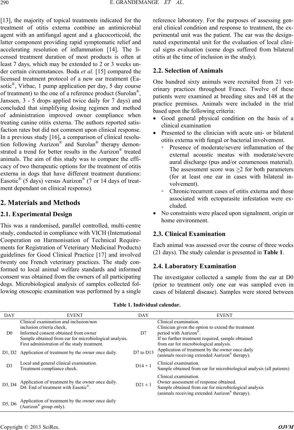 E. GRANDEMANGE ET AL. 290 [13], the majority of topical treatments indicated for the treatment of otitis externa combine an antimicrobial agent with an antifungal agent and a glucocorticoid, the latter component providing rapid symptomatic relief and accelerating resolution of inflammation [14]. The li- censed treatment duration of most products is often at least 7 days, which may be extended to 2 or 3 weeks un- der certain circumstances. Boda et al. [15] compared the licensed treatment protocol of a new ear treatment (Ea- sotic®, Virbac, 1 pump application per day, 5 day course of treatment) to the one of a reference product (Surolan®, Janssen, 3 - 5 drops applied twice daily for 7 days) and concluded that simplifying dosing regimen and method of administration improved owner compliance when treating canine otitis externa. The authors reported satis- faction rates but did not comment upon clinical response. In a previous study [16], a comparison of clinical resolu- tion following Aurizon® and Surolan® therapy demon- strated a trend for better results in the Aurizon® treated animals. The aim of this study was to compare the effi- cacy of two therapeutic options for the treatment of otitis externa in dogs that have different treatment durations: Easotic® (5 days) versus Aurizon® (7 or 14 days of treat- ment dependant on clinical response). 2. Materials and Methods 2.1. Experimental Design This was a randomised, parallel controlled, multi-centre study, conducted in compliance with VICH (International Cooperation on Harmonisation of Technical Require- ments for Registration of Veterinary Medicinal Products) guidelines for Good Clinical Practice [17] and involved twenty one French veterinary practices. The study con- formed to local animal welfare standards and informed consent was obtained from the owners of all participating dogs. Microbiological analysis of samples collected fol- lowing otoscopic examination was performed by a single reference laboratory. For the purposes of assessing gen- eral clinical condition and response to treatment, the ex- perimental unit was the patient. The ear was the design- nated experimental unit for the evaluation of local clini- cal signs evaluation (some dogs suffered from bilateral otitis at the time of inclusion in the study). 2.2. Selection of Animals One hundred sixty animals were recruited from 21 vet- erinary practices throughout France. Twelve of these patients were examined at breeding sites and 148 at the practice premises. Animals were included in the trial based upon the following criteria: Good general physical condition on the basis of a clinical examination Presented to the clinician with acute uni- or bilateral otitis externa with fungal or bacterial involvement. ◦ Presence of moderate/severe inflammation of the external acoustic meatus with moderate/severe aural discharge (pus and/or cerumenous material). The assessment score was >2 for both parameters (for at least one ear in cases with bilateral in- volvement). ◦ Chronic/recurrent cases of otitis externa and those associated with ectoparasite infestation were ex- cluded. No constraints were placed upon signalment, origin or home environment. 2.3. Clinical Examination Each animal was assessed over the course of three weeks (21 days). The study calendar is presented in Table 1. 2.4. Laboratory Examination The investigator collected a sample from the ear at D0 (prior to treatment only one ear was sampled even in cases of bilateral disease). Samples were stored between Table 1. Individual calendar. DAY EVENT DAY EVENT D0 Clinical examination and inclusion/non inclusion criteria check. Informed consent obtained from owner Sample obtained from ear for microbiological analysis. First administration of the study treatment. D7 Clinical examination. Clinician given the option to extend the treatment period with Aurizon®. If no further treatment required, sample obtained from ear for microbiological analysis. D1, D2 Application of treatment by the owner once daily. D7 to D13Application of treatment by the owner once daily (animals receiving extended Aurizon® therapy). D3 Local and general clinical examination. Treatment compliance check. D14 + 1Clinical examination. Sample obtained from ear for microbiological analysis (all patients) D3, D4 Application of treatment by the owner once daily. D4: End of treatment with Easotic®. D21 ± 1 Clinical examination. Owner assessment of response obtained. Sample obtained from ear for microbiological analysis (animals receiving extended Aurizon® therapy). D5, D6 Application of treatment by the owner once daily (Aurizon® group only). Copyright © 2013 SciRes. OJVM 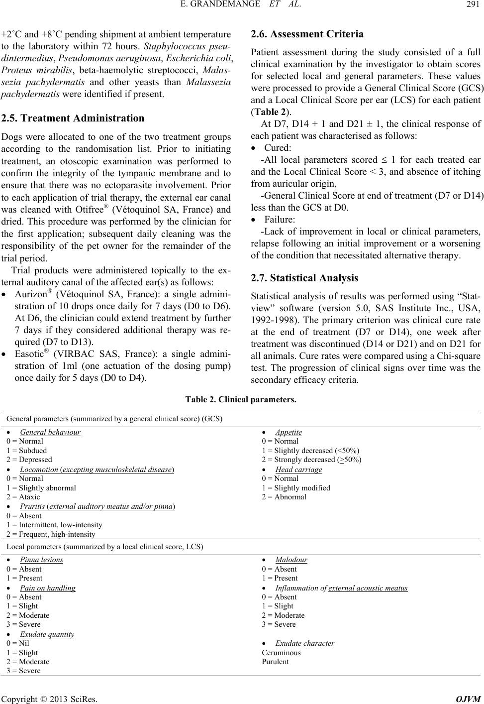 E. GRANDEMANGE ET AL. 291 +2˚C and +8˚C pending shipment at ambient temperature to the laboratory within 72 hours. Staphylococcus pseu- dintermedius, Pseudomonas aeruginosa, Escherichia coli, Proteus mirabilis, beta-haemolytic streptococci, Malas- sezia pachydermatis and other yeasts than Malassezia pachydermatis were identified if present. 2.5. Treatment Administration Dogs were allocated to one of the two treatment groups according to the randomisation list. Prior to initiating treatment, an otoscopic examination was performed to confirm the integrity of the tympanic membrane and to ensure that there was no ectoparasite involvement. Prior to each application of trial therapy, the external ear canal was cleaned with Otifree® (Vétoquinol SA, France) and dried. This procedure was performed by the clinician for the first application; subsequent daily cleaning was the responsibility of the pet owner for the remainder of the trial period. Trial products were administered topically to the ex- ternal auditory canal of the affected ear(s) as follows: Aurizon® (Vétoquinol SA, France): a single admini- stration of 10 drops once daily for 7 days (D0 to D6). At D6, the clinician could extend treatment by further 7 days if they considered additional therapy was re- quired (D7 to D13). Easotic® (VIRBAC SAS, France): a single admini- stration of 1ml (one actuation of the dosing pump) once daily for 5 days (D0 to D4). 2.6. Assessment Criteria Patient assessment during the study consisted of a full clinical examination by the investigator to obtain scores for selected local and general parameters. These values were processed to provide a General Clinical Score (GCS) and a Local Clinical Score per ear (LCS) for each patient (Table 2). At D7, D14 + 1 and D21 ± 1, the clinical response of each patient was characterised as follows: Cured: -All local parameters scored 1 for each treated ear and the Local Clinical Score < 3, and absence of itching from auricular origin, -General Clinical Score at end of treatment (D7 or D14) less than the GCS at D0. Failure: -Lack of improvement in local or clinical parameters, relapse following an initial improvement or a worsening of the condition that necessitated alternative therapy. 2.7. Statistical Analysis Statistical analysis of results was performed using “Stat- view” software (version 5.0, SAS Institute Inc., USA, 1992-1998). The primary criterion was clinical cure rate at the end of treatment (D7 or D14), one week after treatment was discontinued (D14 or D21) and on D21 for all animals. Cure rates were compared using a Chi-square test. The progression of clinical signs over time was the secondary efficacy criteria. Table 2. Clinical parameters. General parameters (summarized by a general clinical score) (GCS) General behaviour 0 = Normal 1 = Subdued 2 = Depressed Appetite 0 = Normal 1 = Slightly decreased (<50%) 2 = Strongly decreased (>50%) Locomotion (excepting musculoskeletal disease) 0 = Normal 1 = Slightly abnormal 2 = Ataxic Head carriage 0 = Normal 1 = Slightly modified 2 = Abnormal Pruritis (ext ernal auditory meatus and/or pinna) 0 = Absent 1 = Intermittent, low-intensity 2 = Frequent, high-intensity Local parameters (summarized by a local clinical score, LCS) Pinna lesions 0 = Absent 1 = Present Malodour 0 = Absent 1 = Present Pain on handling 0 = Absent 1 = Slight 2 = Moderate 3 = Severe Inflammation of external acoustic meat us 0 = Absent 1 = Slight 2 = Moderate 3 = Severe Exudate quantity 0 = Nil 1 = Slight 2 = Moderate 3 = Severe Exudate character Ceruminous Purulent Copyright © 2013 SciRes. OJVM 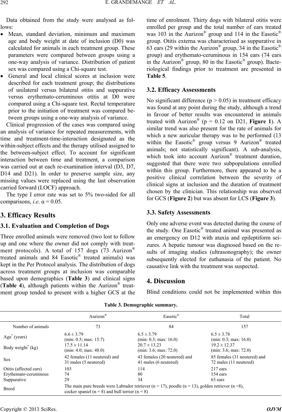 E. GRANDEMANGE ET AL. 292 Data obtained from the study were analysed as fol- lows: Mean, standard deviation, minimum and maximum age and body weight at date of inclusion (D0) was calculated for animals in each treatment group. These parameters were compared between groups using a one-way analysis of variance. Distribution of patient sex was compared using a Chi-square test. General and local clinical scores at inclusion were described for each treatment group; the distributions of unilateral versus bilateral otitis and suppurative versus erythemato-ceruminous otitis at D0 were compared using a Chi-square test. Rectal temperature prior to the initiation of treatment was compared be- tween groups using a one-way analysis of variance. Clinical progression of the cases was compared using an analysis of variance for repeated measurements, with time and treatment-time-interaction designated as the within-subject effects and the therapy utilised assigned to the between-subject effect. To account for significant interaction between time and treatment, a comparison was carried out at each re-examination interval (D3, D7, D14 and D21). In order to preserve sample size, any missing values were replaced using the last observation carried forward (LOCF) approach. The type I error rate was set to 5% two-sided for all comparisons, i.e. α = 0.05. 3. Efficacy Results 3.1. Evaluation and Completion of Dogs Three enrolled animals were removed (two lost to follow up and one where the owner did not comply with treat- ment protocols). A total of 157 dogs (73 Aurizon® treated animals and 84 Easotic® treated animals) was kept in the Per Protocol analysis. The distribution of dogs across treatment groups at inclusion was comparable based upon demographics (Table 3) and clinical signs (Table 4), although patients within the Aurizon® treat- ment group tended to present with a higher GCS at the time of enrolment. Thirty dogs with bilateral otitis were enrolled per group and the total number of ears treated was 103 in the Aurizon® group and 114 in the Easotic® group. Otitis externa was characterised as suppurative in 63 ears (29 within the Aurizon® group, 34 in the Easotic® group) and erythemato-ceruminous in 154 ears (74 ears in the Aurizon® group, 80 in the Easotic® group). Bacte- riological findings prior to treatment are presented in Table 5. 3.2. Efficacy Assessments No significant difference (p > 0.05) in treatment efficacy was found at any point during the study, although a trend in favour of better results was encountered in animals treated with Aurizon® (p = 0.12 on D21, Figure 1). A similar trend was also present for the rate of animals for which a new auricular therapy was to be performed (13 within the Easotic® group versus 9 Aurizon® treated animals; not statistically significant). A sub-analysis, which took into account Aurizon® treatment duration, suggested that there were two subpopulations enrolled within this group. Furthermore, there appeared to be a positive clinical correlation between the severity of clinical signs at inclusion and the duration of treatment chosen by the clinician. This relationship was observed for GCS (Figure 2) but was absent for LCS (Figure 3). 3.3. Safety Assessments Only one adverse event was detected during the course of the study. One Easotic® treated animal was presented as an emergency on D12 with ataxia and epileptiform sei- zures. A hepatic tumour was diagnosed based on the re- sults of imaging studies (ultrasonography); the owner subsequently elected for euthanasia of the patient. No causative link with the treatment was suspected. 4. Discussion Blind conditions could not be implemented within this Table 3. Demographic summary. Aurizon® Easotic® Total Number of animals 73 84 157 Age* (years) 6.6 ± 3.79 (min: 0.5; max: 15.7) 6.5 ± 3.79 (min: 0.3; max: 16.0) 6.5 ± 3.78 (min: 0.3; max: 16.0) Body weight* (kg) 17.5 ± 11.14 (min: 4.0; max: 48.0) 20.7 ± 13.23 (min: 3.6; max: 72.0) 19.2 ± 12.37 (min: 3.6; max: 72.0) Sex 42 females (11 neutered) and 31 males (5 neutered) 43 females (20 neutered) and 41 males (6 neutered) 85 females (31 neutered) and 72 males (11 neutered) Otitis (affected ears) Erythemato-ceruminous Suppurative 103 74 29 114 80 34 217 ears 154 ears 63 ears Breed The main pure breeds were Labrador retriever (n = 17), poodle (n = 13), golden retriever (n =8), cocker spaniel (n = 8) and bull terrier (n = 8) Copyright © 2013 SciRes. OJVM 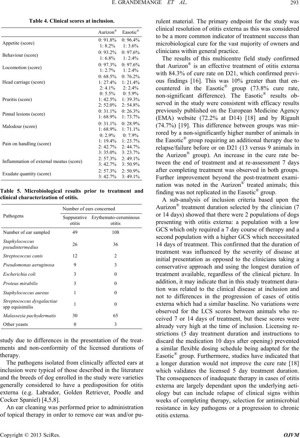 E. GRANDEMANGE ET AL. 293 Table 4. Clinical scores at inclusion. Aurizon® Easotic® Appetite (score) 0: 91.8% 1: 8.2% 0: 96.4% 1: 3.6% Behaviour (score) 0: 93.2% 1: 6.8% 0: 97.6% 1: 2.4% Locomotion (score) 0: 97.3% 1: 2.7% 0: 97.6% 1: 2.4% Head carriage (score) 0: 68.5% 1: 27.4% 2: 4.1% 0: 76.2% 1: 21.4% 2: 2.4% Pruritis (score) 0: 5.5% 1: 42.5% 2: 52.0% 0: 5.9% 1: 39.3% 2: 54.8% Pinnal lesions (score) 0: 31.1% 1: 68.9% 0: 26.3% 1: 73.7% Malodour (score) 0: 31.1% 1: 68.9% 0: 28.9% 1: 71.1% Pain on handling (score) 0: 2.9% 1: 19.4% 2: 42.7% 3: 35.0% 0: 7.9% 1: 23.7% 2: 44.7% 3: 23.7% Inflammation of external meatus (score) 2: 57.3% 3: 42.7% 2: 49.1% 3: 50.9% Exudate quantity (score) 2: 57.3% 3: 42.7% 2: 50.9% 3: 49.1% Table 5. Microbiological results prior to treatment and clinical characterization of otitis. Number of ears concerned Pathogens Suppurative otitis Erythemato-ceruminous otitis Number of ear sampled 49 108 Staphylococcus pseudintermedius 26 36 Streptococcus canis 12 2 Pseudomonas aeru gi n os a 9 3 Escherichia coli 3 0 Proteus mirabilis 3 0 Staphylococcus aureus 1 0 Streptococcus dysgalactiae spp equisimilis 1 0 Malassezia pachydermatis 30 65 Other yeasts 0 3 study due to differences in the presentation of the treat- ments and non-conformity of the licensed durations of therapy. The pathogens isolated from clinically affected ears at inclusion were typical of those described in the literature and the breeds of dog enrolled in the study were varieties generally considered to have a predisposition for otitis externa (e.g. Labrador, Golden Retriever, Poodle and Cocker Spaniel) [4,5,8]. An ear cleaning was performed prior to administration of topical therapy in order to remove ear wax and/or pu- rulent material. The primary endpoint for the study was clinical resolution of otitis externa as this was considered to be a more common indicator of treatment success than microbiological cure for the vast majority of owners and clinicians within general practice. The results of this multicentre field study confirmed that Aurizon® is an effective treatment of otitis externa with 84.3% of cure rate on D21, which confirmed previ- ous findings [16]. This was 10% greater than that en- countered in the Easotic® group (73.8% cure rate, non-significant difference). The Easotic® results ob- served in the study were consistent with efficacy results previously published on the European Medicine Agency (EMA) website (72.2% at D14) [18] and by Rigault (74.7%) [19]. This difference between groups was mir- rored by a non-significantly higher number of animals in the Easotic® group requiring an additional therapy due to relapse/failure before or on D21 (13 versus 9 animals in the Aurizon® group). An increase in the cure rate be- tween the end of treatment and at re-assessment 7 days after completing treatment was observed in both groups. Further improvement beyond the post-treatment exami- nation was noted in the Aurizon® treated animals; this finding was not replicated in the Easotic® group. A sub-analysis of inclusion criteria based upon the Aurizon® treatment duration selected by the clinician (7 or 14 days) showed that there were 2 populations of dogs presenting with otitis externa: a population with a low GCS which only required a 7 day course of therapy and a second population with a higher GCS which necessitated 14 days of treatment. This confirmed that the duration of treatment was influenced by the severity of disease at initial presentation as opposed to the clinicians taking a conservative approach and using the longest duration of treatment available, regardless of the clinical picture. In addition, it may indicate that in this study treatment dura- tion was related to the clinical disease at inclusion and not to differences in the progression of cases of otitis externa which had a similar baseline. No variations were observed for the LCS scores between animals who re- ceived 7 or 14 days of treatment, but these scores were already very high at the time of inclusion. Licensing re- strictions (5 day treatment duration and instructions to discard the medication 10 days after opening) prevented a similar flexible dosing schedule being adapted for the Easotic® group. Furthermore, studies have indicated that a longer duration would not improve the cure rate [18] which validates the licensed 5 day treatment duration. The consequences of inadequate therapy in cases of otitis externa are largely dependant upon the underlying aeti- ology but can include relapse of clinical signs within weeks of completing therapy, selection for antimicrobial resistance in key pathogens or a progression to chronic otitis externa. Copyright © 2013 SciRes. OJVM 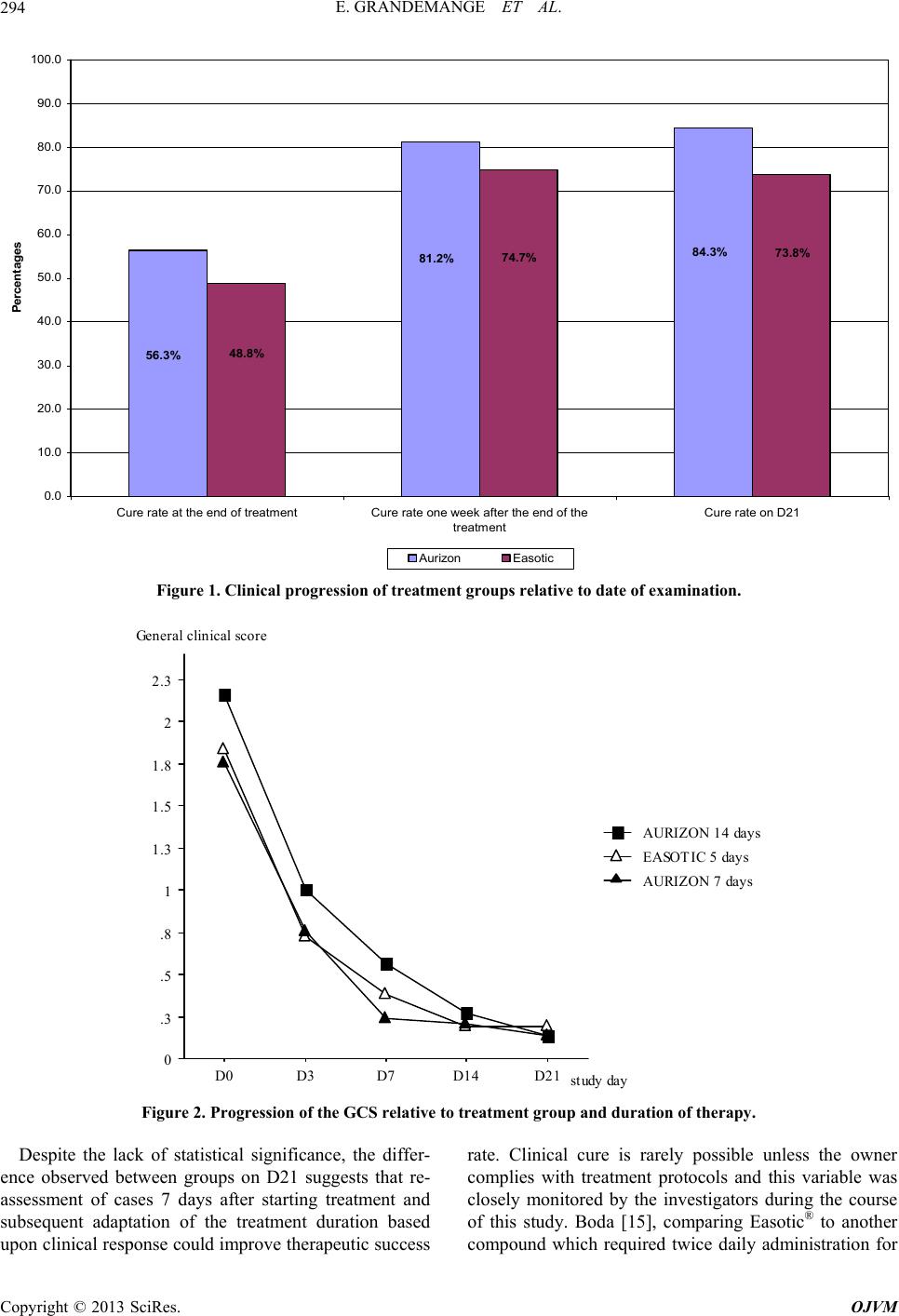 E. GRANDEMANGE ET AL. 294 56.3% 81.2% 84.3%73.8% 48.8% 74.7% 0.0 10.0 20.0 30.0 40.0 50.0 60.0 70.0 80.0 90.0 100.0 Cure rate at the end of treatmentCure rate one week after the end of the treatment Cure rate on D21 Percentages Aurizon Easotic Figure 1. Clinical progression of treatment groups relative to date of examination. 0 .3 .5 .8 1 1.3 1.5 1.8 2 2.3 D0D3D7D14 D21 AURIZON 7 days EASOTIC 5 days AURIZON 14 days study day General clinical score Figure 2. Progression of the GCS relative to treatment group and duration of therapy. Despite the lack of statistical significance, the differ- ence observed between groups on D21 suggests that re- assessment of cases 7 days after starting treatment and subsequent adaptation of the treatment duration based upon clinical response could improve therapeutic success rate. Clinical cure is rarely possible unless the owner complies with treatment protocols and this variable was closely monitored by the investigators during the course of this study. Boda [15], comparing Easotic® to another ompound which required twice daily administration for c Copyright © 2013 SciRes. OJVM 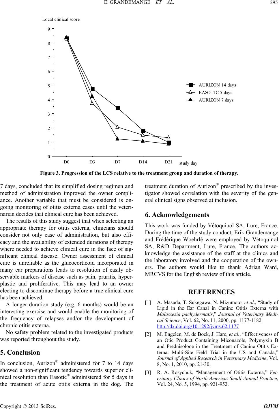 E. GRANDEMANGE ET AL. 295 0 1 2 3 4 5 6 7 8 9 D0D3 D7 D14 D2 1 AURIZON 7 days EASOTIC 5 days AURIZON 14 days study day Local clinical score Figure 3. Progression of the LCS relative to the treatment group and duration of therapy. 7 days, concluded that its simplified dosing regimen and method of administration improved the owner compli- ance. Another variable that must be considered is on- going monitoring of otitis externa cases until the veteri- narian decides that clinical cure has been achieved. The results of this study suggest that when selecting an appropriate therapy for otitis externa, clinicians should consider not only ease of administration, but also effi- cacy and the availability of extended durations of therapy where needed to achieve clinical cure in the face of sig- nificant clinical disease. Owner assessment of clinical cure is unreliable as the glucocorticoid incorporated in many ear preparations leads to resolution of easily ob- servable markers of disease such as pain, pruritis, hyper- plastic and proliferative. This may lead to an owner electing to discontinue therapy before a true clinical cure has been achieved. A longer duration study (e.g. 6 months) would be an interesting exercise and would enable the monitoring of the frequency of relapses and/or the development of chronic otitis externa. No safety problem related to the investigated products was reported throughout the study. 5. Conclusion In conclusion, Aurizon® administered for 7 to 14 days showed a non-significant tendency towards superior cli- nical resolution than Easotic® administered for 5 days in the treatment of acute otitis externa in the dog. The treatment duration of Aurizon® prescribed by the inves- tigator showed correlation with the severity of the gen- eral clinical signs observed at inclusion. 6. Acknowledgements This work was funded by Vétoquinol SA, Lure, France. During the time of the study conduct, Erik Grandemange and Frédérique Woehrlé were employed by Vétoquinol SA, R&D Department, Lure, France. The authors ac- knowledge the assistance of the staff at the clinics and the laboratory involved and the cooperation of the own- ers. The authors would like to thank Adrian Ward, MRCVS for the English review of this article. REFERENCES [1] A. Masuda, T. Sukegawa, N. Mizumoto, et al., “Study of Lipid in the Ear Canal in Canine Otitis Externa with Malassezia pachydermatis,” Journal of Veterinary Medi- cal Science, Vol. 62, No. 11, 2000, pp. 1177-1182. http://dx.doi.org/10.1292/jvms.62.1177 [2] M. Engelen, M. de Bock, J. Hare, et al., “Effectiveness of an Otic Product Containing Miconazole, Polymyxin B and Prednisolone in the Treatment of Canine Otitis Ex- terna: Multi-Site Field Trial in the US and Canada,” Journal of Applied Research in Veterinary Medicine, Vol. 8, No. 1, 2010, pp. 21-30. [3] R. A. Rosychuk, “Management of Otitis Externa,” Vet- erinary Clinics of North America: Small Animal Practice, Vol. 24, No. 5, 1994, pp. 921-952. Copyright © 2013 SciRes. OJVM  E. GRANDEMANGE ET AL. 296 [4] H. M. Hayes Jr., L. W. Pickle and G. P. Wilson, “Effects of Ear Type and Weather on the Hospital Prevalence of Canine Otitis Externa,” Research in Veterinary Science, Vol. 42, No. 3, 1987, pp. 294-298. [5] E. J. Rosser Jr., “Causes of Otitis Externa,” Veterinary Clinics of North America: Small Animal Practice, Vol. 34, No. 2, 2004, pp. 459-468. http://dx.doi.org/10.1016/j.cvsm.2003.10.006 [6] J. J. Campbell, K. S. Coyner, S. C. Rankin, et al., “Eva- luation of Fungal Flora in Normal and Diseased Canine Ears,” Veterinary Dermatology, Vol. 21, No. 6, 2010, pp. 619-625. http://dx.doi.org/10.1111/j.1365-3164.2010.00927.x [7] J. R. August, “Otitis Externa. A Disease of Multifactorial Etiology,” Veterinary Clinics of North America: Small Animal Practi c e, Vol. 18, No. 4, 1988, pp. 731-742. [8] J. J. Kowalski, “The Microbial Environment of the Ear Canal in Health and Disease,” Veterinary Clinics of North America: Small Animal Practice, Vol. 18, No. 4, 1988, pp. 743-754. [9] M. N. Saridomichelakis, R. Farmaki, L. S. Leontides, et al., “Aetiology of Canine Otitis Externa: A Retrospective Study,” Veterinary Dermatology, Vol. 18, No. 5, 2007, pp. 341-347. http://dx.doi.org/10.1111/j.1365-3164.2007.00619.x [10] H. Zamankhan Malayeri, S. Jamshidi and T. Zahraei Sale- hi, “Identification and Antimicrobial Susceptibility Pat- terns of Bacteria Causing Otitis Externa in Dogs,” Vet- erinary Research Communications, Vol. 34, No. 5, 2010, pp. 435-444. http://dx.doi.org/10.1007/s11259-010-9417-y [11] L. S. Jacobson, “Diagnosis and Medical Treatment of Otitis Externa in the Dog and Cat,” Journal of the South African Veterinary Association , Vol. 73, No. 4, 2002, pp. 162-170. http://dx.doi.org/10.4102/jsava.v73i4.581 [12] D. O. Morris, “Medical Therapy of Otitis Externa and Otitis Media,” Veterinary Clinics of North America: Small Animal Practice, Vol. 34, No. 2, 2004, pp. 541-555. http://dx.doi.org/10.1016/j.cvsm.2003.10.009 [13] G. Lehner, C. Sauter Louis and R. S. Mueller, “Repro- ducibility in Ear Cytology in Dogs with Otitis Externa,” Veterinary Record, Vol. 167, No. 1, 2010, pp. 23-26. http://dx.doi.org/10.1136/vr.c3523 [14] E. Bensignor and E. Grandemange, “Comparison of an Antifungal Agent with a Mixture of Antifungal, Antibi- otic and Corticosteroid Agents for the Treatment of Malassezia Species Otitis in Dogs,” Veterinary Record, Vol. 158, No. 6, 2006, pp. 193-195. http://dx.doi.org/10.1136/vr.158.6.193 [15] C. Boda, P. Liege and C. Reme, “Evaluation of Owner Compliance with Topical Treatment of Acute Otitis Ex- terna in Dogs: A Comparative Study of Two Auricular Formulations,” Journal of Applied Research in Veterinary Medicine, Vol. 9, No. 2, 2011, pp. 157-165 [16] S. Rougier, D. Borell, S. Pheulpin, et al., “A Comparative Study of Two Antimicrobial/Anti-Inflammatory Formula- tions in the Treatment of Canine Otitis Externa,” Veteri- nary Dermatology, Vol. 16, No. 5, 2005, pp. 299-307. http://dx.doi.org/10.1111/j.1365-3164.2005.00465.x [17] VICH, “International Co-Operation on Harmonisation of Technical Requirements for Registration of Veterinary Medical Products, Good Clinical Practice,” June 2000 ef- fective 01 July 2001. www.ema.europa.eu/docs/en_GB/document_library/Scie ntific_guideline/2009/10/WC500004343.pdf [18] EMA Easotic European Public Assessment Report. www.ema.europa.eu/docs/en_GB/document_library/EPA R__Scientific_Discussion/veterinary/000140/WC500063 875.pdf [19] D. Rigaut, A. Sanquer, L. Maynard, et al., “Efficacy of a Topical Ear Formulation with a Pump Delivery System for the Treatment of Infectious Otitis Externa in Dogs: a Randomized Controlled Trial,” Journal of Applied Re- search in Veterinary Medicine, Vol. 9, No. 1, 2011, pp. 15-28. Copyright © 2013 SciRes. OJVM |