

Paper Menu >>
Journal Menu >>
 World Journal of Cardiovascular Diseases, 2013, 3, 471-475 WJCD http://dx.doi.org/10.4236/wjcd.2013.37074 Published Online October 2013 (http://www.scirp.org/journal/wjcd/) Plasma natriuretic peptides during supraventricular tachycardia: A study in patients with atrioventricular nodal reentry tachycardia Redi Pecini1*, Steen Pehrson1, Xu Chen1, Anna Margrethe Thøgersen2, Andreas Kjær3, Jesper Hastrup Svendsen1,4,5 1Department of Cardiology, The Heart Centre, Rigshospitalet, University of Copenhagen, Copenhagen, Denmark 2Department of Cardiology, Center for Cardiovascular Research, Århus University Hospital, Aalborg Hospital, Aalborg, Denmark 3Department of Clinical Physiology, Nuclear Medicine and PET & Cluster for Molecular Imaging, Rigshospitalet, University of Co- penhagen, Copenhagen, Denmark 4The Danish National Research Foundation Centre for Cardiac Arrhythmia, Copenhagen, Denmark 5Department of Surgery and Medicine, Faculty of Health Sciences, University of Copenhagen, Copenhagen, Denmark Email: *[email protected] Received 16 August 2013; revised 15 September 2013; accepted 25 September 2013 Copyright © 2013 Redi Pecini et al. This is an open access article distributed under the Creative Commons Attribution License, which permits unrestricted use, distribution, and reproduction in any medium, provided the original work is properly cited. ABSTRACT Aims: To characterize the plasma levels of the atrial natriuretic peptide (ANP) and brain natriuretic pep- tide (BNP) in patients with atrioventricular nodal reentry tachycardia (AVNRT), we measured the plas- ma levels of these peptides before and during ta- chycardia. Methods: We included 10 consecutive pa- tients scheduled for ablation of typical AVNRT with- out structural heart disease. Catheters were inserted in the femoral artery, femoral vein, and coronary sinus (CS) prior to the ablation procedure. Blood samples were drawn before and after 3 min of tachy- cardia to measure plasma levels of ANP and BNP. Right atrial pressure (RAP) was measured at base- line. Results: Of the 10 patients, in three patients it was not possible to induce tachycardia leaving a total of 7 patients available for analysis. Mean age of the seven included patients was 40 ± 12 years (mean ± SD), five were female. ANP levels increased signifi- cantly during tachycardia in the artery (p = 0.0009) and vein (p = 0.003), but only borderline in CS (p = 0.09). BNP levels did not change during tachycardia in any location. Conclusion: ANP levels measured in the peripheral circulation increased acutely during tachycardia due to AVNRT. BNP levels did not in- crease. Keywords: Atrioventricular Nodal Reentry Tachycardia; Atrial Natriuretic Peptide; Brain Natriuretic Peptide 1. INTRODUCTION Brain natriuretic peptide (BNP) and to a lesser extent atrial natriuretic peptide (ANP) have shown to be of value in the diagnosis of heart failure [1,2]. Furthermore, increased levels of BNP have been shown to predict ad- verse events in patients with heart failure [3,4], coronary heart disease [5,6] and mitral regurgitation [7]. The importance of ANP and BNP in patients with tachycardias is less well known. In patients with atrial fibrillation, levels of BNP are increased [8], and the lev- els are higher in patients with persistent and permanent AF than those with paroxysmal AF [9]. In addition, one study of patients with AF found higher levels of BNP in the coronary sinus than in the peripheral circulation. The studies of BNP in patients with supraventricular tachy- cardias are of limited number [10,11]. The measurement of BNP levels in these studies was only done from pe- ripheral blood. As such, the levels of ANP and BNP in patients with supraventricular arrhythmias have not been well charac- terized. For this reason, we undertook the present study in patients with atrioventricular nodal reentry tachycardia (AVNRT) without concomitant structural heart disease and without the influence of pre-study medication. We measured ANP and BNP levels directly at the coronary sinus, as well as from arterial and venous peripheral blood with the purpose of evaluating the possible cardiac release. *Corresponding autho r . Published Online October 2013 in SciRes. http://www.scirp.org/journal/wjcd  R. Pecini et al. / World Journal of Cardiovascular Diseases 3 (2013) 471-475 472 2. METHODS 2.1. Study Population We included consecutive patients referred to our center for ablation of suspected AVNRT. The patients did not have any other concurrent diseases and did not take any medications. They had not had symptoms of arrhythmia for at least one week prior to the ablation procedure. We performed an echocardiography in each patient the day before the ablation procedure. All patients had normal left ventricular ejection fraction (LVEF) and no signs of structural heart disease. Creatinine levels were also nor- mal. The study was carried out in accordance to the decla- ration of Helsinki and was approved by the local ethical committee for Copenhagen, study number KF01-046/04. All patients gave written informed consent. 2.2. Ablation Procedure All antiarrhythmic drugs were withdrawn at least five plasma half-lives prior to the procedure. Two 7 French sheaths were inserted; one in each femoral vein, and a 5 French sheath was positioned in the femoral artery. An additional sheath was also inserted in the coronary sinus. The femoral vein sheaths were used for catheter insertion and for the blood samples. The femoral artery and the coronary sinus sheaths were only used for the purpose of blood sampling. After insertion of the sheaths and positioning of catheters, the right atrial pressure (RAP) measured and then the first series of blood samples was performed. Blood was drawn sequentially in the same order in each patient, first from the coronary sinus (CS), then from the femoral artery and then from the femoral vein. Sampling from each location took approximately 30 seconds. After the first blood sampling (baseline), programmed electri- cal stimulation was used to induce the arrhythmia. If the first electrical programmed stimulation was unsuccessful, a second programmed stimulation was tried after infu- sion of isoprotenerol to achieve an increase in heart rate to about 100 beats per minute. Once the tachycardia oc- curred, it was allowed to continue for at least three min- utes and then the blood sampling was repeated. Thereaf- ter, the arrhythmia was stopped and the ablation of the slow pathway at the atrioventricular node was performed. The right atrial pressure (RAP) was only measured at the baseline. No complications occurred during or after the procedure. 2.3. ANP and BNP Analysis Ten ml of blood was drawn from each sheath, before and during tachycardia (a total of 60 ml) into tubes contain- ing EDTA and 500 μl aprotinin (Trasylol® 500 KIU/ml; Bayer, Leverkusen, Germany) and centrifuged at 2000 × g for 15 min. Then plasma was transferred to glass tubes and immediately frozen and kept at −80˚C until ana- lyzed. BNP was measured by an automated two-site sand- wich immunoassay technique using chemiluminescence (ADVIA Centaur; Siemens Healthcare Diagnostics). The physiologically active C-terminal peptide of BNP was measured (77 - 108). The sensitivity of the assay was 2 pg/ml, and the intra-assay and interassay coefficients of variation were 1.2% and 2.3%, respectively. ANP was measured by an enzyme immunoassay ac- cording to the instructions of the manufacturer (Bio- medica Medicinprodukte GmbH, Vienna, Austria). The lower detection limit for NT-pro-ANP (1-98) was 0.05 nmol/l, and the intra-assay and interassay coefficients of variation were 2% and 4%, respectively. The plasma values of ANP and BNP at the baseline were compared with the values during tachycardia in each patient. 2.4. Statistical Analysis Continuous variables are presented median values and inter-quartile range, and discrete variables are presented as percent. We tested the ANP and BNP values for nor- mal distribution visually with histogram graphs and with the use of Kolmogorov-Smirnov test for normal distribu- tion. Not all the data showed normal distribution. There- fore, we performed logarithmic transformation of the values, after which all values showed normal distribution using the same statistical tests. We used paired t-test for the comparison of ANP and BNP logarithmic values be- fore and during tachycardia. For the correlation analyses, we used the general linear model. A p-value less than 0.05 was considered to indicate statistical significance. 3. RESULTS We initially included 10 patients in the study. They all had episodes with palpitations and 12-lead ECGs per- formed during the episodes had shown short RP tachy- cardia. The arrhythmia could not be induced in three patients. These patients were excluded from the study. In all the inducible patients the induced arrhythmia was the clinically relevant tachycardia. There was no evidence of an accessory pathway between the atria and the ventri- cles. The ablation of the slow pathway for AVNRT at the atrioventricular node was successful in all the patients and the arrhythmia could not be induced thereafter. The baseline data of the seven included patients are presented in Ta b l e 1. Baseline values of ANP and BNP were slightly higher in the coronary sinus, but they were not significantly different from the values in the femoral artery or vein. ANP levels increased significantly during Copyright © 2013 SciRes. WJCD 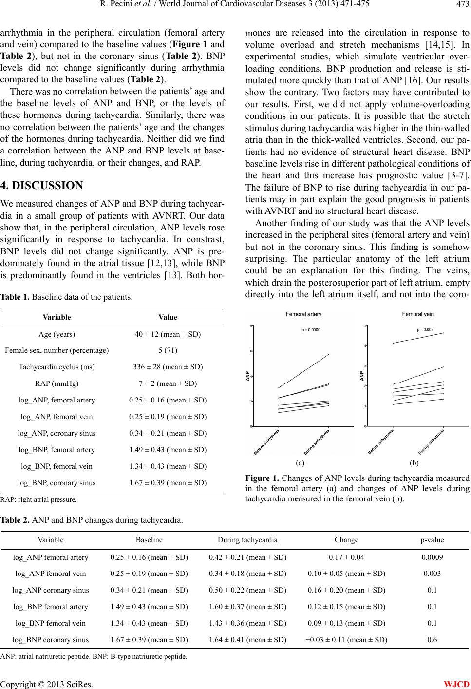 R. Pecini et al. / World Journal of Cardiovascular Diseases 3 (2013) 471-475 Copyright © 2013 SciRes. 473 arrhythmia in the peripheral circulation (femoral artery and vein) compared to the baseline values (Figure 1 and Table 2), but not in the coronary sinus (Table 2). BNP levels did not change significantly during arrhythmia compared to the baseline values (Table 2). mones are released into the circulation in response to volume overload and stretch mechanisms [14,15]. In experimental studies, which simulate ventricular over- loading conditions, BNP production and release is sti- mulated more quickly than that of ANP [16]. Our results show the contrary. Two factors may have contributed to our results. First, we did not apply volume-overloading conditions in our patients. It is possible that the stretch stimulus during tachycardia was higher in the thin-walled atria than in the thick-walled ventricles. Second, our pa- tients had no evidence of structural heart disease. BNP baseline levels rise in different pathological conditions of the heart and this increase has prognostic value [3-7]. The failure of BNP to rise during tachycardia in our pa- tients may in part explain the good prognosis in patients with AVNRT and no structural heart disease. There was no correlation between the patients’ age and the baseline levels of ANP and BNP, or the levels of these hormones during tachycardia. Similarly, there was no correlation between the patients’ age and the changes of the hormones during tachycardia. Neither did we find a correlation between the ANP and BNP levels at base- line, during tachycardia, or their changes, and RAP. 4. DISCUSSION We measured changes of ANP and BNP during tachycar- dia in a small group of patients with AVNRT. Our data show that, in the peripheral circulation, ANP levels rose significantly in response to tachycardia. In constrast, BNP levels did not change significantly. ANP is pre- dominately found in the atrial tissue [12,13], while BNP is predominantly found in the ventricles [13]. Both hor- Another finding of our study was that the ANP levels increased in the peripheral sites (femoral artery and vein) but not in the coronary sinus. This finding is somehow surprising. The particular anatomy of the left atrium could be an explanation for this finding. The veins, which drain the posterosuperior part of left atrium, empty directly into the left atrium itself, and not into the coro- Table 1. Baseline data of the patients. Variable Value Age (years) 40 ± 12 (mean ± SD) Female sex, number (percentage) 5 (71) Tachycardia cyclus (ms) 336 ± 28 (mean ± SD) RAP (mmHg) 7 ± 2 (mean ± SD) log_ANP, femoral artery 0.25 ± 0.16 (mean ± SD) log_ANP, femoral vein 0.25 ± 0.19 (mean ± SD) log_ANP, coronary sinus 0.34 ± 0.21 (mean ± SD) log_BNP, femoral artery 1.49 ± 0.43 (mean ± SD) log_BNP, femoral vein 1.34 ± 0.43 (mean ± SD) log_BNP, coronary sinus 1.67 ± 0.39 (mean ± SD) (a) (b) Figure 1. Changes of ANP levels during tachycardia measured in the femoral artery (a) and changes of ANP levels during tachycardia measured in the femoral vein (b). RAP: right atrial pressure. Table 2. ANP and BNP changes during tachycardia. Variable Baseline During tachycardia Change p-value log_ANP femoral artery 0.25 ± 0.16 (mean ± SD) 0.42 ± 0.21 (mean ± SD) 0.17 ± 0.04 0.0009 log_ANP femoral vein 0.25 ± 0.19 (mean ± SD) 0.34 ± 0.18 (mean ± SD) 0.10 ± 0.05 (mean ± SD) 0.003 log_ANP coronary sinus 0.34 ± 0.21 (mean ± SD) 0.50 ± 0.22 (mean ± SD) 0.16 ± 0.20 (mean ± SD) 0.1 log_BNP femoral artery 1.49 ± 0.43 (mean ± SD) 1.60 ± 0.37 (mean ± SD) 0.12 ± 0.15 (mean ± SD) 0.1 log_BNP femoral vein 1.34 ± 0.43 (mean ± SD) 1.43 ± 0.36 (mean ± SD) 0.09 ± 0.13 (mean ± SD) 0.1 log_BNP coronary sinus 1.67 ± 0.39 (mean ± SD) 1.64 ± 0.41 (mean ± SD) −0.03 ± 0.11 (mean ± SD) 0.6 A NP: atrial natriuretic peptide. BNP: B-type natriuretic peptide. WJCD  R. Pecini et al. / World Journal of Cardiovascular Diseases 3 (2013) 471-475 474 nary sinus. This amount of blood empties then in the left ventricle. Thus, the majority of this amount of blood cir- culates directly into the periphery, and only a minor amount into the coronary artery system. One other ex- planation could be our sequential method of blood sam- pling, starting in the coronary sinus. In any case, there is little doubt that the increased levels of ANP are directly related to the tachycardia episode. Even in patients with lone atrial fibrillation, baseline BNP levels are increased [8], and these levels are even higher in patients with persistent and permanent atrial fibrillation [9,17,18]. On the other hand, atrial fibrillation is an arrhythmia, which is associated with considerable morbidity and mortality [19]. On the contrary, AVNRT is not associated with increased mortality and the treatment is usually guided by the burden of symptoms. Further- more, recent data have shown that the BNP levels in pa- tients with atrial fibrillation are especially increased in the coronary sinus [20]. We did not detect any increase of BNP in blood samples from coronary sinus. This is an- other fundamental difference between these two arrhy- thmias. Whether this difference in BNP levels can ex- plain the difference in the morbidity between atrial fib- rillation and AVNRT is unsure. However, we know that in other cardiac pathologies, morbidity and mortality risk is closely related to the levels of BNP [4-7]. 5. LIMITATIONS Our number of patients was limited. For this reason, we may have overlooked changes in BNP levels due to in- adequate power. It is also possible that if we had repeated the measurements of BNP later on we might have been able to detect a change in the BNP levels. 6. CONCLUSION Peripheral plasma levels of ANP but not BNP rose acutely in response to tachycardia in patients with AVNRT. This finding is in contrast to patients with chronic atrial fibrillation indicating the different pathophysiology in- volved in the two conditions. 7. ACKNOWLEDGMENTS Special thanks to Jens Peter Goetze MD, for his valuable comments on the manuscript. REFERENCES [1] Maisel, A.S., Krishnaswamy, P., Nowak, R.M., et al. (2002) Rapid measurement of B-type natriuretic peptide in the emergency diagnosis of heart failure. Ne w Engla nd Journal of Medicine, 347, 161-167. http://dx.doi.org/10.1056/NEJMoa020233 [2] McDonagh, T.A., Robb, S.D., Murdoch, D.R., et al. (1998) Biochemical detection of left-ventricular systolic dysfunction. Lancet, 351, 9-13. http://dx.doi.org/10.1016/S0140-6736(97)03034-1 [3] Berger, R., Huelsman, M., Strecker, K., et al. (2002) B- type natriuretic peptide predicts sudden death in patients with chronic heart failure. Circulation, 105, 2392-2397. http://dx.doi.org/10.1161/01.CIR.0000016642.15031.34 [4] Anand, I.S., Fisher, L.D., Chiang, Y.T., et al. (2003) Changes in brain natriuretic peptide and norepinephrine over time and mortality and morbidity in the Valsartan Heart Failure Trial (Val-HeFT). Circulation, 107, 1278- 1283. http://dx.doi.org/10.1161/01.CIR.0000054164.99881.00 [5] de Lemos, J.A., Morrow, D.A., Bentley, J.H., et al. (2001) The prognostic value of B-type natriuretic peptide in pa- tients with acute coronary syndromes. New England Journal of Medicine, 345, 1014-1021. http://dx.doi.org/10.1056/NEJMoa011053 [6] Schnabel, R., Lubos, E., Rupprecht, H.J., et al. (2006) B-type natriuretic peptide and the risk of cardiovascular events and death in patients with stable angina: Results from the AtheroGene study. Journal of the American Col- lege of Cardiology, 47, 552-558. http://dx.doi.org/10.1016/j.jacc.2005.09.039 [7] Detaint, D., Messika-Zeitoun, D., Avierinos, J.F., et al. (2005) B-type natriuretic peptide in organic mitral regur- gitation: Determinants and impact on outcome. Circula- tion, 111, 2391-2397. http://dx.doi.org/10.1161/01.CIR.0000164269.80908.9D [8] Li, J. and Wang, L. (2006) B-type natriuretic peptide levels in patients with paroxysmal lone atrial fibrillation. Heart Vessels, 21, 137-140. http://dx.doi.org/10.1007/s00380-005-0884-y [9] Yamada, T., Murakami, Y., Okada, T., et al. (2007) Plas- ma brain natriuretic peptide level after radiofrequency catheter ablation of paroxysmal, persistent, and perma- nent atrial fibrillation. Europace, 9, 770-774. http://dx.doi.org/10.1093/europace/eum157 [10] Chen, L., Wei, T., Zeng, C., et al. (2005) Effect of ra- diofrequency catheter ablation on plasma B-type natri- uretic peptide. Pacing and Clinical Electrophysiology, 28, 200-204. http://dx.doi.org/10.1111/j.1540-8159.2005.09386.x [11] Cakmak, N., Sayar, N., Cakmak, M., et al. (2008) Effects of radiofrequency catheter ablation on myocardial per- formance index and plasma NT-Pro-BNP levels in pa- tients with Wolff-Parkinson-White syndrome. Echocar- diography, 25, 692-698. http://dx.doi.org/10.1111/j.1540-8175.2008.00669.x [12] Kangawa, K. and Matsuo, H. (1984) Purification and complete amino acid sequence of alpha-human atrial na- triuretic polypeptide (alpha-hANP). Biochemical and Bio- physical Research Communications, 118, 131-139. http://dx.doi.org/10.1016/0006-291X(84)91077-5 [13] Ogawa, Y., Nakao, K., Mukoyama, M., et al. (1991) Na- triuretic peptides as cardiac hormones in normotensive and spontaneously hypertensive rats. The ventricle is a major site of synthesis and secretion of brain natriuretic peptide. Circulation Research, 69, 491-500. Copyright © 2013 SciRes. WJCD  R. Pecini et al. / World Journal of Cardiovascular Diseases 3 (2013) 471-475 475 http://dx.doi.org/10.1161/01.RES.69.2.491 [14] de Zeeuw, D., Janssen, W.M. and de Jong, P.E. (1992) Atrial natriuretic factor: Its (patho)physiological signifi- cance in humans. Kidney International, 41, 1115-1133. http://dx.doi.org/10.1038/ki.1992.172 [15] Yoshimura, M., Yasue, H., Okumura, K., et al. (1993) Different secretion patterns of atrial natriuretic peptide and brain natriuretic peptide in patients with congestive heart failure. Circulation, 87, 464-469. http://dx.doi.org/10.1161/01.CIR.87.2.464 [16] Nakagawa, O., Ogawa, Y., Itoh, H., et al. (1995) Rapid transcriptional activation and early mRNA turnover of brain natriuretic peptide in cardiocyte hypertrophy. Evi- dence for brain natriuretic peptide as an “emergency” cardiac hormone against ventricular overload. Kidney In- ternational, 96, 1280-1287. http://dx.doi.org/10.1172/JCI118162 [17] Therkelsen, S.K., Groenning, B.A., Kjaer, A., et al. (2008) ANP and BNP in atrial fibrillation before and after car- dioversion and their relationship to cardiac volume and function. International Journal of Cardiology, 127, 396- 399. http://dx.doi.org/10.1016/j.ijcard.2007.04.059 [18] Nilsson, B., Goetze, J.P., Chen, X., et al. (2009) Increased NT-pro-B-type natriuretic peptide independently predicts outcome following catheter ablation of atrial fibrillation. Scandinavian Journal of Clinical & Laboratory Investi- gation, 69, 843-850. http://dx.doi.org/10.3109/00365510903318209 [19] Camm, A.J., Kirchhof, P., Lip, G.Y., et al. (2010) Guide- lines for the management of atrial fibrillation: The Task Force for the Management of Atrial Fibrillation of the European Society of Cardiology (ESC). Europace, 12, 1360-1420. http://dx.doi.org/10.1093/europace/euq350 [20] Gould, P.A., Gula, L.J., Bhayana, V., et al. (2010) Char- acterization of cardiac brain natriuretic peptide release in patients with paroxysmal atrial fibrillation undergoing left atrial ablation. Circulation: Arrhythmia and Electro- physiology, 3, 18-23. http://dx.doi.org/10.1161/CIRCEP.108.831586 Copyright © 2013 SciRes. WJCD |