

Paper Menu >>
Journal Menu >>
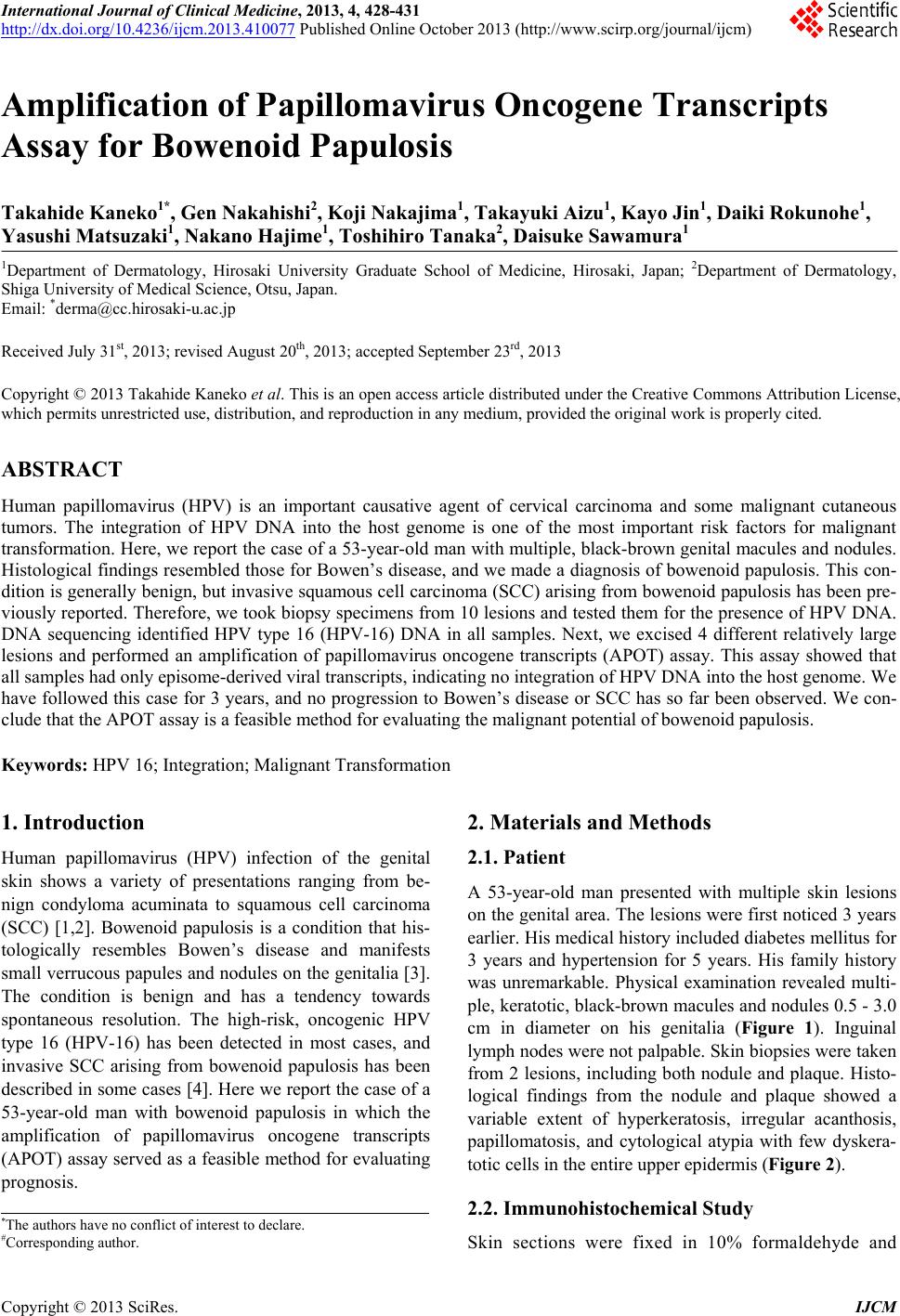 International Journal of Clinical Medicine, 2013, 4, 428-431 http://dx.doi.org/10.4236/ijcm.2013.410077 Published Online October 2013 (http://www.scirp.org/journal/ijcm) Amplification of Papillomavirus Oncogene Transcripts Assay for Bowenoid Papulosis Takahide Kaneko1*, Gen Nakahishi2, Koji Nakajima1, Takayuki Aizu1, Kayo Jin1, Daiki Rokunohe1, Yasushi Matsuzaki1, Nakano Hajime1, Toshihiro Tanaka2, Daisuke Sawamura1 1Department of Dermatology, Hirosaki University Graduate School of Medicine, Hirosaki, Japan; 2Department of Dermatology, Shiga University of Medical Science, Otsu, Japan. Email: *[email protected] Received July 31st, 2013; revised August 20th, 2013; accepted September 23rd, 2013 Copyright © 2013 Takahide Kaneko et al. This is an open access article distributed under the Creative Commons Attribution License, which permits unrestricted use, distribution, and reproduction in any medium, provided the original work is properly cited. ABSTRACT Human papillomavirus (HPV) is an important causative agent of cervical carcinoma and some malignant cutaneous tumors. The integration of HPV DNA into the host genome is one of the most important risk factors for malignant transformation. Here, we report the case of a 53-year-old man with multiple, black-b rown genital macules and nodules. Histological findings resembled those for Bowen’s disease, and we made a diagnosis of bowenoid papulosis. This con- dition is generally benign, but invasive squamous cell carcinoma (SCC) arising from bowenoid papulosis has been pre- viously reported. Therefore, we took biopsy specimens from 10 lesions and tested them for the presence of HPV DNA. DNA sequencing identified HPV type 16 (HPV-16) DNA in all samples. Next, we excised 4 different relatively large lesions and performed an amplification of papillomavirus oncogene transcripts (APOT) assay. This assay showed that all samples had only episome-derived viral transcripts, indicating no integration of HPV DNA into the host genome. We have followed this case for 3 years, and no progression to Bowen’s disease or SCC has so far been observed. We con- clude that the APOT assay is a feasible method for evaluating the malignant potential of bowenoid papulosis. Keywords: HPV 16; Integration; Malignant Transformation 1. Introduction Human papillomavirus (HPV) infection of the genital skin shows a variety of presentations ranging from be- nign condyloma acuminata to squamous cell carcinoma (SCC) [1,2]. Bowenoid papulosis is a condition that his- tologically resembles Bowen’s disease and manifests small verrucous papules and nodules on the genitalia [3]. The condition is benign and has a tendency towards spontaneous resolution. The high-risk, oncogenic HPV type 16 (HPV-16) has been detected in most cases, and invasive SCC arising from bowenoid papulosis has been described in some cases [4]. Here we report the case of a 53-year-old man with bowenoid papulosis in which the amplification of papillomavirus oncogene transcripts (APOT) assay served as a feasible method for evaluating prognosis. 2. Materials and Methods 2.1. Patient A 53-year-old man presented with multiple skin lesions on the genital area. The lesions were first noticed 3 years earlier. His medical history included diabetes mellitus for 3 years and hypertension for 5 years. His family history was unremarkable. Physical examination revealed multi- ple, keratotic, black-brown macules and nodules 0.5 - 3.0 cm in diameter on his genitalia (Figure 1). Inguinal lymph nodes were not palpable. Skin biopsies were taken from 2 lesions, including both nodule and plaque. Histo- logical findings from the nodule and plaque showed a variable extent of hyperkeratosis, irregular acanthosis, papillomatosis, and cytological atypia with few dyskera- totic cells in the entire upper epidermis (Figure 2). 2.2. Immunohistochemical Study *The authors have no conflict of interest to declare. #Corresponding author. Skin sections were fixed in 10% formaldehyde and Copyright © 2013 SciRes. IJCM 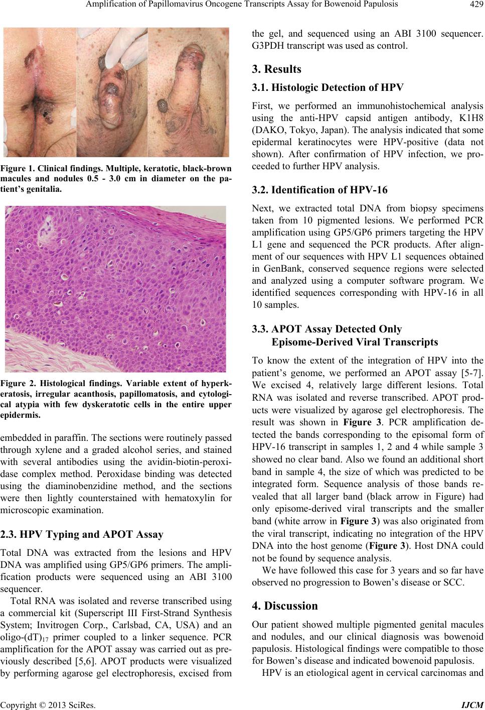 Amplification of Papillomavirus Oncogene Transcripts Assay for Bowenoid Papulosis 429 Figure 1. Clinical findings. Mu ltiple, keratotic, black-brow n macules and nodules 0.5 - 3.0 cm in diameter on the pa- tient’s genitalia. Figure 2. Histological findings. Variable extent of hyperk- eratosis, irregular acanthosis, papillomatosis, and cytologi- cal atypia with few dyskeratotic cells in the entire upper epidermis. embedded in paraffin. The sections were routinely passed through xylene and a graded alcohol series, and stained with several antibodies using the avidin-biotin-peroxi- dase complex method. Peroxidase binding was detected using the diaminobenzidine method, and the sections were then lightly counterstained with hematoxylin for microscopic examination. 2.3. HPV Typing and APOT Assay Total DNA was extracted from the lesions and HPV DNA was amplified using GP5/GP6 primers. The ampli- fication products were sequenced using an ABI 3100 sequencer. Total RNA was isolated and reverse transcribed using a commercial kit (Superscript III First-Strand Synthesis System; Invitrogen Corp., Carlsbad, CA, USA) and an oligo-(dT)17 primer coupled to a linker sequence. PCR amplification for the APOT assay was carried out as pre- viously described [5,6]. APOT products were visualized by performing agarose gel electrophoresis, excised from the gel, and sequenced using an ABI 3100 sequencer. G3PDH transcript was used as control. 3. Results 3.1. Histologic Detection of HPV First, we performed an immunohistochemical analysis using the anti-HPV capsid antigen antibody, K1H8 (DAKO, Tokyo, Japan). The analysis indicated that some epidermal keratinocytes were HPV-positive (data not shown). After confirmation of HPV infection, we pro- ceeded to further HPV analysis. 3.2. Identification of HPV-16 Next, we extracted total DNA from biopsy specimens taken from 10 pigmented lesions. We performed PCR amplification using GP5/GP6 primers targeting the HPV L1 gene and sequenced the PCR products. After align- ment of our sequences with HPV L1 sequences obtained in GenBank, conserved sequence regions were selected and analyzed using a computer software program. We identified sequences corresponding with HPV-16 in all 10 samples. 3.3. APOT Assay Detected Only Episome-Derived Viral Transcripts To know the extent of the integration of HPV into the patient’s genome, we performed an APOT assay [5-7]. We excised 4, relatively large different lesions. Total RNA was isolated and reverse transcribed. APOT prod- ucts were visualized by agarose gel electrophoresis. The result was shown in Figure 3. PCR amplification de- tected the bands corresponding to the episomal form of HPV-16 transcript in samples 1, 2 and 4 while sample 3 showed no clear band. Also we found an addition al short band in sample 4, the size of which was predicted to be integrated form. Sequence analysis of those bands re- vealed that all larger band (black arrow in Figure) had only episome-derived viral transcripts and the smaller band (white arrow in Figure 3) was also originated from the viral transcript, indicating no integration of the HPV DNA into the host genome (Figure 3). Host DNA could not be found by sequence analysis. We have followed this case for 3 years and so far have observed no progression to Bowen’s disease or SCC. 4. Discussion Our patient showed multiple pigmented genital macules and nodules, and our clinical diagnosis was bowenoid papulosis. Histological findings were compatible to tho se for Bowen’s disease and indicated bowenoid papulosis. HPV is an etiological agent in cervical carcinomas and Copyright © 2013 SciRes. IJCM 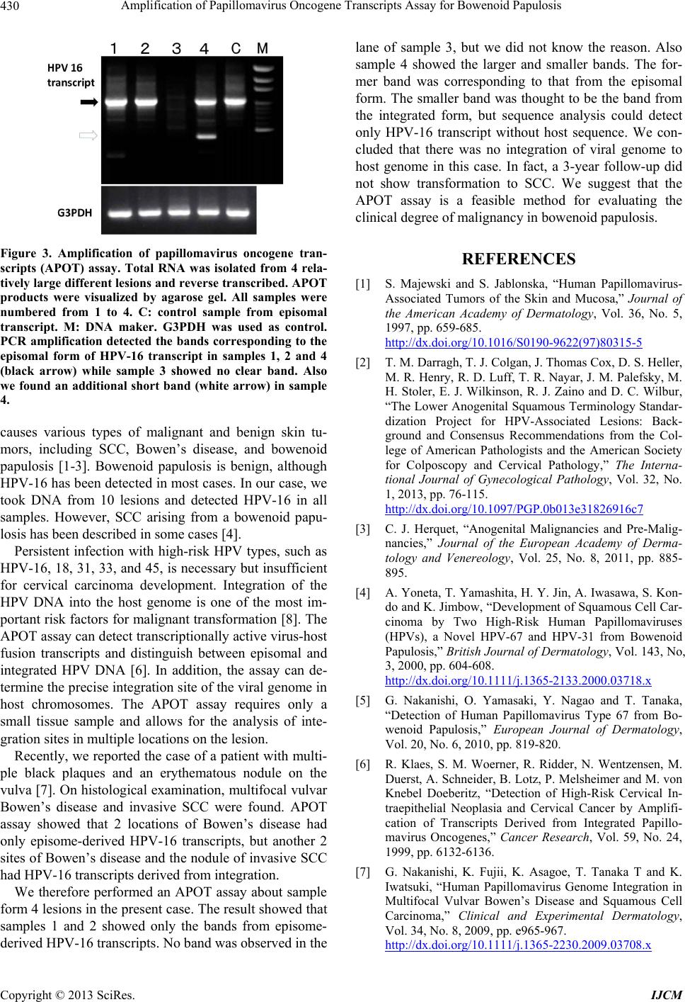 Amplification of Papillomavirus Oncogene Transcripts Assay for Bowenoid Papulosis 430 Figure 3. Amplification of papillomavirus oncogene tran- scripts (APOT) assay. Total RNA was isolated from 4 rela- tively large different lesions and reverse transcribed. APOT products were visualized by agarose gel. All samples were numbered from 1 to 4. C: control sample from episomal transcript. M: DNA maker. G3PDH was used as control. PCR amplification detected the bands corresponding to the episomal form of HPV-16 transcript in samples 1, 2 and 4 (black arrow) while sample 3 showed no clear band. Also we found an additional short band (white arrow) in sample 4. causes various types of malignant and benign skin tu- mors, including SCC, Bowen’s disease, and bowenoid papulosis [1-3]. Bowenoid papulosis is benign, although HPV-16 has been detected in most cases. In our case, we took DNA from 10 lesions and detected HPV-16 in all samples. However, SCC arising from a bowenoid papu- losis has been described in some cases [4]. Persistent infection with high-risk HPV types, such as HPV-16, 18, 31, 33, and 45, is necessary but insufficient for cervical carcinoma development. Integration of the HPV DNA into the host genome is one of the most im- portant risk factors for malignant transformation [8]. The APOT assay can detect transcriptionally activ e virus-host fusion transcripts and distinguish between episomal and integrated HPV DNA [6]. In addition, the assay can de- termine the precise integ ration site of the viral genome in host chromosomes. The APOT assay requires only a small tissue sample and allows for the analysis of inte- gration sites in multiple locations on the lesion. Recently, we reported the case of a patient with multi- ple black plaques and an erythematous nodule on the vulva [7]. On histological exa mination, multifocal vulvar Bowen’s disease and invasive SCC were found. APOT assay showed that 2 locations of Bowen’s disease had only episome-derived HPV-16 transcripts, but another 2 sites of Bowen’s disease and the nodule of invasive SCC had HPV-16 transcripts derived from integration. We therefore performed an APOT assay about sample form 4 lesions in the present case. The result showed that samples 1 and 2 showed only the bands from episome- derived HPV-16 tra nscr ipts. No band was obser ved in the lane of sample 3, but we did not know the reason. Also sample 4 showed the larger and smaller bands. The for- mer band was corresponding to that from the episomal form. The smaller band was thought to be the band from the integrated form, but sequence analysis could detect only HPV-16 transcript without host sequence. We con- cluded that there was no integration of viral genome to host genome in this case. In fact, a 3-year follow-up did not show transformation to SCC. We suggest that the APOT assay is a feasible method for evaluating the clinical degree of malignancy in bowenoid papulosis. REFERENCES [1] S. Majewski and S. Jablonska, “Human Papillomavirus- Associated Tumors of the Skin and Mucosa,” Journal of the American Academy of Dermatology, Vol. 36, No. 5, 1997, pp. 659-685. http://dx.doi.org/10.1016/S0190-9622(97)80315-5 [2] T. M. Darragh, T. J. Colgan, J. Thomas Cox, D. S. Heller, M. R. Henry, R. D. Luff, T. R. Nayar, J. M. Palefsky, M. H. Stoler, E. J. Wilkinson, R. J. Zaino and D. C. Wilbur, “The Lower Anogenital Squamous Terminology Standar- dization Project for HPV-Associated Lesions: Back- ground and Consensus Recommendations from the Col- lege of American Pathologists and the American Society for Colposcopy and Cervical Pathology,” The Interna- tional Journal of Gynecological Pathology, Vol. 32, No. 1, 2013, pp. 76-115. http://dx.doi.org/10.1097/PGP.0b013e31826916c7 [3] C. J. Herquet, “Anogenital Malignancies and Pre-Malig- nancies,” Journal of the European Academy of Derma- tology and Venereology, Vol. 25, No. 8, 2011, pp. 885- 895. [4] A. Yoneta, T. Yamashita, H. Y. Jin, A. Iwasawa, S. Kon- do and K. Jimbow, “Development of Squamous Cell Car- cinoma by Two High-Risk Human Papillomaviruses (HPVs), a Novel HPV-67 and HPV-31 from Bowenoid Papulosis,” British Journal of Dermatology, Vol. 143, No, 3, 2000, pp. 604-608. http://dx.doi.org/10.1111/j.1365-2133.2000.03718.x [5] G. Nakanishi, O. Yamasaki, Y. Nagao and T. Tanaka, “Detection of Human Papillomavirus Type 67 from Bo- wenoid Papulosis,” European Journal of Dermatology, Vol. 20, No. 6, 2010, pp. 819-820. [6] R. Klaes, S. M. Woerner, R. Ridder, N. Wentzensen, M. Duerst, A. Schneider, B. Lotz, P. Melsheimer and M. von Knebel Doeberitz, “Detection of High-Risk Cervical In- traepithelial Neoplasia and Cervical Cancer by Amplifi- cation of Transcripts Derived from Integrated Papillo- mavirus Oncogenes,” Cancer Research, Vol. 59, No. 24, 1999, pp. 6132-6136. [7] G. Nakanishi, K. Fujii, K. Asagoe, T. Tanaka T and K. Iwatsuki, “Human Papillomavirus Genome Integration in Multifocal Vulvar Bowen’s Disease and Squamous Cell Carcinoma,” Clinical and Experimental Dermatology, Vol. 34, No. 8, 2009, pp. e965-967. http://dx.doi.org/10.1111/j.1365-2230.2009.03708.x Copyright © 2013 SciRes. IJCM  Amplification of Papillomavirus Oncogene Transcripts Assay for Bowenoid Papulosis Copyright © 2013 SciRes. IJCM 431 [8] I. Kraus, C. Driesch, S. Vinokurova, E. Hovig, A. Sch- neider, M. von Knebel Doeberitz and M. Duerst, “The Majority of Viral-Cellular Fusion Transcripts in Cervical Carcinomas Cotranscribe Cellular Sequences of Known or Predicted Genes,” Cancer Research, Vol. 68, No. 7, 2008, pp. 2514-2522. http://dx.doi.org/10.1158/0008-5472.CAN-07-2776 |