

Paper Menu >>
Journal Menu >>
 Vol.2, No.7, 422-426 (2013) Case Reports in Clinical Medicine http://dx.doi.org/10.4236/crcm.2013.27111 Copyright © 2013 SciRes. OPEN ACCESS Benign retrorectal tumours: Contribution of laparoscopic approach Victor J. Ovejero-Gomez1*, M. V. Bermudez-Garcia2, L. Martin-Cuesta3, A. Güezmes4, J. Villalba2, A. Ingelmo1, J. M. Bajo-Arenas2 1Department of Surgery, Hospital Sierrallana, Cantabria, Spain; *Correspondin g Author: [email protected] 2Department of Gynaecology, Hospital Sierrallana, Cantabria, Spain 3Department of Radiology, Hospital Sierrallana, Cantabria, Spain 4Department of Pathology, Hospital Sierrallana, Cantabria, Spain Received 30 July 2013; revised 29 August 2013; accepted 9 September 2013 Copyright © 2013 Victor J. Ovejero-Gomez et al. This is an open access article distributed under the Creative Commons Attribution License, which permits unrestricted use, distribution, and reproduction in any medium, provided the original work is properly cited. ABSTRACT Introduction: Presacral tumours are extremely rare entities as the asymptomatic retrorectal mass, although its clinical presentation includes infectious complications and signs of malignant degeneration. Magnetic resonance imaging is the most efficient imaging study for its diagno- sis. The treatment of choi ce is complete surgical excision and traditional approaches are mainly through abdominal, posterior and perineal ap- proach, depending on anatomical characteris- tics of the lesion. Laparoscopic excision of these retrorectal lesions has been reported in a few cases. Patients and methods: We report two cases of 38 and 24-year-old women who com- plained of anorectal symptoms and were diag- nosed as retrorectal tumour by imaging studies. One of them was infected. Both cases were re- sected by means of laparoscopic techniques. There was no surgical complication and they were discharged on the 3rd and 4th postoperative day, respectively. Histopathologic findings re- vealed benign cystic teratoma in both cases. A fol l o w-u p a fter 3 6 m on th s sho wed no recurrence. Our surgical endoscopic technique and a brief review of perioperative cares are presented and discussed. Discussion: Laparoscopic excision could be indicated in selected retrorectal tu- mours and a great surgeon’s resolve is always required. A meticulous dissection must be per- formed in order to identify and preserve vital structures. It’s only absolute contraindications seemed to be the suspicion of malignancy and operative inexperience. Conclusions: The com- plete laparoscopic removal of presacral lesions is a responsible surgical procedure which offers advantages from the sanitary and aesthetic point of view. This new endoscopic indication could be considered probably as a better tech- nique than open approach in selected patients. Keywords: Retrorectal; Tumour; Laparoscopy; Surgical Technique 1. INTRODUCTION Retrorectal tumours are rare entities with a low inci- dence and usually have a congenital origin. They can be a cystic or solid mass and are divided into benign, mostly, and malignant masses [1]. The possibility of malignity is increased in younger patients although a solid mass is a higher associated risk factor. Most cases are asymptomatic but its semeiology is re- lated to presacral location, size and infection. Pain is the most common symptom in adult patients [2] but diges- tive and urinary disturbances and fistulas are possible. The first diagnostic study should be focused on iden- tifying the extraluminal nature of the lesion by an endo- anal exploration. Magnetic resonance imaging is the ra- diological study of choice, currently, because it offers more information than CT scan about tumoral characters and neighbouring anatomical structures [3,4]. Imaging diagnosis is one of the main bases to elaborate a right surgical planning. The definitive diagnosis leads to a complete resection of the lesion even though it was asymptomatic and sus- picion of benignity. Preoperative biopsy is not recom- mended [5] due to the risk of septic complications and tumoral seeding. We present our experience in two presacral masses 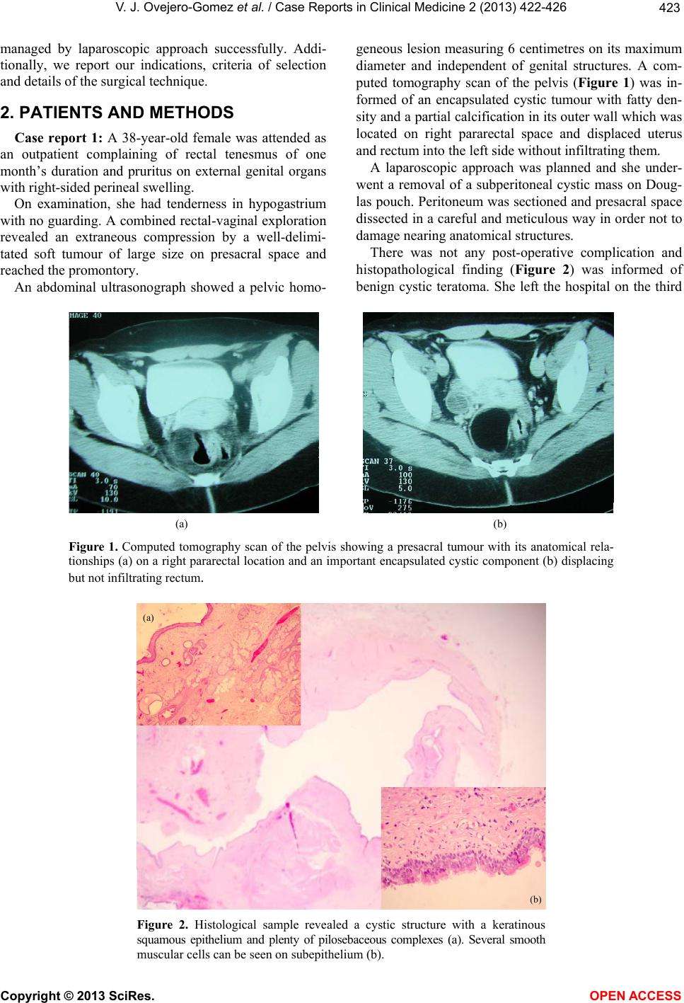 V. J. Ovejero-Gomez et al. / Case Reports in Clinical Medic ine 2 (2013) 422-426 Copyright © 2013 SciRes. OPEN ACCESS 423 managed by laparoscopic approach successfully. Addi- tionally, we report our indications, criteria of selection and details of the surgical technique. 2. PATIENTS AND METHODS Case report 1: A 38-year-old female was attended as an outpatient complaining of rectal tenesmus of one month’s duration and pruritus on external genital organs with right-sided perineal swell i ng. On examination, she had tenderness in hypogastrium with no guardin g. A combined rectal-vaginal exploration revealed an extraneous compression by a well-delimi- tated soft tumour of large size on presacral space and reached the promontory. An abdominal ultrasonograph showed a pelvic homo- geneous lesion measuring 6 centimetres on its maximum diameter and independent of genital structures. A com- puted tomography scan of the pelvis (Figure 1) was in- formed of an encapsulated cystic tumour with fatty den- sity and a partial calcification in its outer wall which was located on right pararectal space and displaced uterus and rectum into the left side without infiltrating them. A laparoscopic approach was planned and she under- went a removal of a subperitoneal cystic mass on Doug- las pouch. Peritoneum was sectioned and presacral space dissected in a careful and meticulous way in order not to damage nearing anatomical structures. There was not any post-operative complication and histopathological finding (Figure 2) was informed of benign cystic teratoma. She left the hospital on the third (a) (b) Figure 1. Computed tomography scan of the pelvis showing a presacral tumour with its anatomical rela- tionships (a) on a right pararectal location and an important encapsulated cystic component (b) displacing but not infiltrating rectum. (a) (b) Figure 2. Histological sample revealed a cystic structure with a keratinous squamous epithelium and plenty of pilosebaceous complexes (a). Several smooth muscular cells can be seen on subepithelium (b). 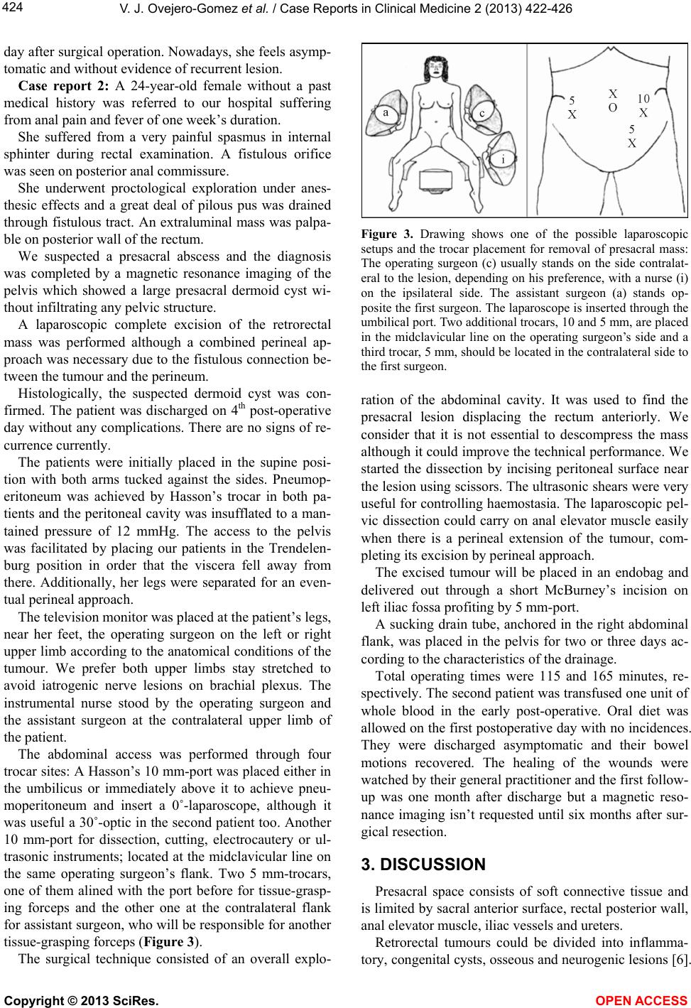 V. J. Ovejero-Gomez et al. / Case Reports in Clinical Medicine 2 (2013) 422-426 Copyright © 2013 SciRes. OPEN ACCESS 424 day after surgical operation. Nowadays, she feels asymp- tomatic and without evidence of recurrent lesion. Case report 2: A 24-year-old female without a past medical history was referred to our hospital suffering from anal pain and fever of one week’ s duration. She suffered from a very painful spasmus in internal sphinter during rectal examination. A fistulous orifice was seen on posterior anal commissure. She underwent proctological exploration under anes- thesic effects and a great deal of pilous pus was drained through fistulous tract. An extraluminal mass was palpa- ble on posterior wall of the rectum. We suspected a presacral abscess and the diagnosis was completed by a magnetic resonance imaging of the pelvis which showed a large presacral dermoid cyst wi- thout infiltrating an y pelvic structure. A laparoscopic complete excision of the retrorectal mass was performed although a combined perineal ap- proach was necessary due to the fistulous connection be- tween the tumour and the perineum. Histologically, the suspected dermoid cyst was con- firmed. The patient was discharged on 4th post-operative day without any complicatio ns. There are no signs of re- currence currently. The patients were initially placed in the supine posi- tion with both arms tucked against the sides. Pneumop- eritoneum was achieved by Hasson’s trocar in both pa- tients and the peritoneal cav ity was insufflated to a man- tained pressure of 12 mmHg. The access to the pelvis was facilitated by placing our patients in the Trendelen- burg position in order that the viscera fell away from there. Additionally, her legs were separated for an even- tual perineal approach. The television monitor was placed at the patient’s legs, near her feet, the operating surgeon on the left or right upper limb according to the anatomical conditions of the tumour. We prefer both upper limbs stay stretched to avoid iatrogenic nerve lesions on brachial plexus. The instrumental nurse stood by the operating surgeon and the assistant surgeon at the contralateral upper limb of the patient. The abdominal access was performed through four trocar sites: A Hasson’s 10 mm-port was placed either in the umbilicus or immediately above it to achieve pneu- moperitoneum and insert a 0˚-laparoscope, although it was useful a 30˚-optic in the second patient too. Another 10 mm-port for dissection, cutting, electrocautery or ul- trasonic instruments; located at the midclavicular line on the same operating surgeon’s flank. Two 5 mm-trocars, one of them alined with the port before for tissue-grasp- ing forceps and the other one at the contralateral flank for assistant surgeon, who will be responsible fo r another tissue-grasping forceps (Figure 3). The surgical technique consisted of an overall explo- Figure 3. Drawing shows one of the possible laparoscopic setups and the trocar placement for removal of presacral mass: The operating surgeon (c) usually stands on the side contralat- eral to the lesion, depending on his preference, with a nurse (i) on the ipsilateral side. The assistant surgeon (a) stands op- posite the first surgeon. The laparoscope is inserted through the umbilical port. Two additional trocars, 10 and 5 mm, are placed in the midclavicular line on the operating surgeon’s side and a third trocar, 5 mm, should be located in the contralateral side to the first surgeon. ration of the abdominal cavity. It was used to find the presacral lesion displacing the rectum anteriorly. We consider that it is not essential to descompress the mass although it could improve the technical performance. We started the dissection by incising peritoneal surface near the lesion using scissors. Th e ultrasonic shears were very useful for controlling haemostasia. The laparoscopic pel- vic dissection could carry on anal elevator muscle easily when there is a perineal extension of the tumour, com- pleting its excision by perineal approach. The excised tumour will be placed in an endobag and delivered out through a short McBurney’s incision on left iliac fossa profiting by 5 mm-port. A sucking drain tube, anchored in the right abdominal flank, was placed in the pelvis for two or three days ac- cording to the characteristics of the drainage. Total operating times were 115 and 165 minutes, re- spectively. The second patien t was transfused one unit of whole blood in the early post-operative. Oral diet was allowed on the first postoperative day with no incidences. They were discharged asymptomatic and their bowel motions recovered. The healing of the wounds were watched by their general practitioner and the first follow- up was one month after discharge but a magnetic reso- nance imaging isn’t requested until six months after sur- gical resection. 3. DISCUSSION Presacral space consists of soft connective tissue and is limited by sacral anterior surface, rectal posterior wall, anal elevator muscle, iliac vessels and ureters. Retrorectal tumours could be divided into inflamma- tory, congenital cysts, osseous and neurogenic lesions [6].  V. J. Ovejero-Gomez et al. / Case Reports in Clinical Medic ine 2 (2013) 422-426 Copyright © 2013 SciRes. OPEN ACCESS 425 Miscellanea with leiomyoma, fibroma, lipoma, lympho- ma and metastasic and carcinoid tumours are also in- cluded in this location. They can be asymptomatic for a long time but could cause semeiology due to the pressure effect such as the perineal swelling or infectious complications as perineal and rectal fistula, some of them shown in our reported cases. Another possibility is its malignant degeneration [7] which would modify the operating attitude. There is not any standard guidelines in the manage- ment of these tumours due to its low incidence and there has been described several surgical procedures depend- ing on both complications and anatomical conditio ns [8]. The traditional surgical approaches are via the perineal, posterior, abdominal and combined via, depending on location and size lesion [9]. The surgical outcome is good in benign tumours. There has been reported a com- plete resection in all cases of benign tumours and most of malignant cases with a little rate of postoperative com- plications [ 10] . The transanal approach or through intersphincterial resection [11] could be indicated in patients with benign tumours of small size and distal presacral location. One of the less injurious posterior accesses is via the para- sacrococcygeal because it provides with an excellent an- orectal exposure and preservation of the sphincterial me- chanism. Its indication would be preserved to the rest of low tumours. Abdominal approach is the access of choice in large size tumours which exceed the promontory and in suspi- cion of malignancy, a lot of them improving with a com- bined approach because of their size and anatomical dis- posal. Besides, it allows a better vascular and ureteral check. The development of laparoscopic technique has pro- moted an increased usefulness in this kind of pathology recently [9,12]. Some of the advantages of this approach are the exact identification of anatomical situation of the mass and its relationship to other adjoining structures, and the possibility of extending rectal dissection to anal elevator musculature with a small assumed morbility with respect to an open procedure. The pathological cha- racteristics of the lesion or its perineal implication could force a combined access as in the second case. The performance of this kind of minimal aggressive procedure could be difficult because of technical com- plexity and local conditions of the tumours which de- mand a surgical team with a huge experience in laparo- scopic technique. A conversion to laparotomy is an op- tion to be taken into account. This inconvenience is explained by a plane of tumoral dissection difficult to identify and the thickness of wall which determines a meticulous resection in a highly sus- ceptible bleeding area. We advise the practice of ultra- sonic instruments [13] because these masses are used to feed by a rich diffuse network of vessels. On the other hand, the surgical management consists of complete re- section with included wall to avoid a recurrence even though a cystic mass of large size could be a candidate to decompress without spillage piercing abdominal wall, with a sucking tube placed into the lesion until it col- lapses. This strategy makes its manipulation easy and reduces the risk of septic widespread in the pelvic cavity in case of rupture. These local difficulties in identifying neighbourghing anatomical structures can cause the removal to become complicated. We consider the vital importance of identi- fying and preserving urinary bladder and ureters, pelvic vessels and hypogas tri c nerves. In our own opinion, the current only absolute contra- indication in the laparo scopic approach would be a clini- cal suspicion of malignancy. An infested mass should be considered as a relative contraindication in this therapeu - tic attitude. In conclusion, the laparoscopic approach in retrorectal tumours could be a safe and feasible surgical access from the technical point of view and with the same criteria of removal and curing as traditional open resection [14]. Furthermore, its main advantages are an earlier oral tol- erance, a short hospital stay, a low incidence of post-op- erative complications, less pain and excellent cosmesis. Before confirming the indication of this operating ap- proach, it is required putting the test patience and re- solve to the surgical team who must hold a high experi- ence in minimally aggressive techniques. REFERENCES [1] Ghosh, J., Eglinton, T., Frizelle, F.A. and Watson, A.J. (2007) Presacral tumours in adults. Surgeon, 5, 31-38. http://dx.doi.org/10.1016/S1479-666X(07)80109-0 [2] Messick, C.A., Londono, J.M. and Hull, T. (2013) Presac- ral tumors: How do they compare in pediatric and adult patients? Pol Przegl Chir, 85, 253-261. [3] Pappalardo, G., Frattaroli, F.M., Casciani, E., Moles, N., Mascagni, D., Spoletini, D., et al. (2009) Retrorectal tu- mors: The choice of surgical approach based on a new classification. The American Surgeon, 75, 240-248. [4] Coco, C., Manno, A., Ma ttana, C., Verbo, A., Sermoneta, D., Franceschini, G., et al. (2008) Congenital tumors of the retrorectal space in the adult: Report of two cases and review of the literature. Tum ori, 94, 602-607. [5] Vega-Menéndez, D., Quintans-Rodríguez, A., Hernández- Granados, P., Nevado-Santos, M., García-Sabrido, J.L. and Rueda-Orgaz, J.A. (2008) Hamartomas quísticos retrorrectales. Cirugía Española, 83, 53-60. http://dx.doi.org/10.1016/S0009-739X(08)70506-2 [6] Hobson, K.G., Ghaemmaghami, V., Roe, J.P., Goodnight,  V. J. Ovejero-Gomez et al. / Case Reports in Clinical Medicine 2 (2013) 422-426 Copyright © 2013 SciRes. OPEN ACCESS 426 J.E. and Khatri, V.P. (2005) Tumors of the retrorectal space. Diseases of the Colon & Rectum, 48, 1964-1974. http://dx.doi.org/10.1007/s10350-005-0122-9 [7] Maruyama, A., Murabayashi, K., Hayashi, M., et al. (1998) Adenocarcinoma arising in a tailgut cyst: Report of a case. Sur gery Today, 28, 1319-1322. http://dx.doi.org/10.1007/BF02482826 [8] Szyllo, K. and Lesnik, N. (2013) Sacrococcy geal te ratoma— Case report and review of the literature. American Journal of Case Reports, 14, 1-5. [9] Köhler, C., Kühne-Heid, R., Klemm, P., Tozzi, R. and Schneider, A. (2003) Resection of presacral ganglioneu- rofibroma by laparoscopy. Surgical Endoscopy and Other Interventional Techniques, 17, 1499. [10] Lev-Chelouche, D., Gutman, M., Goldman, G., Even-Sa- pir, E., Meller, I., Issakov, J., et al. (2003) Presacral tu- mors: A practical classification and treatment of a unique and heterogeneous group of diseases. Surgery, 133, 473- 478. http://dx.doi.org/10.1067/msy.2003.118 [11] Pescatori, M., Brusciano, L., Binda, G.A. and Serventi, A. (2005) A novel approach for perirrectal tumours: The perianal intersphinteric escisión. International Journal of Colorectal Disease, 20, 72-75. http://dx.doi.org/10.1007/s00384-004-0623-3 [12] Chen, Y., Xu, H., Li, Y., Li, J., Wang, D., Yuan, J. and Liang, Z. (2008) Laparoscopic resection of presacral teratomas. Journal of Minimally Invasive Gynecology, 15, 649-651. http://dx.doi.org/10.1016/j.jmig.2008.06.011 [13] Palanivelu, C., Rangarajan, M., Senthilkumar, R., Ma- dankumar, M.V. and Annapoorni, S. (2008) Laparoscopic and perineal escision of an infected “dumb-bell” shaped retrorectal epidermoid cyst. Journal of Laparoendoscopic & Advanced Surgical Techniques , 18, 88-92. http://dx.doi.org/10.1089/lap.2007.0010 [14] Paul, P.G., Pravinkumar, T. and Sheetal, B. (2012) Sacro- coccygeal neurofibroma: Rare cause for chronic pelvic pain. Journal of Minimally Invasive Gynecology, 19, 517- 520. |