

Paper Menu >>
Journal Menu >>
 Vol.5, No.10, 1706-1711 (2013) Health http://dx.doi.org/10.4236/health.2013.510230 The effect of aerobic training on endothelium-dependent vasodilatation in patients with coronary artery disease who were revascularized and young men Seyed Masoud Seyediyan, Farzaneh Ahmadi*, Babak Hamidian, Ebrahim Hajizadeh, Afshin Rezazadeh, Ahmad Reza Asare, Mohammad Hasan Adel, Mohammad Nourizadeh Department of Cardiology, Medical Joundishapour University of Ahvaz, Atherosclerosis Research Center Jondishapour University of Medical Sciences, Ahvaz, Iran; *Corresponding Author: [email protected] Received 13 April 2013; revised 15 May 2013; accepted 19 June 2013 Copyright © 2013 Seyed Masoud Seyediyan et al. This is an open access article distributed under the Creative Commons Attribution License, which permits unrestricted use, distribution, and reproduction in any medium, provided the original work is properly cited. ABSTRACT Aim: The aim of this study was to determine the effect of training on endothelium-dependent vasodilatation in patients with coronary artery disease (CAD) after revascularization and heal- thy young men. Background: Impaired endothe- lial function has been observed in patients with CAD and those with CAD risk factors. Studies have show n that exercise can enhance endothe- lial function. Methods: This experimental cross- sectional study was conducted on patients with CAD (3 months after CABG and PCI) and students of medical school in 2011. Endothelium depen- dent dilation of the brachial artery was deter- mined by using high-resolution vascular ultra- sonography through flow-mediated vasodilata- tion (FMD) after induction of ischemia, and the data were analyzed using SPSS, dependent t- test and ANCOVA. Findings: The findings show - ed that at baseline, FMD was reduced in revas- cularized patients, when compared with healthy young men, after 8 weeks, and exercise training significantly improved FMD in patients under- went training group [from 4.31 ± 1.45 (SD)% to 6.15 ± 0.773 (SD)%, p < 0.05] and substantially increased the same in healthy young men [from 9.18% ± 1.45% to 11.72% ± 1.72%, p < 0.05] However, in the control group, the FMD remain- ed unchanged, and even after aerobic training, it did not significantly modif y the brachial artery diameter in these group s. Conclusion : Our study demonstrates that endothelial dysfunction per- sisting in CAD patients after revascularization and aerobic training can improve endothelial function in different vascular beds in CAD pa- tients and healthy young men. This may contri- bute to the benefit of reg ular exercise in pr event- ing and restricting cardiovascular disease. Keywords: Endotheli um-Dependent Vasodilatation; Coronary Artery Disease; Aerobic Training; High-Resolution Ultrasound; Flow-Mediated Dilation 1. INTRODUCTION Currently, most patients with angina pectoris have their symptoms controlled with medication or by revas- cularization by PTCA or CABG. Consequently, and with rare exceptions [1], much of the evidence that exercise training improves effort tolerance in patients with angina pectoris was obtained before 1990. Exercise training increases the exercise time to the onset of angina or even eliminates angina entirely by at least two mechanisms. First, exercise training reduces the M.O2 requirements during submaximal exercise [1-3]. Second, exercise train- ing reduces endothelial dysfunction [4-6]. Normal coro- nary arteries dilate with exercise, whereas atherosclerotic coronary arteries often demonstrate endothelial dysfunc- tion evidenced by exercise-induced vasoconstriction. Few large trials have examined the effects of exercise-based cardiac rehabilitation in patients following PTCA [6,7]. There are very few data for coronary artery disease and healthy young men. In a previous study, we demon- strated that in healthy middle age men, 8 weeks of circuit training did not affect vascular function [8], whereas, bra chial artery flow mediated dilation (FMD) was enhanced Copyright © 2013 SciRes. OPEN ACCESS 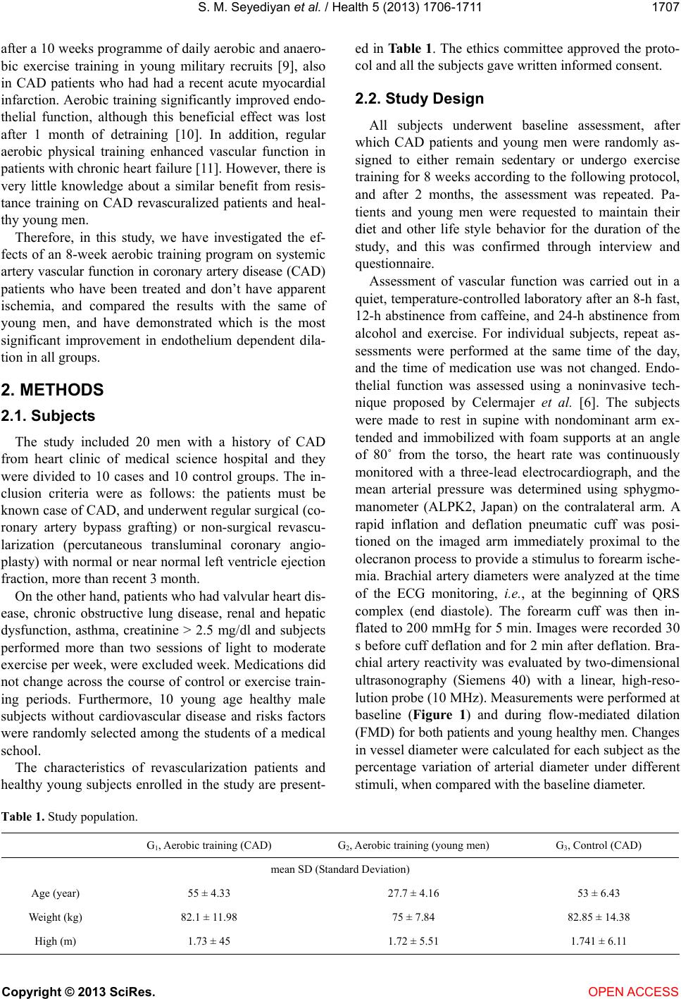 S. M. Seyediyan et al. / Health 5 (2013) 1706-1711 1707 after a 10 weeks programme of daily aerobic and anaero- bic exercise training in young military recruits [9], also in CAD patients who had had a recent acute myocardial infarction. Aerobic training significantly improved endo- thelial function, although this beneficial effect was lost after 1 month of detraining [10]. In addition, regular aerobic physical training enhanced vascular function in patients with chronic heart failure [11]. However, there is very little knowledge about a similar benefit from resis- tance training on CAD revascuralized patients and heal- thy young men. Therefore, in this study, we have investigated the ef- fects of an 8-week aerobic training program on systemic artery vascular function in coronary artery disease (CAD) patients who have been treated and don’t have apparent ischemia, and compared the results with the same of young men, and have demonstrated which is the most significant improvement in endothelium dependent dila- tion in all groups. 2. METHODS 2.1. Subjects The study included 20 men with a history of CAD from heart clinic of medical science hospital and they were divided to 10 cases and 10 control groups. The in- clusion criteria were as follows: the patients must be known case of CAD, and underwent regular surgical (co- ronary artery bypass grafting) or non-surgical revascu- larization (percutaneous transluminal coronary angio- plasty) with normal or near normal left ventricle ejection fraction, more than recent 3 month. On the other hand, patients who had valvular heart dis- ease, chronic obstructive lung disease, renal and hepatic dysfunction, asthma, creatinine > 2.5 mg/dl and subjects performed more than two sessions of light to moderate exercise per week, were excluded week. Medications did not change across the course of control or exercise train- ing periods. Furthermore, 10 young age healthy male subjects without cardiovascular disease and risks factors were randomly selected among the students of a medical school. The characteristics of revascularization patients and healthy young subjects enrolled in the study are present- ed in Table 1. The ethics committee approved the proto- col and all the subjects gave written informed consent. 2.2. Study Design All subjects underwent baseline assessment, after which CAD patients and young men were randomly as- signed to either remain sedentary or undergo exercise training for 8 weeks according to the following protocol, and after 2 months, the assessment was repeated. Pa- tients and young men were requested to maintain their diet and other life style behavior for the duration of the study, and this was confirmed through interview and questionnaire. Assessment of vascular function was carried out in a quiet, temperature-controlled laboratory after an 8-h fast, 12-h abstinence from caffeine, and 24-h abstinence from alcohol and exercise. For individual subjects, repeat as- sessments were performed at the same time of the day, and the time of medication use was not changed. Endo- thelial function was assessed using a noninvasive tech- nique proposed by Celermajer et al. [6]. The subjects were made to rest in supine with nondominant arm ex- tended and immobilized with foam supports at an angle of 80˚ from the torso, the heart rate was continuously monitored with a three-lead electrocardiograph, and the mean arterial pressure was determined using sphygmo- manometer (ALPK2, Japan) on the contralateral arm. A rapid inflation and deflation pneumatic cuff was posi- tioned on the imaged arm immediately proximal to the olecranon process to provide a stimulus to forearm ische- mia. Brachial artery diameters were analyzed at the time of the ECG monitoring, i.e., at the beginning of QRS complex (end diastole). The forearm cuff was then in- flated to 200 mmHg for 5 min. Images were recorded 30 s before cuff deflation and for 2 min after deflation. Bra- chial artery reactivity was evaluated by two-dimensional ultrasonography (Siemens 40) with a linear, high-reso- lution probe (10 MHz). Measurements were performed at baseline (Figure 1) and during flow-mediated dilation (FMD) for both patients and young healthy men. Changes in vessel diameter were calculated for each subject as the percentage variation of arterial diameter under different stimuli, when compared with the baseline diameter. Table 1. Study population. G 1, Aerobic training (CAD) G2, Aerobic training (young men) G3, Control (CAD) mean SD (Standard Deviation) Age (year) 55 ± 4.33 27.7 ± 4.16 53 ± 6.43 Weight (kg) 82.1 ± 11.98 75 ± 7.84 82.85 ± 14.38 High (m) 1.73 ± 45 1.72 ± 5.51 1.741 ± 6.11 Copyright © 2013 SciRes. OPEN ACCESS 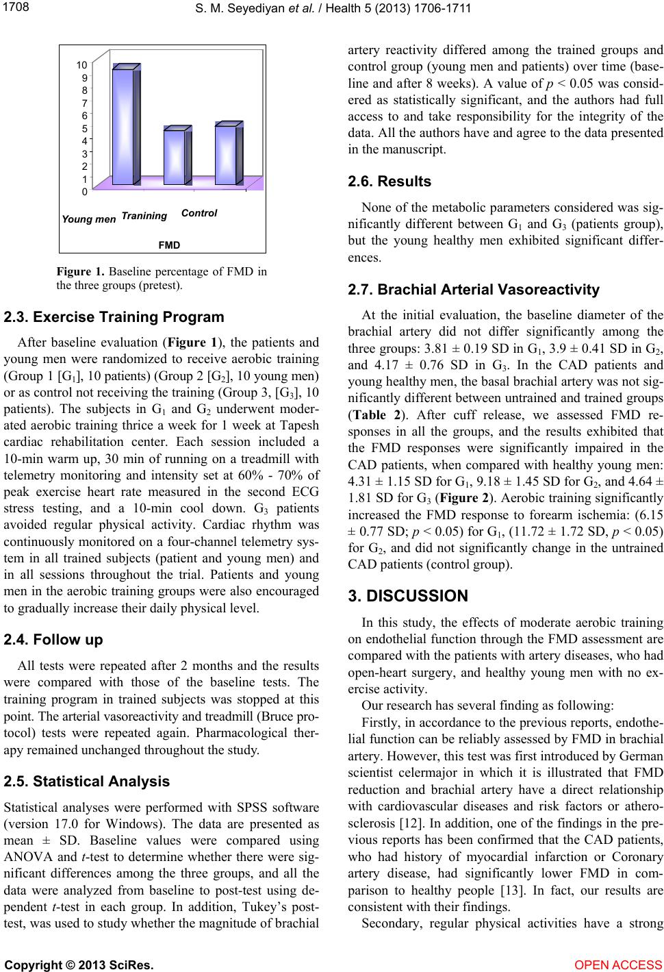 S. M. Seyediyan et al. / Health 5 (2013) 1706-1711 1708 0 1 2 3 4 5 6 7 8 9 10 Young men Tranining Control FMD Figure 1. Baseline percentage of FMD in the three groups (pretest). 2.3. Exercise Training Program After baseline evaluation (Figure 1), the patients and young men were randomized to receive aerobic training (Group 1 [G1], 10 patients) (Group 2 [G2], 10 young men) or as control not receiving the training (Group 3, [G3], 10 patients). The subjects in G1 and G2 underwent moder- ated aerobic training thrice a week for 1 week at Tapesh cardiac rehabilitation center. Each session included a 10-min warm up, 30 min of running on a treadmill with telemetry monitoring and intensity set at 60% - 70% of peak exercise heart rate measured in the second ECG stress testing, and a 10-min cool down. G3 patients avoided regular physical activity. Cardiac rhythm was continuously monitored on a four-channel telemetry sys- tem in all trained subjects (patient and young men) and in all sessions throughout the trial. Patients and young men in the aerobic training groups were also encouraged to gradually increase their daily physical level. 2.4. Follow up All tests were repeated after 2 months and the results were compared with those of the baseline tests. The training program in trained subjects was stopped at this point. The arterial vasoreactivity and treadmill (Bruce pro- tocol) tests were repeated again. Pharmacological ther- apy remained unchanged throughout the study. 2.5. Statistical Analysis Statistical analyses were performed with SPSS software (version 17.0 for Windows). The data are presented as mean ± SD. Baseline values were compared using ANOVA and t-test to determine whether there were sig- nificant differences among the three groups, and all the data were analyzed from baseline to post-test using de- pendent t-test in each group. In addition, Tukey’s post- test, was used to study whether the magnitude of brachial artery reactivity differed among the trained groups and control group (young men and patients) over time (base- line and after 8 weeks). A value of p < 0.05 was consid- ered as statistically significant, and the authors had full access to and take responsibility for the integrity of the data. All the authors have and agree to the data presented in the manuscript. 2.6. Results None of the metabolic parameters considered was sig- nificantly different between G1 and G3 (patients group), but the young healthy men exhibited significant differ- ences. 2.7. Brachial Arterial Vasoreactivity At the initial evaluation, the baseline diameter of the brachial artery did not differ significantly among the three groups: 3.81 ± 0.19 SD in G1, 3.9 ± 0.41 SD in G2, and 4.17 ± 0.76 SD in G3. In the CAD patients and young healthy men, the basal brachial artery was not sig- nificantly different between untrained and trained groups (Table 2). After cuff release, we assessed FMD re- sponses in all the groups, and the results exhibited that the FMD responses were significantly impaired in the CAD patients, when compared with healthy young men: 4.31 ± 1.15 SD for G1, 9.18 ± 1.45 SD for G2, and 4.64 ± 1.81 SD for G3 (Figure 2). Aerobic training significantly increased the FMD response to forearm ischemia: (6.15 ± 0.77 SD; p < 0.05) for G1, (11.72 ± 1.72 SD, p < 0.05) for G2, and did not significantly change in the untrained CAD patients (control group). 3. DISCUSSION In this study, the effects of moderate aerobic training on endothelial function through the FMD assessment are compared with the patients with artery diseases, who had open-heart surgery, and healthy young men with no ex- ercise activity. Our research has several finding as following: Firstly, in accordance to the previous reports, endothe- lial function can be reliably assessed by FMD in brachial artery. However, this test was first introduced by German scientist celermajor in which it is illustrated that FMD reduction and brachial artery have a direct relationship with cardiovascular diseases and risk factors or athero- sclerosis [12]. In addition, one of the findings in the pre- vious reports has been confirmed that the CAD patients, who had history of myocardial infarction or Coronary artery disease, had significantly lower FMD in com- parison to healthy people [13]. In fact, our results are consistent with their findings. Secondary, regular physical activities have a strong Copyright © 2013 SciRes. OPEN ACCESS 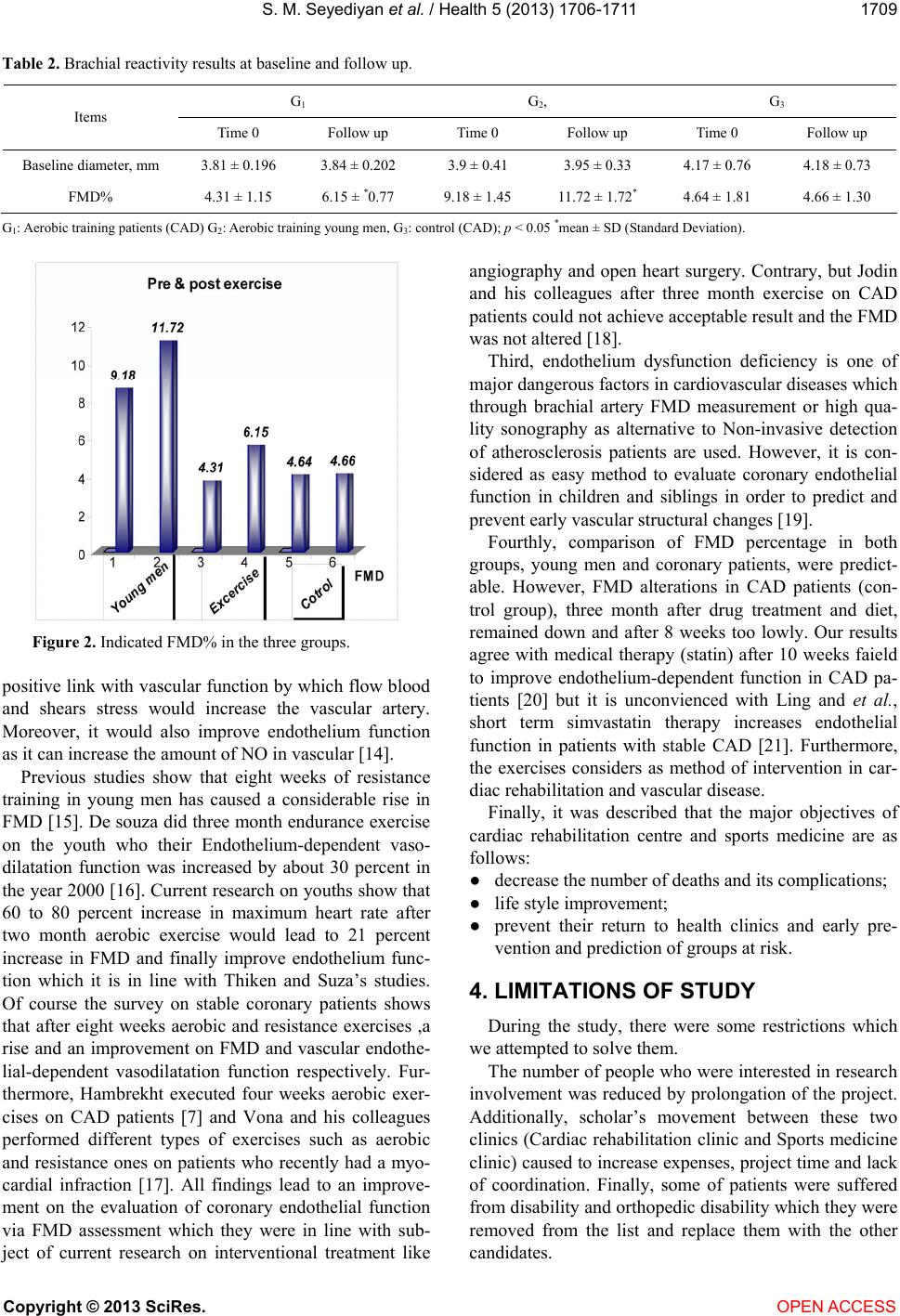 S. M. Seyediyan et al. / Health 5 (2013) 1706-1711 1709 Table 2. Brachial reactivity results at baseline and follow up. G1 G 2, G3 Items Time 0 Follow up Time 0 Follow up Time 0 Follow up Baseline diameter, mm 3.81 ± 0.196 3.84 ± 0.202 3.9 ± 0.41 3.95 ± 0.33 4.17 ± 0.76 4.18 ± 0.73 FMD% 4.31 ± 1.15 6.15 ± *0.77 9.18 ± 1.45 11.72 ± 1.72* 4.64 ± 1.81 4.66 ± 1.30 G1: Aerobic training patients (CAD) G2: Aerobic training young men, G3: control (CAD); p < 0.05 *mean ± SD (Standard Deviation). Figure 2. Indicated FMD% in the three groups. positive link with vascular function by which flow blood and shears stress would increase the vascular artery. Moreover, it would also improve endothelium function as it can increase the amount of NO in vascular [14]. Previous studies show that eight weeks of resistance training in young men has caused a considerable rise in FMD [15]. De souza did three month endurance exercise on the youth who their Endothelium-dependent vaso- dilatation function was increased by about 30 percent in the year 2000 [16]. Current research on youths show that 60 to 80 percent increase in maximum heart rate after two month aerobic exercise would lead to 21 percent increase in FMD and finally improve endothelium func- tion which it is in line with Thiken and Suza’s studies. Of course the survey on stable coronary patients shows that after eight weeks aerobic and resistance exercises ,a rise and an improvement on FMD and vascular endothe- lial-dependent vasodilatation function respectively. Fur- thermore, Hambrekht executed four weeks aerobic exer- cises on CAD patients [7] and Vona and his colleagues performed different types of exercises such as aerobic and resistance ones on patients who recently had a myo- cardial infraction [17]. All findings lead to an improve- ment on the evaluation of coronary endothelial function via FMD assessment which they were in line with sub- ject of current research on interventional treatment like angiography and open heart surgery. Contrary, but Jodin and his colleagues after three month exercise on CAD patients could not achieve acceptable result and the FMD was not altered [18]. Third, endothelium dysfunction deficiency is one of major dangerous factors in cardiovascular diseases which through brachial artery FMD measurement or high qua- lity sonography as alternative to Non-invasive detection of atherosclerosis patients are used. However, it is con- sidered as easy method to evaluate coronary endothelial function in children and siblings in order to predict and prevent early vascular structural changes [19]. Fourthly, comparison of FMD percentage in both groups, young men and coronary patients, were predict- able. However, FMD alterations in CAD patients (con- trol group), three month after drug treatment and diet, remained down and after 8 weeks too lowly. Our results agree with medical therapy (statin) after 10 weeks faield to improve endothelium-dependent function in CAD pa- tients [20] but it is unconvienced with Ling and et al., short term simvastatin therapy increases endothelial function in patients with stable CAD [21]. Furthermore, the exercises considers as method of intervention in car- diac rehabilitation and vascular disease. Finally, it was described that the major objectives of cardiac rehabilitation centre and sports medicine are as follows: ● decrease the number of deaths and its complications; ● life style improvement; ● prevent their return to health clinics and early pre- vention and prediction of groups at risk. 4. LIMITATIONS OF STUDY During the study, there were some restrictions which we attempted to solve them. The number of people who were interested in research involvement was reduced by prolongation of the project. Additionally, scholar’s movement between these two clinics (Cardiac rehabilitation clinic and Sports medicine clinic) caused to increase expenses, project time and lack of coordination. Finally, some of patients were suffered from disability and orthopedic disability which they were removed from the list and replace them with the other candidates. Copyright © 2013 SciRes. OPEN ACCESS  S. M. Seyediyan et al. / Health 5 (2013) 1706-1711 1710 5. CONCLUSION The present study demonstrates endothelial dysfunc- tion persisting in CAD patients that were revascularized and aerobic exercise can improve vascular endothelial function in CAD patients and healthy young men. 6. ACKNOWLEDGEMENTS The research is taken from the thesis of Dr Hamidian and Dr Haji- zadeh and the financial and educational supports were received from the heart and vascular center of university of medical sciences of Joun- diShapour University and physical education department of Azad Uni- versity of Shushter Branch. REFERENCES [1] Hambrecht, R., Walther, C., Mobius-Winkler, S., et al. (2004) Percutaneous coronary angioplasty compared with exercise training in patients with stable coronary artery disease: A randomized trial. Circulation, 109, 1371. http://dx.doi.org/10.1161/01.CIR.0000121360.31954.1F [2] Leon, A.S., Franklin, B.A., Costa, F., et al. (2005) Car- diac rehabilitation and secondary prevention of coronary heart disease: An American Heart Association scientific statement from the Council on Clinical Cardiology (Sub- committee on Exercise, Cardiac Rehabilitation, and Pre- vention) and the Council on Nutrition, Physical Activity, and Metabolism (Subcommittee on Physical Activity), in collaboration with the American Association of Cardio- vascular and Pulmonary Rehabilitation. Circulation, 111, 369. http://dx.doi.org/10.1161/01.CIR.0000151788.08740.5C [3] Thompson, P.D. (2005) Exercise prescription and pro- scription for patients with coronary artery disease. Circu- lation, 112, 2354. http://dx.doi.org/10.1161/CIRCULATIONAHA.104.5025 91 [4] Hambrecht, R., Wolf, A., Gielen, S., et al. (2000) Effect of exercise on coronary endothelial function in patients with coronary artery disease. The New England Journal of Medicine, 342, 454. http://dx.doi.org/10.1056/NEJM200002173420702 [5] Moyna, N.M. and Thompson, P.D. (2004) The effect of physical activity on endothelial function in man. Acta Physiologica Scandinavica, 180, 113. http://dx.doi.org/10.1111/j.0001-6772.2003.01253.x [6] Hambrecht, R., Adams, V., Erbs, S., et al. (2003) Regular physical activity improves endothelial function in patients with coronary artery disease by increasing phosphoryla- tion of endothelial nitric oxide synthase. Circulation, 107, 3152. http://dx.doi.org/10.1161/01.CIR.0000074229.93804.5C [7] Taylor, R.S., Brown, A., Ebrahim, S., et al. (2004) Exer- cise-based rehabilitation for patients with coronary heart disease: Systematic review and meta-analysis of random- ized controlled trials. American Journal of Medicine, 116, 682. http://dx.doi.org/10.1016/j.amjmed.2004.01.009 [8] Taylor, R. and Green, D. (2001) The effect of combined aerobic and resistance exercise training on vascular func- tion in type 2 diabetes. Journal of the American College of Cardiology, 38, 860-866. [9] Clarkson, P., Montgomery, H.E., Mullen, M.J., Donald, A.E., Powe, A.J., Bull, T., Jubb, M., World, M. and Dean- field, J.E. (1999) Exercise training enhances endothelial function in young men. Journal of the American College of Cardiology, 33, 1379-1385. [10] Vona, M., Rossi, A., Capodaqlio, P., Rizzo, S., Servi, P., De Marchi, M. and Cobelli, F. (2004) impact of physical training and detraining on endothelium-dependent vaso- dilation in patients with recent myocardial infarction. American Heart Journal, 147, 1039-1046. [11] Kobayashi, N., Tsuruya, Y., Iwasawa, T., Ikeda, N., Hashimoto, S., Yasu, T., Ueba, H., Kubo, N., Fujii, M., Kawakami, M. and Saito M. (2003) Exercise training in patients with chronic heart failure improves endothelial function predominantly in the trained extremities. Circ J, 67, 505-510. [12] Celermajor, D.S., Sorensen, K.E., Gooch, V.M., Spieal- halter, D.I., Miller, O.I., Sulivan, I.D., Liovd, I.K. and Deanfield, I.E. (1992) Non-invasive detection of endothe- lial dysfunction in children and adult at risk of athero- sclerosis. Lancet, 340, 1111-1115. [13] Tandon, S., Bhargava, K., Gupta, H., Bansal, M. and Kasliwal, R.R. (2004) None-invasive assessment of en- dothelial function by brachial artery flow mediated vaso- dilatation and its association with coronary artery disease: An Indian perspective. Journal of the Indian Medical As- sociation, 102, 243-246, 251-252. [14] Traub, O. and Berk, B.C. (1998) Laminar shear stress: Mechanisms by which endothelial cells transducer an atheroprotective force. Arteriosclerosis, Thrombosis, and Vascular Biology, 18, 677-685. [15] Tinken, T.M., Thijesen, D.H., Hopkins, N., Dawson, E.A., Cable, N.T. and Green, D.J. (2010) Shear stress mediates endothelial adaptations to exercise training in humans. Hypertension, 55, 312-318. [16] De Souza, C.A., Shapiro, L.E., Clevenger, C.M., Sinenno, F.A., Monahan, K.D., Tanaka, H. and Seals, D.R. (2000) Regular aerobic exercise prevents and restors age-related declines in endothelium-dependent vasodilatation in heal- thy men. Circulation, 102, 1351. [17] Vona, M., Codeluppi, G.M., Iannino, T., Ferrari, E., Bo- gousslavsky, J. and Von Segesser, L.K. (2009) Effects of different types of exercise training followed by detriain- ing on endothelium-dependent dilation in patients with recent myocardial infarction. Circulation, 119, 1601- 1608. [18] Joodin, I., Bussieres, L.M., Tardif, J.C. and Juneau, M. (1999) Effects of a short-term primary prevention pro- gram on endothelium-dependent vasodilation in adults at risk for atherosclerosis. Canadian Journal of Cardiology, 15, 83-88. [19] Clarkson, P., Celermajor, D.S., Powe, A.J., Donland, A.E., Henry, R.M.A. and Deanfiled, J.E. (1997) Endothelium- dependent dilatation is impaired in young healthy sub- jects with a family history of premature coronary disease. Circulation, 96, 3378-3383. Copyright © 2013 SciRes. OPEN ACCESS  S. M. Seyediyan et al. / Health 5 (2013) 1706-1711 Copyright © 2013 SciRes. OPEN ACCESS 1711 [20] Allemnn, Y., Vetter, C., Kartal, N., Eyer, S., Stengel, S.M., Sanger, H. and Hess, O.M. (2005) Effect of mild endur- ance exercise training and pravastatin on peripheral vaso- dilatation of forearm resistance vessels in patients with coronary artery disease. European Journal of Preventive Cardiology, 12, 318-320. [21] Ling, M.C., Ruddy, T.D., de Kemp, R.A., Ukkonen, H., Duchesne, L., Willams, K.A., Mc Pherson, R. and Bean- lands, R. (2005) Early effects of statin therapy on endo- thelial function and microvascular reactivity in patients with coronary artery disease. American Heart Journal, 149, 1137. http://dx.doi.org/10.1016/j.ahj.2005.02.033 |