

Paper Menu >>
Journal Menu >>
 Open Journal of Urology, 2013, 3, 248-252 http://dx.doi.org/10.4236/oju.2013.36046 Published Online October 2013 (http://www.scirp.org/journal/oju) Clinical Analysis of a Coaxial Dilator Set Attached to Needle Puncture for Percutaneous Nephrolithotomy* Evandilson Guenes Campos de Barros1#, Salvador Vilar Correia Lima2, Fabio de Oliveira Vilar3, Eugenio Soares Lustosa4, Roberto Santos Lima4 1Master’s Program of Post Graduation in Surgery, Center of Health Sciences, Federal University of Pernambuco, Pernambuco, Brazil 2Department of Surgery, Federal University of Pernambuco, Pernambuco, Brazil 3Urology Service of the Hospital das Clinicas, Federal University of Pernambuco, Pernambuco, Brazil 4Member of the Brazilian Society of Urology, Recife, Brazil Email: #[email protected] Received August 19, 2013; revised September 17, 2013; accepted September 25, 2013 Copyright © 2013 Evandilson Guenes Campos de Barros et al. This is an open access article distributed under the Creative Com- mons Attribution License, which permits unrestricted use, distribution, and reproduction in any medium, provided the original work is properly cited. ABSTRACT Introduction: Percutaneous approach to the kidney is a very useful alternative in renal surgery which minimizes the morbidity of a variety of procedures. Objectives: To evaluate the efficacy, practicity of a reusable system of metallic coaxial dilator coupled to a special puncture needle to perform lumbar puncture and dilation of the percutaneous tract. Methods: A randomized experimental study was carried out involving 50 individuals. These patients were randomized as follows: Group 1 had the procedure performed with the conventional disposable Amplatz set traditionally used for this procedure. Patients in Group 2 were operated utilizing the new reusab le coaxial set specially designed for th is pur- pose. The following parameters were measured to compare the 2 groups: sex Corporeal Mass Index (CMI), Pre and postoperative hematocrit and hemoglobin. Time interval between the puncture, dilation and access to the renal pelvis and insertion of the nephroscope were also analyzed. Results: There was no statistical difference between the 2 groups concerning hematocrit and hemoglobin changes when comparing pre- with postoperative period. Patients in Group 2 required a significant lower time between puncture and final access to the upper collecting system. Conclusion: The new coaxial dilator set showed to be as safe as the conv entional Amplatz set with the advan tage of reusability and de- crease of tract dilation time. Keywords: Percutaneous Nephrolitho tomy; Metallic Coaxial Dilator; Kidney Stone 1. Introduction Urinary lithiasis is one of the most common diseases af- fecting the urinary tract with an incidence rate ranging from 5% to 15% according to world literature [1]. Offi- cial data from Brazilian health system indicate that over 300.000 hospital admissions occurred between January and May 2012. From this group, 8.79% were related to urinary stone disease [2]. The removal of urinary stones has evolved signifi- cantly in the last 25 years since the introduction of per- cutaneous nephrolithotomy (PNL) ureterorenoscopy (URS ) and extracorporeal shockwave lithotripsy (ESWL). The sophistications introduced to these equipments and dif- ferent sources of energy to destroy stones gave a new insight to the adequate definition of the type of treatment to be applied according to stone size and location and as such represents a significant advance in the treatment of this disease [3,4]. In an attempt to reduce costs and time to perform percutaneous approach to the kidney without increasing morbidity to th e procedure, we propose a new reusable dilating system, which significantly decreases these items and offers new possibilities in performing percutaneous renal surgery. In this procedure, a path is created for percutaneous removal of stones by using an association of principles already used for this purpose. On the other hand, serial or telescopic, pneumatic dila- tors have shown to require greater surgical time and in- creased incidence of bleeding [5]. Most surgeons prefer to use sequential dilators to create access to the kidney. *I declare that all information shown in this study are results of ou r work and there is no plagiarism. #Corresponding author. With the development of more sophisticated devices, it is possible to promote a gateway to the kidney with C opyright © 2013 SciRes. OJU 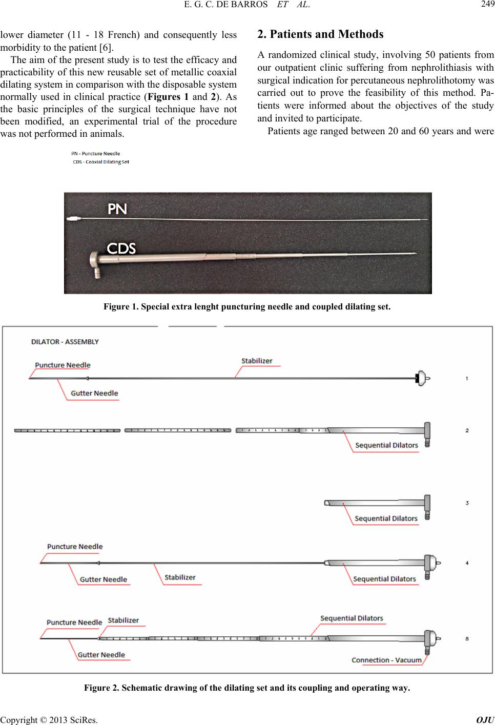 E. G. C. DE BARROS ET AL. 249 lower diameter (11 - 18 French) and consequently less morbidity to the patient [6]. The aim of the present study is to test the efficacy and practicability of this new reusable set of metallic coaxial dilating system in comparison with the disposable system normally used in clinical practice (Figures 1 and 2). As the basic principles of the surgical technique have not been modified, an experimental trial of the procedure was not performed in animals. 2. Patients and Methods A randomized clinical study, involving 50 patients from our outpatient clinic suffering from nephrolithiasis with surgical indication for percutaneous nephrolithotomy was carried out to prove the feasibility of this method. Pa- tients were informed about the objectives of the study and invited to participate. Patients age ranged between 20 and 60 years and were Figure 1. Special extra lenght puncturing ne e dle and coupled dilating set. Figure 2. Schematic drawing of the dilating set and its coupling and operating way. Copyright © 2013 SciRes. OJU 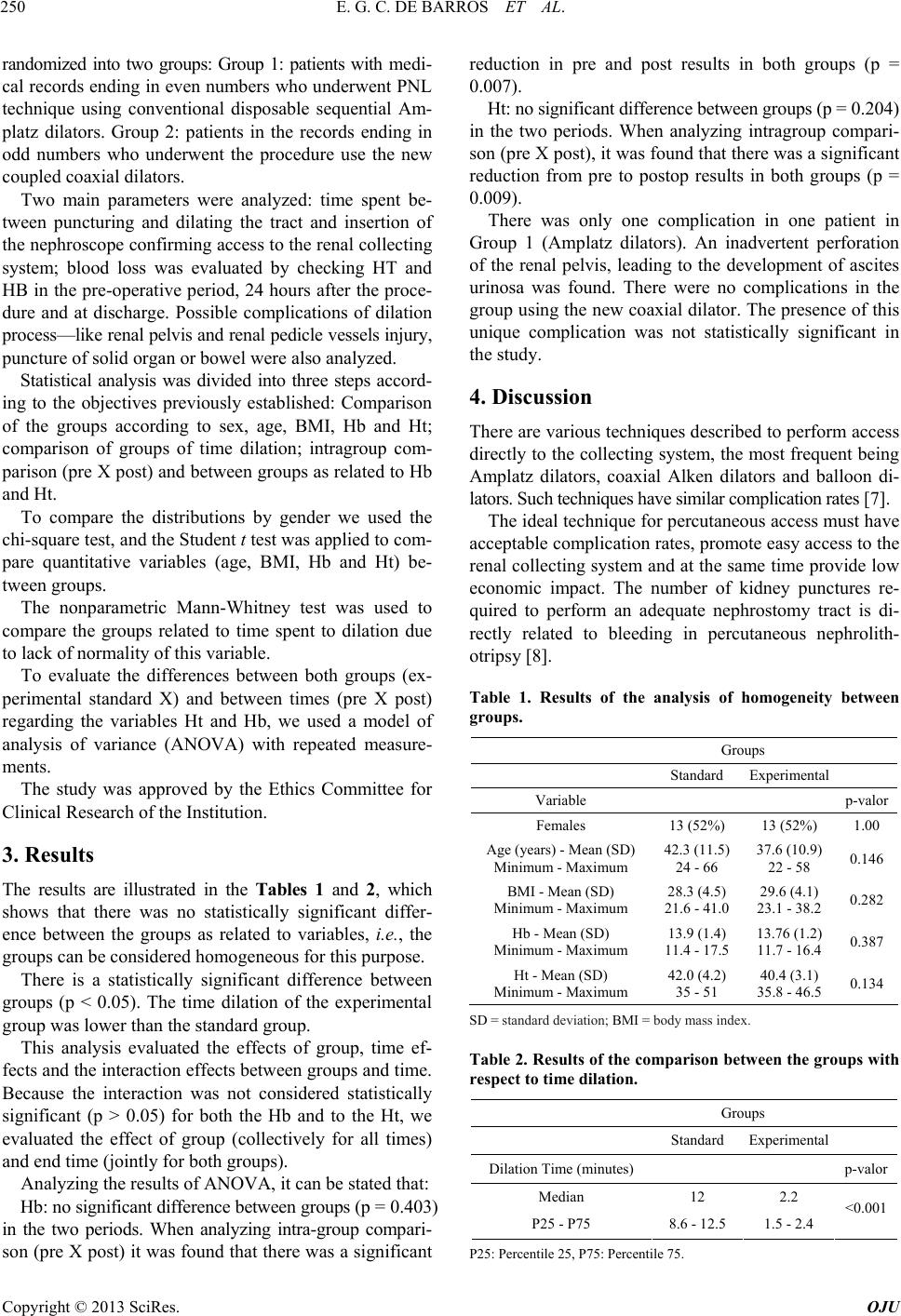 E. G. C. DE BARROS ET AL. 250 randomized into two groups: Group 1: patients with medi- cal records ending in ev en numbers who underwent PN L technique using conventional disposable sequential Am- platz dilators. Group 2: patients in the records ending in odd numbers who underwent the procedure use the new coupled coaxial d ilators. Two main parameters were analyzed: time spent be- tween puncturing and dilating the tract and insertion of the nephroscope confirming access to the renal collecting system; blood loss was evaluated by checking HT and HB in the pre-operative period, 24 hours after the proce- dure and at discharge. Possible complications of dilation process—like renal pelvis and renal pedicle vessels injury, puncture of solid organ or bowel were also analyzed. Statistical analysis was divided into three steps accord- ing to the objectives previously established: Comparison of the groups according to sex, age, BMI, Hb and Ht; comparison of groups of time dilation; intragroup com- parison (pre X post) and between groups as related to Hb and Ht. To compare the distributions by gender we used the chi-square test, and the Student t test was applied to com- pare quantitative variables (age, BMI, Hb and Ht) be- tween groups. The nonparametric Mann-Whitney test was used to compare the groups related to time spent to dilation due to lack of normality of this variable. To evaluate the differences between both groups (ex- perimental standard X) and between times (pre X post) regarding the variables Ht and Hb, we used a model of analysis of variance (ANOVA) with repeated measure- ments. The study was approved by the Ethics Committee for Clinical Research of the Institution. 3. Results The results are illustrated in the Tables 1 and 2, which shows that there was no statistically significant differ- ence between the groups as related to variables, i.e., the groups can be cons i dere d h o mogeneous for this pu rp ose. There is a statistically significant difference between groups (p < 0.05). The time dilation of the experimental group was lower than the standard group. This analysis evaluated the effects of group, time ef- fects and the interaction effects between groups and time. Because the interaction was not considered statistically significant (p > 0.05) for both the Hb and to the Ht, we evaluated the effect of group (collectively for all times) and end time (jointly for both groups). Analyzing the results of ANOVA, it can be stated that: Hb: no significant difference between groups (p = 0. 4 0 3 ) in the two periods. When analyzing intra-group compari- son (pre X post) it wa s found that there was a significant reduction in pre and post results in both groups (p = 0.007). Ht: no significant difference between groups (p = 0.204) in the two periods. When analyzing intragroup compari- son (pre X post), it w as found that there was a significant reduction from pre to postop results in both groups (p = 0.009). There was only one complication in one patient in Group 1 (Amplatz dilators). An inadvertent perforation of the renal pelvis, leading to the development of ascites urinosa was found. There were no complications in the group using the new coaxial dilato r. The presence of this unique complication was not statistically significant in the study. 4. Discussion There are various techniques described to perform access directly to the collecting system, the most frequent being Amplatz dilators, coaxial Alken dilators and balloon di- lators. Such techniques have similar complication rates [7]. The ideal technique for percutaneous access must have acceptable complication rates, promote easy access to the renal collecting system and at the same time provide low economic impact. The number of kidney punctures re- quired to perform an adequate nephrostomy tract is di- rectly related to bleeding in percutaneous nephrolith- otripsy [8] . Table 1. Results of the analysis of homogeneity between groups. Groups Standard Experimental Variable p-valor Females 13 (52%) 13 (52%) 1.00 Age (years) - Mean (SD) Min imum - Ma xi mum 42.3 (11.5) 24 - 66 37.6 (10.9) 22 - 58 0.146 BMI - Mean (SD) Min imum - Ma xi mum 28.3 (4.5) 21.6 - 41.0 29.6 (4.1) 23.1 - 38.20.282 Hb - Mean (SD) Min imum - Ma xi mum 13.9 (1.4) 11.4 - 17.5 13.76 (1.2) 11.7 - 16.40.387 Ht - Mean (SD) Min imum - Ma xi mum 42.0 (4.2) 35 - 51 40.4 (3.1) 35.8 - 46.50.134 SD = standard deviation; BMI = body mass index. Table 2. Results of the comparison between the groups with respect to tim e dilation. Groups Standard Experimental Dilation Time (minut es) p-valor Median 12 2.2 P25 - P75 8.6 - 12.5 1.5 - 2.4 <0.001 P25: Percentile 25, P75: Percentile 75. Copyright © 2013 SciRes. OJU  E. G. C. DE BARROS ET AL. 251 The preference for the comparative study between the coaxial dilator and coupled sequential dilators refers to the failure rate of inflatable dilators (17%). This rate rises to 25% when applied to patients with previous surgery [9]. Pneumatic dilators have not shown better results than the Amplatz dilators concerning decreased surgical time and decreased postoperative complications and a higher cost [10]. According to data collected, there was homogeneity between groups A and B, related to gender, age, body mass index (BMI), and hematological: hemoglobin (Hb) and hematocrit (Ht). The concern about having homogeneous groups refers to the possibility of difficulty in performing the proce- dure on specific groups, mainly related to BMI. This confirmation supports the reduction rates of research biases. Kessaris et al. [11] analyzed 2200 percutaneous renal procedures and stated that it was not a risk factor for renal bleeding: age, sex, number of punctures renal and procedure duration. The measurement of efficacy in performing the ne- phrostomy tract was defined as the time required estab- lishing the path after percutaneous puncture of the col- lecting system and insertion of the nephroscope. The time dilation was lower in the group that used the new coaxial dilator: The mean time spent with the standard technique 12 was minutes, and 2.2 minutes with the new set (p < 0.001). This can be explained by the elimination of some steps in the dilating processs, which leads to savings in time such as switching stabilizer using a punc- ture needle as a guide; stabilizer safety valve “stop”, which prevents the progression of the device in addition to the limit established by the surgeon, decreasing com- plication rates; sequential dilators and coupled to the system in monoblock; stabilizer removed en bloc and dilator set, maintaining 30 French (F) sheath and guide wire. The level of bleeding caused by the dilato r was similar in both groups, as shown by comparing drop in Hb and Ht between groups and within groups. No patient in both groups required blood transfusion. These data confirm reports of the literature [12]. Michel et al. [13] in a series of 315 cases reported 17.5% of bloo d transfusion. Bleeding is the most common complication and is as- sociated with the number of punctures, stone volume and diameter of the surgical material [8]. All patients under- went only one puncture per treatment session, the di- ameter of the material used was 30 F in the two groups. As described before the bleeding rates are similar to the reports by others [13]. The overall rate of complications in percutaneous ne- phrolithotomy can reach up to 83% [14]; however, the vast majority of these complications are minor, such as bleeding or fever without further repercussions. Major complications occur between 0.4% - 4.7% to sepsis and 0.6% - 4% to renal bleeding needing surgical interven- tion [6]. In the present study, one lesion of the renal pel- vis was observed in the control group. No complications were seen in the study group. When considering costs to perform perctaneous neph- rolitotripsy, although no specific studies were performed specifically considering this item the new set of dialato rs promotes significant savings according to the literature without increasing the surgical risks to patients and maintaining similar efficacy [15]. 5. Conclusion Data obtained in the present stu dy support the idea that a reusable coaxial metallic set may represent a feasible option in the percutaneous approach to the kidney and produces significant savings especially for institutional use without increasing surgical risks. REFERENCES [1] H. Wilkinson, “Clinical Investigation and Management of Patients with Renal Stones,” Annals of Clinical Bio- chemistry, Vol. 38, No. 3, 2001, pp. 180-187. http://dx.doi.org/10.1258/0004563011900623 [2] Ministério da Saúde, “Sistema de Informações Hospi- talares do SUS (SIH/SUS),” Morbidade Hospital do SUS por Local de Internação, Brasil, 2012. http://tabnet.datasus.gov.br/cgi/tabcgi.exe?sih/cnv/niuf.de f [3] T. Akman, M. Binbay, E. Tekinarslan, U. Ozkuvanci, C. Kezer, A. Erbin, Y. Berberoglu and A. Yaser-Musluma- noglu, “Outcomes of Percutaneous Nephrolithotomy in Patients with Solitary Kidney: A Single-Center Experi- ence,” Urology, Vol. 78, No. 2, 2011, pp. 272-276. http://dx.doi.org/10.1016/j.urology.2010.12.029 [4] M. S. Michel, L. Trojan and J. J. Rassweile s, “Complica- tions in Percutaneous Nephrolithotomy,” European Uro- logy, Vol. 51, No. 4, 2007, pp. 899-906. http://dx.doi.org/10.1016/j.eururo.2006.10.020 [5] T. Lopes, K. Sangam, P. Alken, B. S. Barroilhet, C. Saus- sine, L. Shi and J. de la Rossete, “Clinical Reserch Office of the Endourological Society. Percutaneous Nephro- lithotomy Study Group,” Journal of Endourology, Vol. 25, No. 5, 2011, pp. 55-62. http://dx.doi.org/10.1089/end.2010.0488 [6] T. S. Shin, H. J. Cho, S. H. Hong, J. Y. Lee, S. W. Kim and T. K. Hwang, “Complications of Percutaneous Neph- rolithotomy Classified the Modified Clavien Grading System: A Single Centes’s Experience over 16 years,” Korean Journal of Urology, Vol. 52, No. 11, 2011, pp. 769-775. http://dx.doi.org/10.4111/kju.2011.52.11.769 [7] Z. Huang, F. Fu, Z. Zhong, L. Zhang, R. Xu and X. Zhao, “Chinese Minimally Invasive Percutaneous Nephrolitho- tomy for Intrarenal Stones in Patients with Solitary Kid- ney: A Single-Center Experience,” PLoS One, Vol. 7, No. 7, 2012, Article ID: e40577. Copyright © 2013 SciRes. OJU  E. G. C. DE BARROS ET AL. Copyright © 2013 SciRes. OJU 252 http://dx.doi.org/10.1371/journal.pone.0040577 [8] Y. B. Jeog, S. B. Jeon, Y. G. Kim and Y. K. Park, “POS01.97: Comparison of Balloon and Amplatz Fascial Dilation Systems for Percutaneous Tract Dilation in Per- cutaneous Nephrolithotomy,” Urology, Vol. 70, No. 3, 2007, pp. 219-220. [9] M. L. Stoller, J. S. Wolf Jr. and M. A. Lezin, “Estimated Blood Loss and Transfusion Rate Associated with Percu- taneous Nephrolithotomy,” Journal of Urology, Vol. 132, No. 1, 1994, pp. 1977-1981. [10] A. Yamaguchi, A. Skolaricos, N. P. Buchholz, G. B. Chomón, M. Gasso, P. Saba, S. Nakada and J. de la Ros- sete, “Operating Times and Bleeding Complications in Percutaneous Nephrolithotomy: A Comparissonnof Tract Dilation Methods in 5.537 Patients in the Clinical Re- search Office of the Endourological Society Percutaneous Nephrolithotomy Global Study,” Journal—Endourolo- gical Society , Vol. 25, No. 6, 2011, pp. 933-9. http://dx.doi.org/10.1089/end.2010.0606 [11] D. N. Kessaris, G. C. Bellman, N. P. Pardalidis and A. G. Smith, “Management of Hemorrhage after Percutaneous Renal Surgery,” Journal of Urology, Vol. 153, No. 3, 1995, pp. 604-608. http://dx.doi.org/10.1016/S0022-5347(01)67659-6 [12] C. H. Shen, M. C. Cheng, C. T. Lin, Y. C. Jou and P. C. Chen, “Innovative Metal Dilators for Percutaneous Neph- rostomy Tract: Report 546 Cases,” Urology, Vol. 70, No. 3, 2007, pp. 418-21. http://dx.doi.org/10.1016/j.urology.2007.03.083 [13] M. S. Michel, L. Trojan, J. J. Rasswe iles, “Complications in Percutaneous Nephrolithotomy. European Urology, Vol. 51, No. 4, 2007, pp. 899-906. http://dx.doi.org/10.1016/j.eururo.2006.10.020 [14] G. Vallencien, R. Capdeville, B. Veillon, M. Charton and J. M. Brisset, “Colonic Perforation during Percutaneous Nephrolithotomy,” Journal of Urology, Vol. 134, No. 6, 1985, pp. 1185-1187. [15] T. Lopes, K. Sangam, P. Alken, Silva, B. S. Barroilhet, C. Saussine, S. Lei and J. de la Rosette, “The Clinical Re- search Office of the Endourological Society Percutaneous Nephrolithotomy Global Study: Tract Dilation Compari- sons in 5537 Patients,” Journal of Endourology, Vol. 25, No. 5, 2011, pp. 755-762. http://dx.doi.org/10.1089/end.2010.0488 List of Abreviations PNL: Percutaneous nephrolithotomy URS: Ureterorenoscopy ESWL: Extracorporeal shockwave lithotripsy BMI: Body mass index Hb: Hemogl o bin Ht: Hematocrit ANOVA: Analysis of varianc e F: French |