

Paper Menu >>
Journal Menu >>
 Open Journal of Orthopedics, 2013, 3, 261-268 http://dx.doi.org/10.4236/ojo.2013.36049 Published Online October 2013 (http://www.scirp.org/journal/ojo) 261 Differences in Survival Rates between Different Patterns of Unstable Pertrochanteric Femoral Fractures* Amir Herman1,2#, Yair Landau1, Alexander Hazanov3, Tal Segev4,5, Ran Thein1, Nachshon Shazar1 1Department of Orthopedic Surgery, Chaim Sheba Medical Center, Tel-Hashomer, Israel; 2Talpiot Medical Leadership Program, Chaim Sheba Medical Center, Tel-Hashomer, Israel; 3Department of Anesthesiology, Chaim Sheba Medical Center, Tel-Hashomer, Israel; 4Department of Vascular Surgery, Chaim Sheba Medical Center, Tel-Hashomer, Israel; 5Sackler Faculty of Medicine, Tel Aviv University, Tel Aviv, Israel. Email: #[email protected] Received August 6th, 2013; revised September 6th, 2013; accepted September 21st, 2013 Copyright © 2013 Amir Herman et al. This is an open access article distributed under the Creative Commons Attribution License, which permits unrestricted use, distribution, and reproduction in any medium, provided the original work is properly cited. ABSTRACT Background: Unstable pertrochanter femur fractures are common in orthopedic practice. They pose a surgical chal- lenge in both reduction and fixation. The fixation devices used are based on hip intramedullary nailing with femur head lag screw or blade. The aim of this paper is to compare different types of unstable pertrochanter fractures. Materials & Methods: We retrospectively reviewed 386 unstable pertrochanter femur fractures surgically treated in our hospital from 2000 to 2009. These included 62 (16.1%) unstable pertrochanter fractures with fractured lesser trochanter (31.A2-2, 31.A2-3); 63 (16.3%) reverse oblique fractures (31.A3-1), 51 (13.2%) transverse fractures (31.A3-2), 145 (37.6%) comminuted fractures (31.A3-3) and 65 (16.8%) subtrochanter fractures. We compared survival rates between fracture types. The clinical characteristics, surgery immediate outcome (e.g., tip apex distance, reduction quality), and long term results, i.e., complications were also compared between fracture types. Results: Survival analysis showed that the fracture types can be grouped into low and high risk fracture types. The former group included, reverse oblique and comminuted fractures. Lesser trochanter, transverse and subtrochanter fractures were included in the high risk group. The survival estimates for five years were 64.6% and 49.3% for the low and high risk fracture types, respectively (p value = 0.008). Multivariate survival analysis showed that the hazard ratio for the high risk fracture group was 1.9 (95% CI = 1.37 - 2.67). No differences were found between unstable pertrochanter femur fractures with regards to clinical and epidemiology characteristics. Optimal tip apex distance (TAD) of less than 25 mm was found in 66.7%, 57.1% and 66.7% of lesser trochanter, reverse oblique and sutrochanter fractures, respectively. TAD of less than 25 mm was found in 81.2% of both transverse and comminuted fractures (p value = 0.032). No statistically significant differ- ence was found between fracture types, in regards to complication or revision rates. Conclusions: Survival rates were higher in patients suffering from reverse oblique or comminuted pertrochanteric fractures. No differences were found between fracture types, in regards to clinical and other outcome parameters. Keywords: Intertrochanteric Fractures; Survival; Fracture Classification 1. Introduction Pertrochanteric fractures are among the most widely treated orthopedic injuries. Their annual incidence is ex- pected to reach 500,000 by 2040, in the US alone [1]. About half of this is estimated to be unstable factures [2]. The AO/OTA classification system divides pertrochan- teric fractures into several groups: simple pertrochanteric (31.A1), pertrochanteric with lesser trochanter fracture (31.A2) and complex pertrochanteric fractures (31.A3) [3]. Each of these three groups is further divided into three groups, consisting of a total of nine different fracture patterns. These patterns are commonly divided into stable pertrochanteric fractures (31.A1 fracture group and 31.A2- 1 subtype) and unstable pertrochanteric fractures (31.A2- 2 and 31.A2-3 subtypes and 31.A3 fracture group) [4-6]. Fixation devices available for pertrochanteric fractures vary from intra-medullary devices and extra-medullary plate both with femur head screw. The extra-medullary *Conflict of interest statement: No benefit in any form has been or will b e received from a commercial party related directly or indirectly to the subject of this manuscript. #Corresponding author. Copyright © 2013 SciRes. OJO  Differences in Survival Rates between Different Patterns of Unstable Pertrochanteric Femoral Fractures 262 device was shown to have fewer complications in stable pertrochanteric fractures. Patients with unstable pertro- chanteric fractures were shown to benefit from intrame- dullary fixation devices such as the Cephalomedullary nail, Gamma nail, Y nail among other [5,7-20]. Subtrochanteric fractures are defined as fractures with- in the immediate 5 cm distal to the lesser trochanter. As opposed to femur shaft fractures, these fractures require fixation of the femoral head-neck-shaft complex. Frac- tures of the subtrochanteric region often are fixed by in- tramedulary proximal femur fixation devices. As such these factures can be included as part of the unstable per- subtrochanteric fracture patterns [21]. Several authors described their experience of different unstable pertrochanteric fractures. These include a recent review by Lundy focusing on subtrochanteric fractures and a description of open reduction in treating subtro- chanteric fractures [2]. Other authors focused on reverse oblique or transverse fracture types [6]. We are unaware of any work comparing the survival among different sub- types of unstable pertrochanteric fractures. In this report we compare between the different pat- terns of unstable per-subtrochanteric fractures. We com- pare the survival rates, epidemiology, reduction achieved, screw placement, complications and revision rates be- tween the different fracture types. 2. Patients & Methods The study presented is a retrospective study based on na- tional mortality registry and our hospital admission and outpatient-clinic files. The study was approved by our institute’s internal review board (IRB) ethics committee. Since this is a retrospective study, patients’ informed con- sent was not required by the IRB. Between 2000 and 2009, 386 unstable pertrochanteric fractures were operated in our institute. These consisted of 62 (16.1%) pertrochanteric fractures in which the les- ser trochanter was fractured and the fracture was defined as unstable (31.A2-2 and 31.A2-3). There were 63 (16.3%) reverse oblique fractures (31.A3-1), 51 (13.2%) transver- es pertrochanteric fractures (31.A3-2) and 145 (37.6%) of comminuted fractures (31.A3-3). Subtrochanteric frac- tures included 65 (16.8%) fractures. The Fractures had been fixed by means of the Targon proximal femur (Targon PF) device (Aesculap, Tuttlin- gen, Germany) or with the antirotation trochanteric nail- ing system (ATN) device (dePuy, Warsaw, IN, USA). Both of which are double screw intramedullary fixation devices. All surgeries were performed in accordance to standard surgery technique and the manufacturer’s rec- ommendations. The radiology computerized achieve was used for clas- sification of fractures according to the OTA/AO classifi- cation system [3]. Patterns were classified by two inde- pendent researchers (A.O and Y.L). The senior author (N.S) was consulted whenever consensus was not rea- ched. Radiology measurements were performed includ- ing the tip apex distance and placement of the center of the lag screw within the head-neck interface “Safe-zone” (defined as the second quarter from the bottom) [22]. Acceptable reduction was considered as translation of less than 20 mm in any plane as measured by the medial cortex in anterior-posterior radiography view or anterior cortex on axial radiography view. Statistical analysis was performed by an experienced biostatistician (A.H.). Data analysis was conducted using SPSS© 16 (SPSS©, Chicago, IL). Categorical data are presented as frequency count (percent of available data). Comparisons of categorical variables between fracture types were performed using either the chi-square test or the Fisher’s exact test. The latter was used when expec- ted count in any cell was less or equal five. Continuous variables are presented as mean (±standard deviation). Comparisons of continuous variables among fracture pat- terns were performed using the Kruskal-Wallis test. Com- parisons of paired data, mainly the increase in mobility aids before and one year after surgery, were performed using the Wilcoxon rank sign test. Survival data are pre- sented using the Kaplan-Meier survival curves. Kaplan- Meier survival estimates were used for one and five years survival along with 95% confidence interval (95% CI). Comparisons between survival curves were done by the log-rank test. Data were extracted by reviewing of the patients’ ad- mission and out-patients clinic electronic files. Compli- cations, comorbidities, use of walking aids and recovery parameters were extracted from the hospital records. The national mortality registry was consulted for mortality status and date. Patients that were not registered as de- ceased were considered as censured at the date of the in- quiry. Multivariate survival analysis was performed with the Cox proportional hazards model. The independent co- variates in the model were fracture group (according to high or low risk fracture type), ASA score and Age group —up to 60 years old (46 pts, 11.9%), 60 - 80 years old (158 pts, 40.7%), above 80 year old (182 pts, 47.1%). Results are reported as hazard ratios and their 95% confidence in- tervals. Hazard ratio should be interpreted as relative risk, so that hazard ratio above one means excessive risk. Data were not available for all patients in all variables measured. The numbers of patients used for analysis of each variable are given within the summary tables. In each analysis frequencies and percents were calculated from available data. For example, for epidemiologic and clini- cal presentation parameters, percents were calculated from Copyright © 2013 SciRes. OJO  Differences in Survival Rates between Different Patterns of Unstable Pertrochanteric Femoral Fractures Copyright © 2013 SciRes. OJO 263 the entire patients’ population. Complications and revi- sion rates are calculated from a subset of patients which included patients that either had a complication or com- pleted at least one year of follow-up and showed signs of union on X-ray radiography. 3. Results Between 2000 and 2009, 386 unstable pertrochanteric fractures were operated in our institute. These included 95 (24.6%) fractures in men and 291 (75.4%) fractures in women. Mean age at surgery was 76.16 years (±15.2). Mean American society of anesthesiologists (ASA) score was 2.75 (±0.61), where 124 patients (32.2%) and 225 patients (58.4%) had ASA scores of 2 and 3, respectively. Patients with transverse fractures (31.A3-2) had higher ASA scores (p value = 0.026). Mechanism of injury was low energy fall in 175 fractures (87.1%). Initial treatment at the emergency ward included skeletal traction in 145 fractures (71.8%). Skeletal traction was performed more often in reverse oblique, comminuted or subtrochanter frac- tures. This difference was found to be statistically signi- ficant (p value = 0.001) Epidemiological and clinical data according to fracture types are presented in Table 1. Of the 386 fractures treated, 254 fractures (65.8%) were treated by the Targon PF © device, while 132 frac- tures (34.3%) were fixed using the ATN © device. No statistically significant difference was found between the fracture types. The nail length was found to be longer (300 mm - 340 mm) in fixation of subtrochanteric and comminuted fractures (Table 2). Immediate surgical outcome varied between fracture types. Tip Apex Distance (TAD) was found to be below 25 mm in about 81% of comminuted and transverse frac- Table 1. Demographic and clinical characteristics (Total = 386 patients). Fractured Lesser Trochanter (A2.2 & A2.3) N = 62 Reverse Oblique (A3.1) N = 63 Transverse (A3.2) N = 51 Comminuted (A3.3) N = 145 Subtrochanteric N = 65 P value Gender Male Female 20 (32.3%) 42 (67.7%) 11 (17.5%) 52 (82.5%) 9 (17.6%) 42 (82.4%) 35 (24.1%) 110 (75.9%) 20 (30%) 45 (69.2%) 0.173 Age 78.9 (±12.5) 79.0 (±9.8) 77.5 (±14.2) 75.8 (±14.8) 70.4 (±21.1) 0.243 Side Left Right 32 (51.6%) 30 (48.4%) 32 (50.8%) 31 (49.2%) 29 (56.9%) 22 (43.1%) 78 (53.8%) 67 (46.2%) 34 (52.3%) 31 (47.7%) 0.971 ASA score 2.69 (±0.64) 2.78 (±0.60) 3.00 (±0.566) 2.68 (±0.61) 2.72 (±0.50) 0.026 Above one year follow-up 26 (41.9%) 26 (41.3%) 19 (37.3%) 72 (49.7%) 22 (34.4%) 0.253 Skeletal traction (N = 202) 3 (7%) 11 (40.7%) 5 (17.9%) 25 (32.9%) 13 (46.4%) 0.001 Diabetes Mellitus (N = 201) 10 (23.8%) 4 (14.8%) 5 (17.9%) 19 (25.0%) 5 (17.9%) 0.764 Mechanism of injury (N = 202) Low energy (fall) High energy 37 (86%) 6 (14%) 25 (92.6%) 2 (7.4%) 24 (85.7%) 4 (14.3%) 68 (89.5%) 8 (10.5%) 21 (77.8%) 6 (22.2%) 0.327 Table 2. Surgery and post operative outcome parameters. Fractured Lesser Trochanter (A2.2 & A2.3) N = 62 Reverse Oblique (A3.1) N = 63 Transverse (A3.2) N = 51 Comminuted (A3.3) N = 145 Subtrochanteric N = 65 P value IM Nail Type Targon PF AT N 34 (53.9%) 29 (46.1%) 49 (77.8%) 14 (22.2%) 28 (54.9%) 23 (45.1%) 96 (66.2%) 49 (33.8%) 47 (73.4%) 17 (26.6%) 0.13 Nail length Standard (200 - 240 mm) Long (300 - 340 mm) 56 (90.3%) 6 (9.7%) 58 (92.1%) 5 (7.9%) 48 (94.1%) 3 (5.9%) 109 (75.2%) 36 (24.8%) 12 (19.0%) 51 (81.0%) 0.0001 Tip Apex distance ≤ 25 mm (N = 235) 28 (66.7%) 24 (57.1%) 29 (81.2%) 69 (81.2%) 20 (66.7%) 0.032 Lag screw within “safe-zone” (N = 378) 53 (85.5%) 50 (80.6%) 42 (87.5%) 120 (84.5%) 49 (76.6%) 0.506 Reduction not achieved (N = 383) 6 (9.8%) 17 (27.0%) 10 (19.6%) 35 (24.1%) 12 (19.0%) 0.134 Mobility points change from pre to 1 year post surgery (N = 60) 1.66 (±1.23) 1.77 (±1.30) 2.14 (±1.06) 1.90 (±1.33) 0.75 (±1.35) 0.174 Walking 1 year after surgery (N = 77) 72% - 93.5% 15 (100%) 8 (88.9%) 11 (100%) 25 (89.3%) 13 (92.9%) 0.566  Differences in Survival Rates between Different Patterns of Unstable Pertrochanteric Femoral Fractures 264 tures. The TAD was below 25 mm in only 57% - 66% of lesser trochanter, reveres oblique and subtrochanteric fractures. This difference was found to be statistically significant (p value = 0.032). However, 76% to 87% of the lag screws were placed within the head-neck inter- face “safe-zone”. This difference was not found to be sta- tistically significant (p value = 0.506). Reduction was not achieved in 7.6% patients. No statistically significant dif- ference was found between fracture types (p value = 0.386). Seventy two patients (93.5%) walked of 77 patients whose mobility status was recorded one year after sur- gery. Mobility aids used as recorded by the Parker score increased by a mean of 1.6 points (±1.3) one year after surgery. Sixty nine percent of the patients used a walking frame one year after surgery (Figure 1). No statistically significant difference was found in regards to mobility status between fracture types (Table 2). One hundred and ninety one patients had either at least one year of follow-up with documented fracture union or any complication. Common complications included 14 (7.3%) fractures that had cutout, 10 fractures (5.2%) that had hardware failure and 7 fractures fixed with internal rotation above 30 degrees. Less common complications included deep wound infection, secondary loss of reduc- tion, and fractures non-unions, with rates of 5 (2.6%), 5 (2.6%) and 3 (1.6%) fractures, respectively. No statisti- cally significant difference was found between fracture types (Table 3). Thirty two patients (16.8%) of the aforementioned 191 patients had revision surgery. Common revision types included 14 (7.3%) hardware removals, 7 (3.7%) arthro- Figure 1. Walking aids score: where no aids, one aid, two aids, walking frame and wheel chair received a score of 1 - 5 in an ascending order. Mean increase in ambulation score from preoperative to 1 year after surgery was 1.63 points (±1.32). This difference was found to be statistically significant (p value = 0.0001). No statistically significant difference was found between fracture patterns. Table 3. Complications. Total N = 191 Fractured Lesser Trochanter (A2.2 & A2.3) N = 34 Reverse Oblique (A3.1) N = 31 Transverse (A3.2) N = 23 Comminuted (A3.3) N = 78 Subtrochanteric N = 25 P value Cut out—14 pts (7.3%) 3 (8.8%) 2 (6.5%) 4 (17.4%) 5 (6.4%) --- 0.232 Hardware failure—10 pts (5.2%) 4 (11.7%) 2 (6.5%) --- 4 (5.2%) --- 0.278 Nonunion—3 pts (1.6%) 1 (2.9%) --- 1 (4.3%) --- 1 (4.0%) 0.16 Deep wound infection —5 pts (2.6%) --- --- --- 4 (5.1%) 1 (4.0%) 0.508 Secondary loss of reduction 5 pts (2.6%) 1 (2.9%) 1 (3.2%) --- 1 (1.3%) 1 (4.0%) 0.674 Internal rotation—7 pts (3.7%) 3 (8.8%) 2 (6.5%) --- 1 (1.3%) 1 (4.0%) 0.172 Other—3 pts (1.5%) 1 (2.9%) --- --- 1 (1.3%) 1 (4.0%) 0.564 Total complications—47 pts (24.6%) 13 (38.2%) 7 (22.6%) 5 (21.7%) 16 (20.5%) 6 (24.0%) 0.369 Table 3 includes surgical complications and revision surgery. Other complications include one patient with pain that required revision, 1 patient with fracture at the lower end of the fixation device, and one patient with superior placement of the fixation device that required revision. No cases of deep vein thrombosis were reported. Copyright © 2013 SciRes. OJO  Differences in Survival Rates between Different Patterns of Unstable Pertrochanteric Femoral Fractures 265 plasties and 5 (2.6%) rotational corrections. No statisti- cally significant difference was found between fracture types (Table 4). One year and five years survival of the entire cohort were 79.6% (95% CI of 75.1% - 84.2%) and 48.9% (95% CI of 42.3% - 56.4%), respectively. Comparing survival between specific inter-trochanteric fractures, no statisti- cally significant difference was found (p value = 0.07, Table 5). The fractures were grouped into high and low risk fracture types, according to survival. The low risk fracture types included reverse oblique fractures (31.A3- 1) and comminuted fractures (31.A3-3). High risk frac- ture types included lesser trochanter unstable fractures (31.A2-2 and 31.A2-3), transverse fractures (31.A3-2) and subtrochanteric fractures (32 either A, B or C). One year and five years survival for the low risk fracture types were 86% (95% CI 81.4% - 90.8%) and 64.6% (95% CI 57.4% - 72.6%), respectively. One year and five years survival for the high risk fracture types were 76.4% (95% CI, 70.4% - 82.9%) and 49.3% (95% CI, 41.0% - 59.2%). This difference was found to be statistically sig- nificant (p value = 0.008, Figure 2). Multivariate analysis by the Proportional hazards mo- del showed that the high risk fracture group was asso- ciated with higher mortality risk. Setting the low risk fracture group to have hazard ratio of one (as baseline) the hazard ratio for the high risk fracture group was 1.9 (95% CI = 1.37 - 2.67). This difference was found to be statistically significant (p value = 0.0001). The hazard rate associated with age 60 - 80 was 17.7 (95% CI = 4.3 - 73.1). The hazard rate associated with age above 80 years old was 19.6 (95% CI = 4.7 - 80.7). The hazard rate as- sociated with each point of the ASA score was 1.152 (95% CI = 0.88 - 1.5). This was not found to be statisti- cally significant (p value = 0.29). 4. Discussion In this manuscript, it was shown that survival rates dif- fered between different types of unstable pertrochanteric fractures. These fractures could be divided into low and high risk fracture types. The low risk group includes: re- verse oblique and comminuted fractures. The high risk fracture types include: lesser trochanter, transverse and subtrochanteric fractures. No difference in clinical or epi- demiologic characteristics was found in order to explain the difference in survival. No difference was found in complication and revision rates that could also elucidate the survival difference. In previous works, other authors focused mainly on Table 4. Revisions. Total N = 191 pts Fractured Lesser Trochanter (A2.2 & A2.3) N = 34 Reverse Oblique (A3.1) N = 31 Transverse (A3.2) N = 23 Comminuted (A3.3) N = 78 Subtrochanteric N = 25 P value Total Hip replacement —3 pts (1.6%) 2 (5.9%) --- 1 (4.3%) --- --- 0.086 Hemiarthroplasty—4 pts (2.1%) --- --- --- 4 (5.1%) --- 0.441 Exchange nail—4 pts (2.1%) --- --- 1 (4.3%) 2 (2.6%) 1 (4.0%) 0.533 Hardware removal—11 pts (5.7%) 3 (8.8%) 3 (9.4%) 1 (4.3%) 4 (5.1%) --- 0.552 Nail removal and plating—3 pts (1.6%) --- --- 2 (8.7%) --- 1 (4.0%) 0.018 Rotation correction—5 pts (2.6%) 2 (5.9%) 1 (3.2%) --- 1 (1.3%) 1 (4.0%) 0.455 Soft tissue revision due to infection 1 pt (0.5%) --- --- --- 1 (1.3%) --- 1.00 Total revisions—32 pts (16.8%) 7 (20.6%) 4 (12.9%) 5 (21.7%) 12 (15.4%) 4 (16.0%) 0.876 Table 5. Survival estimates (95% confidence intervals). Total = 386 patients Fractured Lesser Trochanter (A2.2 & A2.3) N = 62 Reverse Oblique (A3.1) N = 63 Transverse (A3.2) N = 51 Comminuted (A3.3) N = 145 Subtrochanteric N = 65 Six months 95.2% (90% - 100%) 93.5% (87.6% - 99.9%)88.2% (79.8% - 97.5%)91% (86.5% - 95.8%) 80% (70.8% - 90.3%) One year 77.4% (67.6% - 88.5%) 90.3% (83.3% - 98%)74.5% (63.4% - 87.5%)84.1% (78.4% - 90.3%) 76.9% (67.3% - 87.9%) Five years 55.7% (42% - 73.9%) 63.2% (50.9% - 78.4%)37.9% (22.8% - 62.8%)64.5% (56% - 74.3%) 50.5% (38.5% - 66.3%) P value for survival curves comparing all the fracture types = 0.07. Copyright © 2013 SciRes. OJO 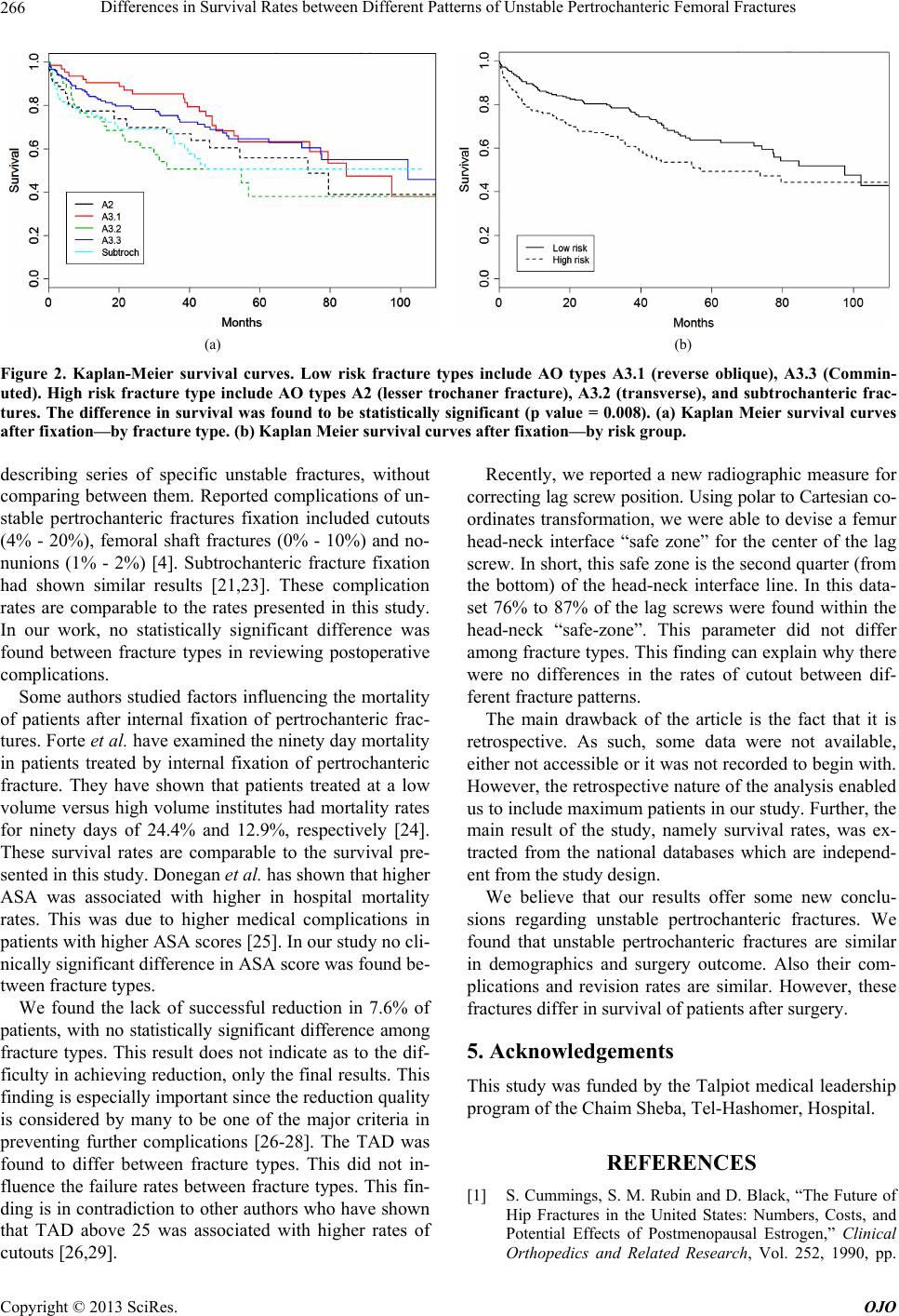 Differences in Survival Rates between Different Patterns of Unstable Pertrochanteric Femoral Fractures 266 (a) (b) Figure 2. Kaplan-Meier survival curves. Low risk fracture types include AO types A3.1 (reverse oblique), A3.3 (Commin- uted). High risk fracture type include AO types A2 (lesser trochaner fracture), A3.2 (transverse), and subtrochanteric frac- tures. The difference in survival was found to be statistically significant (p value = 0.008). (a) Kaplan Meier survival curves after fixation—by fracture type. (b) Kaplan Meier survival curves after fixation—by risk group. describing series of specific unstable fractures, without comparing between them. Reported complications of un- stable pertrochanteric fractures fixation included cutouts (4% - 20%), femoral shaft fractures (0% - 10%) and no- nunions (1% - 2%) [4]. Subtrochanteric fracture fixation had shown similar results [21,23]. These complication rates are comparable to the rates presented in this study. In our work, no statistically significant difference was found between fracture types in reviewing postoperative complications. Some authors studied factors influencing the mortality of patients after internal fixation of pertrochanteric frac- tures. Forte et al. have examined the ninety day mortality in patients treated by internal fixation of pertrochanteric fracture. They have shown that patients treated at a low volume versus high volume institutes had mortality rates for ninety days of 24.4% and 12.9%, respectively [24]. These survival rates are comparable to the survival pre- sented in this study. Donegan et al. has shown that higher ASA was associated with higher in hospital mortality rates. This was due to higher medical complications in patients with higher ASA scores [25]. In our study no cli- nically significant difference in ASA score was found be- tween fracture types. We found the lack of successful reduction in 7.6% of patients, with no statistically significant difference among fracture types. This result does not indicate as to the dif- ficulty in achieving reduction, only the final results. This finding is especially important since the reduction quality is considered by many to be one of the major criteria in preventing further complications [26-28]. The TAD was found to differ between fracture types. This did not in- fluence the failure rates between fracture types. This fin- ding is in contradiction to other authors who have shown that TAD above 25 was associated with higher rates of cutouts [26,29]. Recently, we reported a new radiographic measure for correcting lag screw position. Using polar to Cartesian co- ordinates transformation, we were able to devise a femur head-neck interface “safe zone” for the center of the lag screw. In short, this safe zone is the second quarter (from the bottom) of the head-neck interface line. In this data- set 76% to 87% of the lag screws were found within the head-neck “safe-zone”. This parameter did not differ among fracture types. This finding can explain why there were no differences in the rates of cutout between dif- ferent fracture patterns. The main drawback of the article is the fact that it is retrospective. As such, some data were not available, either not accessible or it was not recorded to begin with. However, the retrospective nature of the analysis enabled us to include maximum patients in our study. Further, the main result of the study, namely survival rates, was ex- tracted from the national databases which are independ- ent from the study design. We believe that our results offer some new conclu- sions regarding unstable pertrochanteric fractures. We found that unstable pertrochanteric fractures are similar in demographics and surgery outcome. Also their com- plications and revision rates are similar. However, these fractures differ in survival of patients after surgery. 5. Acknowledgements This study was funded by the Talpiot medical leadership program of the Chaim Sheba, Tel-Hashomer, Hospital. REFERENCES [1] S. Cummings, S. M. Rubin and D. Black, “The Future of Hip Fractures in the United States: Numbers, Costs, and Potential Effects of Postmenopausal Estrogen,” Clinical Orthopedics and Related Research, Vol. 252, 1990, pp. Copyright © 2013 SciRes. OJO  Differences in Survival Rates between Different Patterns of Unstable Pertrochanteric Femoral Fractures 267 163-166. [2] K. Koval, G. B. Aharonoff, A. S. Rokito, T. Lyon and J. D. Zuckerman, “Patients with Femoral Neck and Inter- trochanteric Fractures: Are They the Same? Clinical Or- thopedics and Related Research, Vol. 330, 1996, pp. 166- 172. http://dx.doi.org/10.1097/00003086-199609000-00020 [3] Fracture and Dislocation Classification Compendium, “Or- thopaedic Trauma Association Classification, Database and Outcomes Committee,” Journal of Orthopedic Trauma, Vol. 20, No. 10 (S1), 2007, pp. 31-42. [4] D. Lindskog and M. R. Baumgaertner, “Unstable Intertro- chanteric Hip Fractures in the Elderly,” Journal of the American Academy of Orthopaedic Surgeons, Vol. 12, No. 3, 2004, pp. 189-190. [5] M. Parker and H. H. Handoll, “Gamma and Other Cepha- locondylic Intramedullary Nails versus Extramedullary Im- plants for Extracapsular Hip Fractures in Adults,” Coch- rane Database Systematic Review, Vol. 3, 2008, p. CD93. [6] C. Sadowski, A. Lübbeke, M. Saudan, N. Riand, R. Stern and P. Hoffmeyer, “Treatment of Reverse Oblique and Transverse Intertrochanteric Fractures with Use of an In- tramedullary Nail or a 95˚ Screw-Plate: A Prospective, Randomized Study,” Journal of Bone and Joint Surgery (Am), Vol. 84, No. 3, 2002, pp. 372-381. [7] S. Bridle, A. D. Patel, M. Bircher and P. T. Calvert, “Fi- xation of Intertrochanteric Fractures of the Femur, a Ran- domized Prospective Comparison of the Gamma Nail and the Dynamic Hip Screw,” Journal of Bone and Joint Sur- gery (Br), Vol. 73-B, No. 2, 1991, pp. 330-334. [8] B. Giraud, E. Dehoux, N. Jovenin, K. Madi, A. Harisboure, G. Usandizaga, et al., “Pertrochanteric Fractures: A Rando- mized Prospective Study Comparing Dynamic Screw Plate and Intramedullary fixation,” Revue de Chirurgie Ortho- pedique et Reparatrice de l’Appareil Moteur, Vol. 91, No. 8, 2005, pp. 732-736. http://dx.doi.org/10.1016/S0035-1040(05)84484-8 [9] D. Hardy, P. Y. Descamps, P. Krallis, L. Fabeck, P. Smets, C. L. Bertens and P. E. Delince, “Use of an Intramedul- lary Hip-Screw Compared with a Compression Hip- Screw with a Plate for Intertrochanteric Femoral Frac- tures: A Prospective, Randomized Study of One Hundred Patients,” Journal of Bone and Joint Surgery (Am), Vol. 80, No. 5, 1998, pp. 618-630. [10] K. Leung, W. S. So, W. Y. Shen and P. W. Hui, “Gamma Nails and Dynamic Hip Screws for Peritrochanteric Frac- tures: A Randomized Prospective Study in Elderly Pa- tients,” Journal of Bone and Joint Surgery (Am), Vol. 74-B, No. 3, 1992, pp. 345-351. [11] J. Madsen, L. Næss, A. K. Aune, A. Alho, A. Ekeland and K. Strømsøe, “Dynamic Hip Screw with Trochanteric Stabilizing Plate in the Treatment of Unstable Proximal Femoral Fractures: A Comparative Study with the Gam- ma Nail and Compression Hip Screw,” Journal of Ortho- pedic Trauma, Vol. 12, No. 4, 1998, pp. 241-248. http://dx.doi.org/10.1097/00005131-199805000-00005 [12] C. Mainds and R. J. Newman, “Implant Failures in Pa- tients with Proximal Fractures of the Femur Treated with a Sliding Screw Device,” Injury, Vol. 20, No. 2, 1989, pp. 98-109. http://dx.doi.org/10.1016/0020-1383(89)90151-4 [13] E. Osnes, C. M. Lofthus, J A. Falch, et al., “More Post- Operative Femoral Fractures with the Gamma Nail than the Sliding Screw Plate in the Treatment of Trochanteric Fractures,” Acta Orthopedica Scandinavica, Vol. 72, No. 3, 2001, pp. 252-256. http://dx.doi.org/10.1080/00016470152846574 [14] J. Pajarinen, J. Lindahl, O. Michelsson, V. Savolainen and E. Hirvensalo, “Pertrochanteric Femoral Fractures Treated with a Dynamic Hip Screw or a Proximal Femo- ral Nail; a Randomised Study Comparing Postoperative Rehabilitation,” Journal of Bone and Joint Surgery (Br), Vol. 87, No. 1, 2005, pp. 76-81. [15] S. Papasimos, C. M. Koutsojannis, A. Panagopoulos, P. Megas and E. Lambiris, “A Randomised Comparison of AMBI, TGN and PFN for Treatment of Unstable Trochan- teric Fractures,” Archives of Orthopaedic and Trauma Surgery, Vol. 125, No. 7, 2005, pp. 462-468. http://dx.doi.org/10.1007/s00402-005-0021-5 [16] P. Radford, M. Needoff and J. K. Webb, “A Prospective Randomized Comparison of the Dynamic Hip Screw and the Gamma Locking Nail,” Journal of Bone and Joint Surgery (Br), Vol. 75-B, No. 5, 1993, pp. 789-793. [17] C. Rogmark, L. Flensburg and H. Fredin, “Undisplaced Femoral Neck Fractures—No Problems? A Consecutive Study of 224 Patients Treated with Internal Fixation,” In- jury, Vol. 40, No. 3, 2009, pp. 274-276. [18] M. Saudan, A. Lubbeke, C. Sadowski, N. Riand, R. Stern and P. Hoffmeyer, “Pertrochanteric Fractures: Is There an Advantage to an Intramedullary Nail? A Randomized, Pro- spective Study of 206 Patients Comparing the Dynamic Hip Screw and Proximal Femoral Nail,” Journal of Or- thopaedic Trauma, Vol. 16, No. 6, 2002, pp. 386-393. http://dx.doi.org/10.1097/00005131-200207000-00004 [19] I. Schipper, E. W. Steyerberg, R. M. Castelein, F. H. W. M. van der Heijden, P. T. den Hoed, A. J. H. Kerver and A. B. van Vugt, “Treatment of Unstable Trochanteric Frac- tures: Randomised Comparison of the Gamma Nail and the Proximal Femural Nail,” Journal of Bone and Joint Surgery (Br), Vol. 86-B, No. 1, 2004, pp. 86-94. [20] R. Simmermacher, A. M. Bosch and C. H. Van der Wer- ken, “The AO/ASIF-Proximal Femoral Nail (PFN): A New Device for the Treatment of Unstable Proximal Fe- mur Fractures,” Injury, Vol. 30, No. 5, 1990, pp. 327-332. http://dx.doi.org/10.1016/S0020-1383(99)00091-1 [21] D. Lundy, “Subtrochanteric Femoral Fractures,” Journal of the American Academy of Orthopaedic Surgeons, Vol. 15, No. 11, 2007, pp. 663-671. [22] A. L. F. Afsari, E. Lindvall, A. Infante, H. C. Sagi and G. J. Haidukewych, “Clamp-Assisted Reduction of High Sub- trochanteric Fractures of the Femur,” Journal of Bone and Joint Surgery (Am), Vol. 92, No. S1 (Part 2), 2009, pp. 1913-1918. http://dx.doi.org/10.2106/JBJS.H.01563 [23] M. Baumgaertner, S. L. Curtin, D. M. Lindskog and J. M. Keggi, “The Value of the Tip-Apex Distance in Predict- ing Failure of Fixation of Peritrochanteric Fractures of the Hip,” Journal of Bone and Joint Surgery (Am), Vol. 77, No. 7, 1995, pp. 1058-1064. [24] M. Forte, B. A. Virnig, M. F. Swiontkowski, M. Bhandari, Copyright © 2013 SciRes. OJO  Differences in Survival Rates between Different Patterns of Unstable Pertrochanteric Femoral Fractures Copyright © 2013 SciRes. OJO 268 R. Feldman, L. E. Eberly and R. L. Kane, “Ninety-Day Mortality after Intertrochanteric Hip Fracture: Provider Volume Matter?” Journal of Bone and Joint Surgery (Am), Vol. 92, No. 4, 2010, pp. 799-806. http://dx.doi.org/10.2106/JBJS.H.01204 [25] D. J. Donegan, A. N. Gay, K. Baldwin, E. E. Morales, J. L. Esterhai and S. Mehta, “Use of Medical Comorbidities to Predict Complications after Hip Fracture Surgery in the Elderly,” Journal of Bone and Joint Surgery (Am), Vol. 92, No. 4, 2010, pp. 807-813. http://dx.doi.org/10.2106/JBJS.I.00571 [26] T. Barton, R. Gleeson, C. Topliss, R. Greenwood, W. J. Harries and T. J. S. Chesser, “A Comparison of the Long Gamma Nail with the Sliding Hip Screw for the Treat- ment of AO/OTA 31-A2 Fractures of the Proximal Part of the Femur: A Prospective Randomized Trial,” Journal of Bone and Joint Surgery (Am), Vol. 92, No. 4, 2010, pp. 792-798. http://dx.doi.org/10.2106/JBJS.I.00508 [27] T. Davis, J. L. Sher, A. Horsman, M. Simpson, B. B. Por- ter and R. G. Checketts, “Intertrochanteric Femoral Frac- tures—Mechanical Failure after Intrenal Fixation,” Jour- nal of Bone and Joint Surgery (Br), Vol. 72-B, No. 1, 1990, pp. 26-31. [28] S. Larsson, S. Friberg and L. Hansson, “Trochanteric Frac- tures, Influence of Reduction and Implant Position on Im- paction and Complications,” Clinical Orthopedics and Re- lated Research, Vol. 259, 1990, pp. 130-139. [29] M. Güvena, U. Yavuzb, B. Kadıogluc, B. Akmand, V. Kı- lıncoglue, K. Ünayc and F. Altıntas, “Importance of Screw Position in Intertrochanteric Femoral Fractures Treated by Dynamic Hip Screw,” Orthopaedics & Traumatology: Surgery & Research, Vol. 96, No. 1, 2010, pp. 21-27. |