

Paper Menu >>
Journal Menu >>
 Open Journal of Orthopedics, 2013, 3, 253-257 http://dx.doi.org/10.4236/ojo.2013.36047 Published Online October 2013 (http://www.scirp.org/journal/ojo) 253 Long Bone Non-Unions and Malunions: Risk Factors and Treatment Outcomes in Calabar, Southern Nigeria Ikpeme A. Ikpeme1*, Nkese E. Mkpanam2, Innocent E. Abang1, Ngim E. Ngim1, Anthony M. Udosen1 1Department of Orthopaedics & Traumatology, University of Calabar, Calabar, Nigeria; 2Department of Community Medicine, Uni- versity of Calabar, Calabar, Nigeria. Email: *[email protected] Received August 28th, 2013; revised September 25th, 2013; accepted October 1st, 2013 Copyright © 2013 Ikpeme A. Ikpeme et al. This is an open access article distributed under the Creative Commons Attribution Li- cense, which permits unrestricted use, distribution, and reproduction in any medium, provided the original work is properly cited. ABSTRACT Background: Diaphyseal non-unions and malunions constitute significant morbidities in fracture care. Fracture treat- ment modalities seek to restore anatomic orientation and functional rehabilitation as soon as possible after a fracture incident. Malunions and non unions present a treatment challenge with the need for prolonged hospitalization, multiple surgical interventions and economic burden. In the developing world, traditional bonesetting practices are popular and these often result in a host of preventable complications. The added socioeconomic costs of treating these complications present a considerable strain on the resources of these already fragile economies and households. Aim: To document the risk factors, treatment options and outcomes for diaphyseal non-unions and malunions in our environment. Patients and Methods: Fifty-two consecutive patients comprising 37 non-unions and 15 malunions who presented in the ortho- paedic unit of a tertiary hospital in Southern Nigeria were evaluated. Information sought included biodata, location of pathology, type of incident fracture, local risk factors including traditional bonesetting; treatment options and final out- comes. Information obtained was analyzed using SPSS version 20 (IBM, New York). Results are presented in simple frequency tables. Results: There were 34 males and 18 females (M:F = 1.9:1) with a mean age of 38.76 ± 14.55 years. There were 37 non-unions and 15 malunions. The femur was the commonest site of pathology in 21 (40.4%) cases, and among the non-unions, the atrophic variety was the commonest type (n = 26; 70.3%). The mean fracture-to-surgery interval was 11.35 ± 7.95 months and traditional bonesetting was the commonest risk factor (n = 36; 69.2%). Plate and screw Osteosynthesis with bonegraft augmentation was the commonest treatment modality and the overall union rate was 94%. Conclusion: Traditional bonesetting plays a major role in the health seeking behaviour of many African so- cieties. The complications are varied and add to the overall socioeconomic burden of fracture care in these developing economies. Identification of traditional bonesetting practices as an important risk factor should translate into a focus on these practices in preventive public health decisions in fracture care. Continuing public health education backed by po- litical will and can potentially drive a paradigm shift in health seeking attitudes in the developing world. Keywords: Non-Union; Malunions; Traditional Bonesetting; Resource-Poor Economy 1. Introduction Long bones serve to support the trunk providing a stable framework for propulsion and facilitate pre-hension, reach and grasp, functions which are important in the homo erectus. Non-unions are estimated to occur in 1% - 10% of humeral shaft fractures treated non-operatively, 10% - 15% of humeral fractures treated operatively, 0.9% of femoral shaft fractures treated with modern intrame- dullary nailing techniques and 2% to 10% of all tibial fractures in the United States [1-3], and result in a large number of therapeutic interventions with significant so- cio-economic costs [1-4]. The factors that cause non un- ions may be considered as those inherent in the fracture, patient (host) factors and surgical (treatment) factors. They include the involved bone and bony region injured, the degree of soft tissue injury, patient’s age, the presence of co-morbidities, smoking and non-steroidal anti-inflam- matory drug (NSAID) abuse. Unstable fixation, excessive iatrogenic stripping of the periosteum, infection, malnu- trition, chronic alcoholism and injudicious interventions by traditional bonesetters are other risk factors for non- unions [1,5,6-8]. The definition of non-union has undergone an evolution. Prior to 1998, the definition of the condition was time- *Corresponding author. Copyright © 2013 SciRes. OJO  Long Bone Non-Unions and Malunions: Risk Factors and Treatment Outcomes in Calabar, Southern Nigeria 254 bound as failure of union in a fracture ≥9 months post injury with no observable progressive signs of healing for at least 3 months [9]. The prevention of prolonged morbidity and recognition of the need for early interven- tion necessitated a review of the definition to be the ab- sence of progressive signs of bone healing on radio- graphs over 3 consecutive months [4,9,10]. Some regions still however define non-union as a failure to heal within the time boundaries of injury and treatment [6,7]. The ti- bia is the most commonly affected bone [1,4,10], owing to its subcutaneous location and relative lack of muscle cover except in the postero-lateral aspect. Non-unions are classified in different ways, each with a therapeutic and prognostic significance. Atrophic non- unions have traditionally been ascribed to failure of hea- ling biology, while hypertrophic non-unions are said to occur in the presence of excessive movement or infection in a setting of appropriate healing biological factors. The- rapeutically therefore, while bonegrafts are always nec- essary in atrophic non-unions, stabilization with compres- sion and reduction of excessive movements may be all that is required in hypertrophic non-unions. In terms of their anatomic location, Non-unions may be diaphyseal or metaphyseal. Diaphyseal non-unions have a reduced bio- logic potential compared to metaphyseal non-unions, but are amenable to a wider range of treatment/stabilization options [1]. Non-unions may also be classified as aseptic or infected. While the goal of management of aseptic non-union is the promotion of stability and union, the goal of treatment of an infected non-union is to first con- vert it into a non-infected non-union and then treat the fracture. Acceptable reduction in the tibia is characterized as greater than 50% cortical contact, less than 100 angula- tion in any plane, less than 50 valgus or varus tilt, less than 100 of anterior or posterior angulation, less than 100 of rotation and less than 10 mm leg length discrepancy. Fracture site distraction is not tolerated because a 5 mm distraction may increase healing time to 8 - 12 months [11]. The treatment of diaphyseal non-unions involves crea- ting fresh bone ends and restoration of marrow continuity, stable fixation with compression of the non-union and augmentation of bone healing using biologic and non- biologic agents like low-intensity pulsed ultrasound sti- mulation, electrical bone growth stimulation, bone grafts and bonegraft substitutes with osteobiological agents. A malunion occurs when a fracture has healed in a non-anatomic or unacceptable attitude with respect to alignment, length and angulation, often with significant functional impairment especially in the lower limbs. Ge- nerally, greater than 15 mm shortening, 10 [12]. In the upper limb, malunions often present more of a cosmetic than functional nuisance. Generally, malun- ions frequently result from conservative treatment of frac- tures, failure to adhere to physicians’ instructions, ill-ad- vised weight bearing and injudicious interventions by tra- ditional bonesetters. The treatment of long bone malun- ion aims to correct translational, rotational and angular deformities and achieve a cosmetically and functionally accepted limb. This often involves osteoclasis, open re- duction and internal fixation using intramedullary rods or plate and screw assemblies and various types of osteoto- mies. Understanding the biology of fracture healing is es- sential and bonegraft augmentation may be required. The aim of this study is to document the risk factors, treat- ment options and outcomes for long bone diaphyseal non- union and malunions in our environment. 2. Materials and Methods A study of 52 consecutive patients with 15 malunions and 37 non-unions is over a 2-year period. Patients were evaluated for age, sex, location of pathology, type of in- cident fracture, risk factors for malunions and non-union, type of non-union, treatment options and final outcomes. Information obtained was analysed using SPSS statistics, Version 20 (IBM Corp, New York). 3. Results There were 52 cases of long bone diaphyseal malunions and non-unions in this series. There were 34 males and 18 females (M:F = 1.9:1) with a mean age of 38.76 ± 14.55 years. There were a total of 15 malunions and 37 non-unions. Of the non-unions, there were 26(70.3%) atrophic, 6(16.2) hypertrophic and 5 septic (13.5%) va- rieties. The incident fracture was closed in 41 cases and open in 11 patients. Forty-two (80.7%) patients had at least secondary level education while 10(19.3%) had pri- mary level or no education. Thirty-eight (73.1%) patients were employed in the public or private sector, 13(25%) were students and 1 patient (1.9%) was unemployed. The mean fracture-surgery interval was 11.35 ± 7.95 months and the femur was the commonest site of pathology in 21(40.4%) patients followed by the tibia in 16(30.8%) patients. Union was achieved in 47(90.4%) patients after the first surgical intervention. Five patients (9.6%) requir- ed more than one surgical intervention, and out of these, union was subsequently achieved in two. Non-union per- sisted in three patients. Two out of these 3 cases were in- fected non-unions and 1 was atrophic. The overall union rate was therefore 94% (Table 1). In terms of risk factors, traditional bonesetting was the commonest being present in 36(69.2%) patients followed by open fractures in 6(11.5%) patients (Table 2). Plate 0 varus or val- gus angulations, 100 recurvatum, 100 internal rotation and 150 external rotation are unacceptable for tibial fractures Copyright © 2013 SciRes. OJO 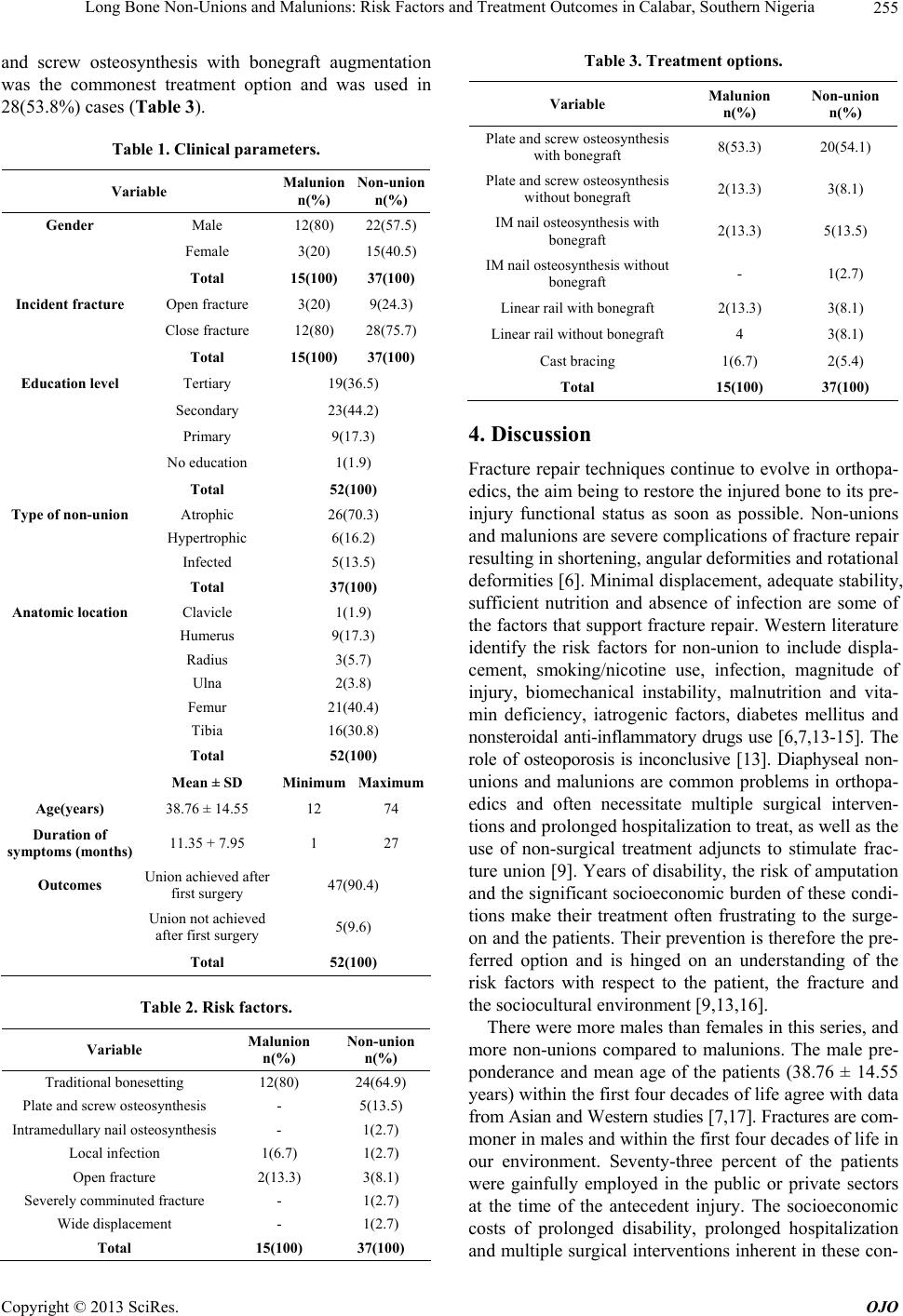 Long Bone Non-Unions and Malunions: Risk Factors and Treatment Outcomes in Calabar, Southern Nigeria 255 and screw osteosynthesis with bonegraft augmentation was the commonest treatment option and was used in 28(53.8%) cases (Table 3). Table 1. Clinical parameters. Variable Malunion n(%) Non-union n(%) Gender Male 12(80) 22(57.5) Female 3(20) 15(40.5) Total 15(100) 37(100) Incident fracture Open fracture 3(20) 9(24.3) Close fracture 12(80) 28(75.7) Total 15(100) 37(100) Education level Tertiary 19(36.5) Secondary 23(44.2) Primary 9(17.3) No education 1(1.9) Total 52(100) Type of non-union Atrophic 26(70.3) Hypertrophic 6(16.2) Infected 5(13.5) Total 37(100) Anatomic location Clavicle 1(1.9) Humerus 9(17.3) Radius 3(5.7) Ulna 2(3.8) Femur 21(40.4) Tibia 16(30.8) Total 52(100) Mean ± SD Minimum Maximum Age(years) 38.76 ± 14.55 12 74 Duration of symptoms (months) 11.35 + 7.95 1 27 Outcomes Union achieved after first surgery 47(90.4) Union not achieved after first surgery 5(9.6) Total 52(100) Table 2. Risk factors. Variable Malunion n(%) Non-union n(%) Traditional bonesetting 12(80) 24(64.9) Plate and screw osteosynthesis - 5(13.5) Intramedullary nail osteosynthesis - 1(2.7) Local infection 1(6.7) 1(2.7) Open fracture 2(13.3) 3(8.1) Severely comminuted fracture - 1(2.7) Wide displacement - 1(2.7) Total 15(100) 37(100) Table 3. Treatment options. Variable Malunion n(%) Non-union n(%) Plate and screw osteosynthesis with bonegraft 8(53.3) 20(54.1) Plate and screw osteosynthesis without bonegraft 2(13.3) 3(8.1) IM nail osteosynthesis with bonegraft 2(13.3) 5(13.5) IM nail osteosynthesis without bonegraft - 1(2.7) Linear rail with bonegraft 2(13.3) 3(8.1) Linear rail without bonegraft 4 3(8.1) Cast bracing 1(6.7) 2(5.4) Total 15(100) 37(100) 4. Discussion Fracture repair techniques continue to evolve in orthopa- edics, the aim being to restore the injured bone to its pre- injury functional status as soon as possible. Non-unions and malunions are severe complications of fracture repair resulting in shortening, angular deformities and rotational deformities [6]. Minimal displacement, adequate stability, sufficient nutrition and absence of infection are some of the factors that support fracture repair. Western literature identify the risk factors for non-union to include displa- cement, smoking/nicotine use, infection, magnitude of injury, biomechanical instability, malnutrition and vita- min deficiency, iatrogenic factors, diabetes mellitus and nonsteroidal anti-inflammatory drugs use [6,7,13-15]. The role of osteoporosis is inconclusive [13]. Diaphyseal non- unions and malunions are common problems in orthopa- edics and often necessitate multiple surgical interven- tions and prolonged hospitalization to treat, as well as the use of non-surgical treatment adjuncts to stimulate frac- ture union [9]. Years of disability, the risk of amputation and the significant socioeconomic burden of these condi- tions make their treatment often frustrating to the surge- on and the patients. Their prevention is therefore the pre- ferred option and is hinged on an understanding of the risk factors with respect to the patient, the fracture and the sociocultural environment [9,13,16]. There were more males than females in this series, and more non-unions compared to malunions. The male pre- ponderance and mean age of the patients (38.76 ± 14.55 years) within the first four decades of life agree with data from Asian and Western studies [7,17]. Fractures are com- moner in males and within the first four decades of life in our environment. Seventy-three percent of the patients were gainfully employed in the public or private sectors at the time of the antecedent injury. The socioeconomic costs of prolonged disability, prolonged hospitalization and multiple surgical interventions inherent in these con- Copyright © 2013 SciRes. OJO  Long Bone Non-Unions and Malunions: Risk Factors and Treatment Outcomes in Calabar, Southern Nigeria 256 ditions is well documented in different regions of the world and arise from a variety of factors [7,9]. In our set- ting with a fragile economy the involved age bracket is the dominant productive group and the economic costs of their injuries and consequent treatment puts direct pres- sure on the economic viability of their households. This is so because there is no social support and universal in- surance system to help alleviate the financial burden of their treatment. The femur was the commonest site of pathology in this series. This differs from other literature from Asia and the Western world which report the tibia and forearm with the highest rates [7,17-19]. The role of traditional bonesetting in African society is documented in other studies [20-22]. As this study shows, the incident fracture was close in 41(79%) cases. Orthodox fracture repair te- chniques like interlocking nails and plate & screw osteo- synthesis would have been offered to these patients with the documented advantages of stable anatomical recon- struction and early mobilization, and early return to eco- nomically productive activities. However, 36(69.2%) pa- tients chose to be treated by traditional bonesetters and only returned to seek orthopedic surgical care when the complications had developed at a mean fracture-surgery interval of 11.35 ± 7.95 months. Considering that 42(80.7%) of the patients had at least secondary level education and 38(73.1%) of them were gainfully employed, it appears that neither educational level nor socio-economic status have any inhibitory influence on the choice of traditional bonesetting as the option of first choice in patients with fractures in our environment. Lower limb complications with attendant deformity and limb-length inequality ul- timately force the choice and need for orthopaedic surgi- cal intervention. Plate and screw osteosynthesis with bonegraft aug- mentation was the treatment option in the majority of our patients (n = 28; 53.8%). This may be explained by the majority cases being atrophic non-unions. Also, in a so- ciety where orthopaedic surgical care is not the option of first choice among the majority, augmenting the chances of healing at first contact must be seen as an integral part of the first treatment plan. This philosophy seems to be consistent in the developing world [20,23]. Consistency in improved outcomes compared to the outcomes of tra- ditional bonesetting will ultimately sway confidence from age-old suboptimal traditional bonesetting practices that have been sustained by erroneous cultural beliefs in the supernatural powers of the traditional bonesetters. Union was achieved in 47(90.4%) of the patients after the first surgical intervention. Five patients (9.6%) required more than one operative intervention, with union subsequently achieved in two of them. Persistent non-union occurred in 3 patients after the second operative intervention. These may have benefited from such augmentations as low in- tensity pulsed ultrasound stimulation or non-invasive electrical bone growth stimulation. These options are not accessible within our healthcare system currently. Our overall union rate of 94% however compares favourably with the results of other studies from the developing world [7,20,23]. This study focused on risk factors within the local fracture environment. The role of systemic risk factors like smoking/nicotine use, diabetes, NSAIDS use and malnutrition is well documented and addressing these issues is an integral part of fracture treatment protocol in our institution. The role of traditional bonesetting as a risk factor for non-unions and malunions is important be- cause it is a potential focus of public health enlighten- ment intervention in our locality. By the application of herbal formentations, scarifications, ill-informed splint- ing and excessive massage protocols, traditional boneset- ting presents the risk of the creation of local ischaemia, subclinical infections and biomechanical instability in the pathogenesis of malunions and non-unions. 5. Conclusion Diaphyseal non-unions and malunions are associated with significant morbidity especially in the lower limb where limb-length inequality, malrotation and malalignment can cause severe functional deficiencies. The treatment of these conditions also results in a severe economic burden for these patients with such issues as prolonged hospita- lization, multiple surgical interventions and application of expensive adjunctive treatment modalities. Traditional bonesetting plays a significant role in the health care seek- ing behaviour of people in African societies. While cul- tural beliefs and financial consideration continue to fuel its popularity, the long-term complications and overall socioeconomic cost of treating these complications should drive a paradigm shift in health-seeking behaviours in the developing world. However, identifying and addressing this risk factor in the public health enlightenment inter- vention processes is a necessity if these complications are to be prevented in these resource-challenged econo- mies. This study suggests that neither educational level nor gainful employment has addressed healthcare issues fueled by age-long erroneous cultural bias and beliefs. Aggressive health education campaigns, supported by po- litical will, should help drive a paradigm shift in health seeking behaviors and interventions. REFERENCES [1] K. J. Pugh and S. R. Rozbruch, “Non-Unions and Malun- ions,” In: M. R. Baumgaertner and P. Tornetta III, Eds., Orthopaedic Knowledge Update, American Academy of Orthopaedic Surgeons, 2011, pp. 115-130. [2] J. O. Angleu, M. T. Archdeacon, L. K. Cannada and D. H. Jr., “Avoiding Complications in the Treatment of Hume- Copyright © 2013 SciRes. OJO  Long Bone Non-Unions and Malunions: Risk Factors and Treatment Outcomes in Calabar, Southern Nigeria Copyright © 2013 SciRes. OJO 257 ral Fractures,” The Journal of Bone and Joint Surgery (American), Vol. 90, No. 7, 2008, pp. 1580-1589. [3] P. K. Beredjiklian, R. J. Naranja, R. B. Heppenstall, C. T. Brighton and J. L. Esterhai, “Results of Treatment of 111 Patients with Non-Union of Femoral Shaft Fractures,” The University of Pennsylvania Orthopaedic Journal, Vol. 12, 1999, pp. 52-56. [4] D. J. Hak, “Management of Aseptic Tibial Non-Union,” Journal of the American Academy of Orthoapedic Sur- geons, Vol. 19, No. 9, 2011, pp. 563-573. [5] J. Gilles, S. Wallstabe, A. P. Schulz and U. Gerlach, “Is Non-Union of Tibial Shaft Fractures Due to Noncultur- able Bacterial Pathologens? A Clinical Investigation Us- ing PCR and Culture Techniques,” Journal of Orthopae- dic Surgery and Research, Vol. 7, No. 20, 2012, p. 20. www.josr-online.com/content/pdf/1749-799x-7-20.pdf http://dx.doi.org/10.1186/1749-799X-7-20 [6] C.-C. Wu, “Treatment of Long-Bone Fractures, Malun- ions and Non-Unions: Experience at Chang Gung Memo- rial Hospital, Taoyuan, Taiwan,” Chang Gung Medical Journal, Vol. 29, No. 4, 2006, pp. 347-357. [7] Z. U. Malik, K. M. N. T. Ahmed and A. K. S. T. S. Hus- sai, “Analysis of Causes and Treatment Modality in Non- Union of Long Bones Diaphyseal Fractures,” Pakistani Armed Forces Medical Journal, Vol. 61, No. 3, 2011, pp. 433-437. [8] A. M. Udosen, I. A. Ikpeme and N. E. Ngim, “Traditional Bonesetting in Africa: Counting the Cost,” Proceedings of the 5th SICOT/SIROT Annual International Conference, September 2007, Marrakech, Morocco, p. 203. [9] L. S. Phieffer and JA Goulet, “Delayed Unions of the Ti- bia, Instructional Course Lecture,” Journal of Bone and Joint Surgery (American), Vol. 88, No. 1, 2006, pp. 205- 216. [10] N. Wu, Y.-C. Lee, D. S. H. Murray, T. Wilcox and L. Bou- langer, “Economic Burden of Illness among US Patients Experiencing Fracture Non-Union,” Orthopaedics Re- search and Reviews, Vol. 2013, No. 5, 2013, pp. 21-33. http://dx.doi.org/10.2147/ORR.S41123 [11] C. R. Wheeless III, “Malunion of the Tibia,” Wheeless Textbook of Orthopaedics, 2012. www.wheelessonline.com/ortho/malunion_of_the_tibia [12] J. A. Goulet and D. J. Hak, “Non-Unions and Malunions of the Tibia,” In: M. W. Chapman, Ed., Chapman’s Or- thopaedic Surgery, Lippincott Williams & Wilkins, Phila- delphia, 2001, pp. 978-999. [13] V. Perumal and C. S. Roberts, “Factors Contributing to Non-Union of Fractures,” Current Orthopaedics, Vol. 21, No. 4, 2007, pp. 258-261. http://dx.doi.org/10.1016/j.cuor.2007.06.004 [14] J.-J. Yang, L.-C. Lin, K.-H. Chao, S.-Y. Chuang, C.-C. Wu, T.-T. Yeh and Y.-T. Lian, “Risk Factors for Non-Union in Patients with Intracapsular Femoral Neck Fractures Treated with Three Cannulated Screws Placed in Either a Triangle or an Inverted Triangle Configuration,” The Jour- nal of Bone & Joint Surgery, Vol. 95, No. 1, 2013, pp. 61- 69. http://dx.doi.org/10.2106/JBJS.K.01081 [15] M. Lee, “Non-Unions of the Humerus,” Journal of Hand Surgery, Vol. 18, No. 1, 2005, pp. 51-53. [16] I. R. Murray, C. J. Foster and A. E. C. M. Robinson, “Risk Factors for Non-Union after Non-Operative Treatment of Displaced Midshaft Fractures of the Clavicle,” The Jour- nal of Bone & Joint Surgery, Vol. 95, No. 13, 2003, pp. 1153-1158. [17] L. A. Mills and A. H. R. W. Simpson, “The Relative Inci- dence of Fracture Non-Union in the Scottish Population (5.17 Million): A 5-Year Epidemiological Study,” British Medical Journal Open, Vol. 3, No. 2, 2013, pp. 1-8. http://bmjopen.bmj.com/content/3/2/e002276.full.pdf+ht ml http://dx.doi.org/10.1136/bmjopen-2012-002276 [18] D. R. Marsh, S. Shah, J. Elliot and N. Kurdy, “The Illiza- yov Method in Non-Union, Malunion and Infection of Fractures,” The Journal of Bone & Joint Surgery, Vol. 79-B, No. 2, 1997, pp. 273-279. http://dx.doi.org/10.1302/0301-620X.79B2.6636 [19] G. V. Russell, M. L. Graves, M. T. Archdeacon, D. P. Ba- rei, G. A. Brien Jr. and S. E. Porter, “The Clamshell Oste- otomy: A New Technique to Correct Complex Diaphy- seal Malunions,” The Journal of Bone & Joint Surgery (American), Vol. 91, No. 2, 2009, pp. 314-324. http://dx.doi.org/10.2106/JBJS.H.00158 [20] M. Tall, I. Quedraogo, A. N. Kasse, B. J. D. Tekpa, G. Ban- koungou, S. Belem and M. F. Toe, “Femur Malunions Treated with Open Osteotomy and Intramedullary Nailing in Developing Countries,” Orthopaedics & Traumatology: Surgery & Research, Vol. 98, No. 7, 2012, pp. 784-787. [21] A. B. Omololu, S. O. Ogunlade and V. K. Gopaldasani, “The Practice of Traditional Bonesetting: Training Algo- rithm,” Clinical Orthopaedics and Related Research, Vol. 466, No. 10, 2008, pp. 2392-2398. http://dx.doi.org/10.1007/s11999-008-0371-8 [22] A. A. Dada, W. Yinusa and S. O. Giwa, “Review of the Practice of Traditional Bonesetting in Nigeria,” African Health Sciences, Vol. 11, No. 2, 2011, pp. 262-265. [23] A. S. Gavaskar and R. Kumar, “Open Interlocking Nail- ing and Bonegratfing for Neglected Femoral Shaft Frac- tures,” Journal of Orthopaedic Surgery, Vol. 18, No. 1, 2010, pp. 45-49. |