

Paper Menu >>
Journal Menu >>
 American Journal of Molecular Biology, 2013, 3, 248-254 AJMB http://dx.doi.org/10.4236/ajmb.2013.34032 Published Online October 2013 (http://www.scirp.org/journal/ajmb/) TLR5 involvement in attenuated IL-8 production in nuclear decorin silenced oral mucosal dysplastic keratinocytes and squamous cell carcinoma* Nyla Dil1,2#, Abhijit G. Banerjee1,3 1Department of Oral Biology, Faculty of Dentistry, University of Manitoba, Winnipeg, Canada 2Department of Immunology, and Department of Medical Microbiology and Infectious Diseases, Faculty of Medicine, University of Manitoba, Winnipeg, Canada 3Center for Genomic Bio-Medicine and Research Institute, Durg, Chhattisgarh, India Email: #[email protected] Received 7 August 2013; revised 3 September 2013; accepted 27 September 2013 Copyright © 2013 Nyla Dil, Abhijit G. Banerjee. This is an open access article distributed under the Creative Commons Attribution License, which permits unrestricted use, distribution, and reproduction in any medium, provided the original work is properly cited. ABSTRACT Head and neck cancer is one of the most prevalent cancers in the world. Roughly half of these malignan- cies originate from oral mucosa and constitute oral squamous cell carcinomas. Despite many advances in diagnostic and therapeutic regimens, five-year sur- vival rate remains at roughly 50%, indicating the need for in depth understanding of the oral squamous cell carcinoma immunobiology. We have previously shown that in human dysplastic oral keratinocytes (DOK) and malignant squamous cell carcinoma (SCC-25), multifunctional proteoglycan decorin is aberrantly expressed and localized in the nucleus bound to nuclear EGFR. In vitro nuclear decorin knockdown significantly reduced IL-8 and IL8-de- pendent migration, invasion and angiogenesis in these cells. Since toll-like receptor (TLR) signalling leads to IL-8 production, we examined here if these receptors played a role in decorin silencing mediated reduction in IL-8 levels in oral mucosal d ysplast ic keratino cytes and squamous carcinoma cells. Decorin silenced DOK and SCC-25 cells showed a marked diminution of TLR5 mRNA and protein expression compared with respective controls that translated into the loss of func- tion in response to appropriate TLR ligand. In these mucosal oral epithelia , decorin stable kno ckdown sig- nificantly down-regulated IL-8 production following activation with TLR5 ligand flagellin. These data sug- gest that decorin silencing interferes with IL-8 pro- duction, in part, by altering TLR5 expression and signaling in dysplastic and malignant oral epithelia. This study highlights the significance of TLR5 ex- pression and signaling and its plausible interactions with proteoglycans in mucosal cancers. Keywords: Oral Cancer; Mucosal Cancer; Nuclear Decorin; TLR5; IL-8; Flagellin; PAMP; DAMP; SLRP; Proteoglycans 1. BACKGROUND Head and neck squamous cell carcinoma (HNSCC) is a devastating disease that accounts for roughly 1.6 million newly diagnosed cases and 330,000 deaths each year worldwide [1,2]; half of these cancers are localized in the oral cavity and are termed oral squamous cell carcinoma. A multifactorial mucosal cancer, oral squamous cell car- cinoma, is primarily associated with chronic tobacco, alcohol use and betel chewing; however, chronic in- flammation, viral infections (human papillomavirus), genetic predisposition and poor oral hygiene have also been strongly implicated in oral tumorigenesis [3-5]. The majority of inflammatory conditions are triggered by Toll-like receptors (TLRs) which are a large family of at least 11 evolutionarily conserved proteins character- ized by ligand specific activation and complex down- stream signaling [6-8]. TLRs are predominantly ex- pressed on various immune and structural cells where they serve as innate cell surface pathogen sensors and promote local inflammation by triggering the induction of various proinflammatory mediators, including IL-8. Recently, TLR expression or up-regulation has been de- tected in various tumour types, especially in epithelium derived cancers [9-11]. Expression of TLRs varies in different cancerous cell types; however, evidence indi- *Competing Interests: No financial or non-financial competing interests exist. #Corresponding author. OPEN ACCESS  N. Dil, A. G. Banerjee / American Journal of Molecular Biology 3 (2013) 248-254 249 cates that TLR expression is functionally associated with tumorigenesis. It has been suggested that TLR expression might promote malignant transformation of epithelial cells [11,12]. Engagement of TLRs promotes tumor de- velopment and metastasis, protects the cancerous cells from immune attack and induces resistance to apoptosis and chemo-resistance in some malignancies [10,12-14]. In addition to its putative proinflammatory functions, IL-8 is a potent angiogenic factor and plays an important role in cancer progression and metastasis [15-18]. IL-8 is responsible for most of the angiogenic activity induced by human oral mucosal dysplastic and malignant epithe- lial cells [19,20]. Previously, we have shown that in hu- man dysplastic oral keratinocytes (DOK) and malignant squamous carcinoma cells (SCC-25), multifunctional proteoglycan decorin is atypically expressed and local- ized in the nucleus bound to nuclear epidermal growth factor receptor (EGFR) [18,21]. Post-transcriptional nu- clear decorin knock-down resulted in significantly re- duced IL-8 and IL8-dependent migration, invasion and angiogenic potential in these dysplastic and malignant oral mucosal epithelia [18,20]. Since TLR signaling is an upstream event in the majority of cytokines/chemokine expression, in this current study, we examined if TLR pathway is implicated in the aberrant nuclear decorin silencing mediated attenuated IL-8 production. 2. MATERIALS AND METHODS 2.1. Cell Lines and Culture Conditions Oral mucosa origin, premalignant-Dysplastic Oral Kera- tinocytes (DOK) and malignant-Squamous Carcinoma (SCC-25) cell lines were routinely maintained in DMEM/F12 (Hyclone, Logan, Utah) supplemented with 10% Foetal Calf Serum for use as in vitro model in our studies, as described previously [20,22,23]. Silencing of decorin gene expression was achieved using short hairpin RNA (shRNA) technology as described earlier [18]. Briefly, oligonucleotides targeting decorin transcript va- riants A1 and A2 and scrambled sequence control were custom synthesized, annealed, and cloned into shRNA expression vector pGeneClip PuroTM (Promega) by Super Array Bioscience Corporation (Frederick, MD). BLAST queries were performed to ensure that the sequences have no significant homology with any other human genes. Transformation grade shRNAi plasmids were transfected into DOK and SCC-25 cells using Effectene™ transfec- tion reagent following manufacturer’s protocol (Qiagen, Valencia, CA). The stable transfectants were selected for puromycin (Calbiochem, San Diego, CA) resistance at 2.5 µg/ml optimal concentration. Pools of stable trans- fectants (maintained at 1 µg/ml of puromycin) were used in all experiments to avoid clone-specific differences. Decorin knock down was confirmed at transcript and protein level by quantitative real-time reverse transcrip- tion-PCR and Western blotting, respectively. Pooled decorin-shRNA transfected DOK or SCC-25 clones showed more than 80% reduction in decorin mRNA ex- pression and almost complete abrogation of decorin pro- tein expression in nuclear lysates and/or in whole cell lysates when compared to control-shRNA transfected clones or no transfection wild type DOK [18]. Herein, untransfected DOK and SCC-25 cells will be referred to as wild type (WT), scrambled shRNA stable transfectants as control (or Ctrl-shRNA in figures), and decorin shRNA stable transfectants as decorin silenced (or DCN- shRNA in figures). 2.2. Multiplex PCR The transcript expression levels of innate immune TLRs and co-regulatory molecules were quantified in decorin silenced, control, and WT DOK and SCC25 cells using multiplex PCR (MPCR) kit for human signaling receptor set-2 (TLR1, TLR2, TLR3, TLR5, TLR6, TLR9 and CD14) from Maxim Biotech, San Francisco, CA. This set also included housekeeping gene-GAPDH, as internal cDNA loading control in each reaction. MPCR was car- ried out according to the manufacturer’s instructions. Briefly, 1X MPCR buffer, 2.5 units of Taq DNA poly- merase, and cDNA template from DOK and SCC25 cells were mixed in a 25 µl reaction and subjected to 35 cycles of PCR, with denaturing, annealing, and extension tem- peratures at 96˚C, 67˚C, and 70˚C, respectively. Follow- ing MPCR, the DNA amplicons were fractionated elec- trophoretically on 2% agarose gel containing 0.5 µg/ml ethidium bromide. 2.3. Real-Time PCR RNA was extracted from DOK and SCC-25 cells using RNeasy Plus mini kit (Qiagen, Valencia, CA) and 2.5 g of total RNA was used to synthesize cDNA, using Su- prescript III Reverse Transcriptase (Invitrogen, San Diego, CA). Quantitative RT-PCR was performed using QuantiTect SYBR Green PCR kit (Qiagen, Valencia, CA) on the Mini Opticon Real-Time PCR system (BioRad, Hercules, CA) as per manufacturer’s protocol. Quantitative PCR primer pairs were designed for SYBR Green chemistry based detection of amplicons for TLR5 (5’-TGCATTAAGGGGACTAAGCCT-3’, 5’-AAAAGGGAGAACTTTAGGGACT-3’) and GAPDH (5’-ACAGTCAGCCGCATCTTCTT-3’, 5’-GTTAAAAGCAGCCCT G GTGA-3’). GAPDH was used as relative house-keeping gene expression control to normalize for sample variations. 2.4. Western Blot Analysis Cells were rinsed with ice-cold PBS and were lysed in a Copyright © 2013 SciRes. OPEN ACCESS 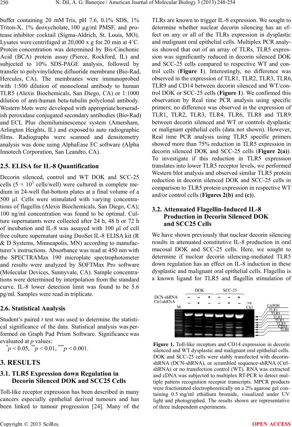 N. Dil, A. G. Banerjee / American Journal of Molecular Biology 3 (2013) 248-254 250 buffer containing 20 mM Tris, pH 7.6, 0.1% SDS, 1% Triton-X, 1% deoxycholate, 100 µg/ml PMSF, and pro- tease inhibitor cocktail (Sigma-Aldrich, St. Louis, MO). Lysates were centrifuged at 20,000 x g for 20 min at 4˚C. Protein concentration was determined by Bis-Cinchonic Acid (BCA) protein assay (Pierce, Rockford, IL) and subjected to 10% SDS-PAGE analysis, followed by transfer to polyvinylidene difluoride membrane (Bio-Rad, Hercules, CA). The membranes were immunoprobed with 1:500 dilution of monoclonal antibody to human TLR5 (Alexis Biochemicals, San Diego, CA) or 1:1000 dilution of anti-human beta-tubulin polyclonal antibody. Western blots were developed with appropriate horserad- ish peroxidase conjugated secondary antibodies (Bio-Rad) and ECL Plus chemiluminescence system (Amersham, Arlington Heights, IL) and exposed to auto radiographic films. Radiographs were scanned and densitometry analysis was done using AlphaEase FC software (Alpha Innotech Corporation, San Leandro, CA). 2.5. ELISA for IL-8 Quantification Decorin silenced, control and WT DOK and SCC-25 cells (5 × 10⁵ cells/well) were cultured in complete me- dium in 24-well flat-bottom plates at a final volume of a 500 µl. Cells were stimulated with varying concentra- tions of flagellin (Alexis Biochemicals, San Diego, CA); 100 ng/ml concentration was found to be optimal. Cul- ture supernatants were collected after 24 h, 48 h or 72 h of incubation and IL-8 was assayed with 100 μl of cell free culture supernatant using DuoSet IL-8 ELISA kit (R & D Systems, Minneapolis, MN) according to manufac- turer’s instructions. Absorbance was read at 450 nm with the SPECTRAMax 190 microplate spectrophotometer and results were analyzed by SOFTMax Pro software (Molecular Devices, Sunnyvale, CA). Sample concentra- tions were determined by interpolation from the standard curve. IL-8 lower detection limit was found to be 5.6 pg/ml. Samples were read in triplicate. 2.6. Statistical Analysis Student’s paired t test was used to determine the statisti- cal significance of the data. Statistical analysis was per- formed on Graph Pad Prism Software. Significance was evaluated at p values: *p < 0.05, **p < 0.01, ***p < 0.001. 3. RESULTS 3.1. TLR5 Expression down Regulation in Decorin Silenced DOK and SCC25 Cells Toll-like receptor expression has been described in many cancers especially epithelial derived tumours and has been linked to tumour progression [24]. Many of the TLRs are known to trigger IL-8 expression. We sought to determine whether nuclear decorin silencing has an ef- fect on any or all of the TLRs expression in dysplastic and malignant oral epithelial cells. Multiplex PCR analy- sis showed that out of an array of TLRs, TLR5 expres- sion was significantly reduced in decorin silenced DOK and SCC-25 cells compared to respective WT and con- trol cells (Figure 1). Interestingly, no difference was observed in the expression of TLR1, TLR2, TLR3, TLR6, TLR9 and CD14 between decorin silenced and WT/con- trol DOK or SCC-25 cells (Figure 1). We confirmed this observation by Real time PCR analysis using specific primers; no difference was observed in the expression of TLR1, TLR2, TLR3, TLR4, TLR6, TLR8 and TLR9 between decorin silenced and WT or controls dysplastic or malignant epithelial cells (data not shown). However, Real time PCR analysis using TLR5 specific primers showed more than 75% reduction in TLR5 expression in decorin silenced DOK and SCC-25 cells (Figure 2(a)). To investigate if this reduction in TLR5 expression translates into lower TLR5 receptor levels, we performed Western blot analysis and observed similar TLR5 protein reduction in decorin silenced DOK and SCC-25 cells in comparison to TLR5 protein expression in respective WT and/or control cells (F igures 2(b) and (c)). 3.2. Attenuated Flagellin-Induced IL-8 Production in Decorin Silenced DOK and SCC25 Cells We have shown previously that nuclear decorin silencing results in attenuated constitutive IL-8 production in oral mucosal DOK and SCC-25 cells. Here, we sought to determine if nuclear decorin silencing-mediated TLR5 down regulation has an effect on IL-8 induction in these dysplastic and malignant oral epithelial cells. Flagellin is a known ligand for TLR5 and flagellin stimulation of Figure 1. Toll-like receptors and CD14 expression in decorin silenced and WT dysplastic and malignant oral epithelial cells. DOK and SCC-25 cells were stably transfected with decorin- shRNA (DCN-shRNA), or scrambled sequence-shRNA (Ctrl- shRNA) or no transfection control (WT). RNA was extracted and cDNA was subjected to multiplex RT-PCR to detect mul- tiple pattern recognition receptor transcripts. MPCR products were fractionated electrophoretically on a 2% agarose gel con- taining 0.5mg/ml ethidium bromide, visualized under UV light and photographed. The results shown are representative of three independent experiments. Copyright © 2013 SciRes. OPEN ACCESS 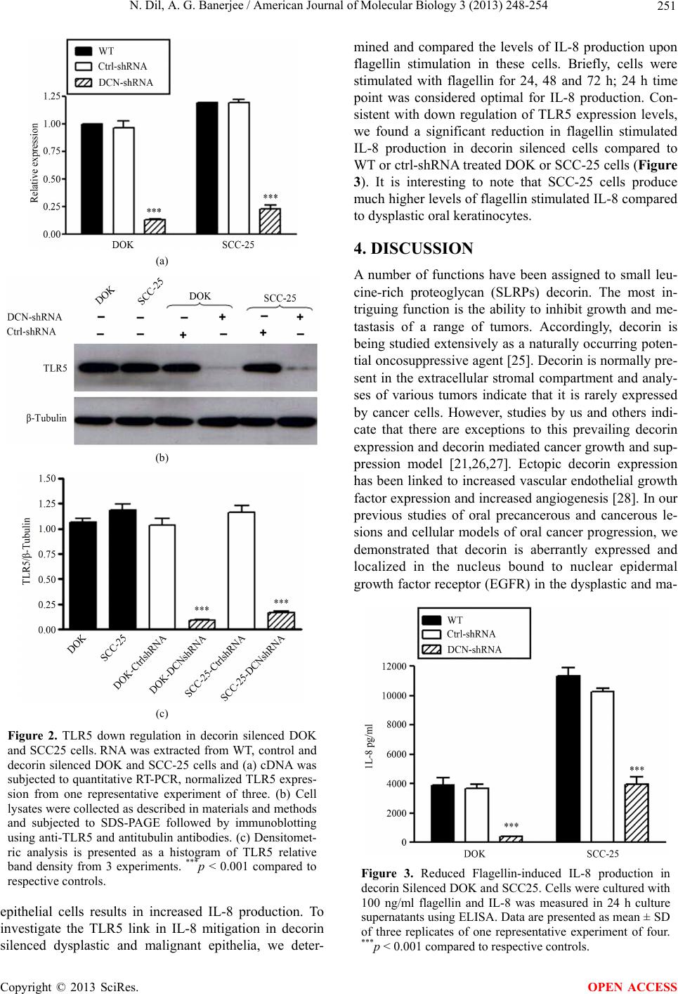 N. Dil, A. G. Banerjee / American Journal of Molecular Biology 3 (2013) 248-254 251 (a) (b) (c) Figure 2. TLR5 down regulation in decorin silenced DOK and SCC25 cells. RNA was extracted from WT, control and decorin silenced DOK and SCC-25 cells and (a) cDNA was subjected to quantitative RT-PCR, normalized TLR5 expres- sion from one representative experiment of three. (b) Cell lysates were collected as described in materials and methods and subjected to SDS-PAGE followed by immunoblotting using anti-TLR5 and antitubulin antibodies. (c) Densitomet- ric analysis is presented as a histogram of TLR5 relative band density from 3 experiments. ***p < 0.001 compared to respective controls. epithelial cells results in increased IL-8 production. To investigate the TLR5 link in IL-8 mitigation in decorin silenced dysplastic and malignant epithelia, we deter- mined and compared the levels of IL-8 production upon flagellin stimulation in these cells. Briefly, cells were stimulated with flagellin for 24, 48 and 72 h; 24 h time point was considered optimal for IL-8 production. Con- sistent with down regulation of TLR5 expression levels, we found a significant reduction in flagellin stimulated IL-8 production in decorin silenced cells compared to WT or ctrl-shRNA treated DOK or SCC-25 cells (Figure 3). It is interesting to note that SCC-25 cells produce much higher levels of flagellin stimulated IL-8 compared to dysplastic oral keratinocytes. 4. DISCUSSION A number of functions have been assigned to small leu- cine-rich proteoglycan (SLRPs) decorin. The most in- triguing function is the ability to inhibit growth and me- tastasis of a range of tumors. Accordingly, decorin is being studied extensively as a naturally occurring poten- tial oncosuppressive agent [25]. Decorin is normally pre- sent in the extracellular stromal compartment and analy- ses of various tumors indicate that it is rarely expressed by cancer cells. However, studies by us and others indi- cate that there are exceptions to this prevailing decorin expression and decorin mediated cancer growth and sup- pression model [21,26,27]. Ectopic decorin expression has been linked to increased vascular endothelial growth factor expression and increased angiogenesis [28]. In our previous studies of oral precancerous and cancerous le- sions and cellular models of oral cancer progression, we demonstrated that decorin is aberrantly expressed and localized in the nucleus bound to nuclear epidermal growth factor receptor (EGFR) in the dysplastic and ma- Figure 3. Reduced Flagellin-induced IL-8 production in decorin Silenced DOK and SCC25. Cells were cultured with 100 ng/ml flagellin and IL-8 was measured in 24 h culture supernatants using ELISA. Data are presented as mean ± SD of three replicates of one representative experiment of four. ***p < 0.001 compared to respective controls. Copyright © 2013 SciRes. OPEN ACCESS 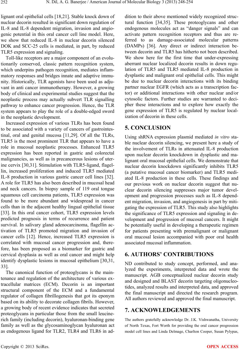 N. Dil, A. G. Banerjee / American Journal of Molecular Biology 3 (2013) 248-254 252 lignant oral epithelial cells [18,21]. Stable knock down of nuclear decorin resulted in significant down regulation of IL-8 and IL-8 dependent migration, invasion and angio- genic potential in this oral cancer cell line model. Here, we show that reduced IL-8 in nuclear decorin silenced DOK and SCC-25 cells is mediated, in part, by reduced TLR5 expression and signaling. Toll-like receptors are a major component of an evolu- tionarily conserved, classic pattern recognition system, which underpins pathogen recognition, mediates inflam- matory responses and bridges innate and adaptive immu- nity. Historically, TLR agonists have been used as adju- vant in anti cancer immunotherapy. However, a growing body of clinical and experimental studies suggest that the neoplastic process may actually subvert TLR signalling pathway to enhance cancer progression. Hence, the TLR system appears to play the role of a double-edged sword in the neoplastic development. Increased expression of various TLRs has been found to be associated with a variety of cancers of gastrointes- tinal, oral and genital mucosa [11,29]. Of all the TLRs, TLR5 is the most prominent TLR that appears to have a role in mucosal neoplastic processes. Enhanced TLR5 expression has been reported in gastric and colorectal malignancies, as well as in precancerous lesions of uter- ine cervix [30,31]. Stimulation with TLR5-ligand, flagel- lin, increased proliferation and induced TLR5 mediated IL-8 production in various gastric cancer cell lines [32]. A role for TLR5 has also been described in mucosal head and neck cancers. In biopsy sample of 119 oral tongue squamous cell carcinoma patients, TLR5 expression was found to be more abundant and widespread in cancer cells than in the adjacent healthy lingual epithelial tissue [33]. In this oral cancer cohort, TLR5 expression levels predicted prognosis in terms of recurrence and patient survival. In salivary gland adenocarcinoma, flagellin ac- tivation of TLR5 promoted migration and invasion of cancer cells [12]. Hence, increased TLR5 expression is correlated with mucosal cancer progression and, there- fore, has been proposed as a biomarker for gastric and cervical dysplasia as well as oral cancer and might help identify dysplastic lesions in mucosal epithelium [30,31, 33]. The canonical function of proteoglycans is the main- tenance and regulation of the architecture of various ex- tracellular matrices (ECM). Decorin is an important structural component of the ECM and a fundamental regulator of collagen fibrillogenesis that got its eponym based on its ability to decorate collagen fibrils. However, a growing body of recent evidence indicates that secreted proteoglycans in particular those from the small leucine- rich family (including decorin), hyaluronan-binding gene family as well as the glycosaminoglycan hyaluronan act as endogenous ligand for TLR2, TLR4 and TLR6 in ad- dition to their above mentioned widely recognized struc- tural function [34,35]. These proteoglycans and other endogenous molecules act as “danger signals” and can activate pattern recognition receptors and thus are re- ferred to as damage-associated molecular patterns (DAMPs) [36]. Any direct or indirect interaction be- tween decorin and TLR5 has hitherto not been described. We show here for the first time that under-expressing aberrant nuclear localized decorin results in down regu- lation of TLR5 and TLR5 mediated IL-8 production in dysplastic and malignant oral epithelial cells. This might be due to nuclear decorin interactions with its binding partner nuclear EGFR (which acts as a transcription fac- tor) or additional interactions with other nuclear and/or cytosolic factors. Further studies are warranted to deci- pher these interactions and to explore how exactly the gene expression of TLR5 is regulated by nuclear local- ization of decorin in these cells. 5. CONCLUSION Using shRNA expression plasmid mediated in vitro sta- ble nuclear decorin silencing, we present here a study of the involvement of TLRs in attenuated IL-8 production upon nuclear decorin knockdown in dysplastic and ma- lignant oral mucosal epithelial cells. We demonstrate that nuclear decorin knockdown significantly inhibits TLR5 (a putative mucosal cancer biomarker) and TLR5 medi- ated IL-8 production in these cells. These findings and our previous work on nuclear decorin suggest that nu- clear decorin silencing suppresses major tumor devel- opment and progression processes such as IL-8 depend- ent migration, invasion, and angiogenesis in part by miti- gating the expression of TLR5. This study also highlights the significance of TLR5 expression and signaling in de- velopment and progression of mucosal cancers. It might be potentially useful in developing a therapeutic regimen for patients presenting with premalignant or malignant oral mucosal lesion accompanied with poor oral health associated mucosal inflammation. 6. AUTHORS’ CONTRIBUTIONS ND contributed to study concept, performed, and ana- lyzed the experiments, interpreted data and wrote the manuscript. AGB conceptualized nuclear decorin study and designed and BLAST decorin targeting oligonucleo- tides, analyzed results and interpreted data, and approved the final manuscript and directed the research program. All authors reviewed and approved the final manuscript. 7. ACKNOWLEDGEMENTS The authors gratefully acknowledge Dr. J.K. Vishwanatha, University of North Texas, Fort Worth for providing the oral cancer progression model cell lines and Linda Delmage, Charlton Cooper, Susan Pylypas, Copyright © 2013 SciRes. OPEN ACCESS  N. Dil, A. G. Banerjee / American Journal of Molecular Biology 3 (2013) 248-254 253 University of Manitoba for technical assistance. This project was sup- ported by research grants form Manitoba Health Research Council and Manitoba Medical Service Foundation and by funds from Department of Oral Biology and Faculty of Dentistry, University of Manitoba. REFERENCES [1] Jemal, A., Siegel, R., Ward, E., Hao, Y.P., Xu J.Q., Mur- ray, T. and Thun, M.J. (2008) Cancer statistics, 2008. CA: A Cancer Journal for Clinicians, 58, 71-96. http://dx.doi.org/10.3322/CA.2007.0010 [2] Jemal, A., Center, M.M., Ward, E. and Thun, M.J. (2009) Cancer occurrence. Methods in Molecular Biology, 471, 3-29. http://dx.doi.org/10.1007/978-1-59745-416-2_1 [3] Parkin, D.M., Bray, F., Ferlay, J. and Pisani, P. (2005) Global cancer statistics, 2002. CA: A Cancer Journal for Clinicians, 55, 74-108. http://dx.doi.org/10.3322/canjclin.55.2.74 [4] Dobrossy, L. (2005) Epidemiology of head and neck cancer: Magnitude of the problem. Cancer and Metasta- sis Reviews, 24, 9-17. http://dx.doi.org/10.1007/s10555-005-5044-4 [5] Ramqvist, T. and Dalianis, T. (2010) Oropharyngeal can- cer epidemic and human papillomavirus. Emerging Infec- tious Diseases, 16, 1671-1677. http://dx.doi.org/10.3201/eid1611.100452 [6] Medzhitov, R., Preston-Hurlburt, P. and Janeway, Jr. C.A. (1997) A human homologue of the Drosophila Toll pro- tein signals activation of adaptive immunity. Nature, 388, 394-397. http://dx.doi.org/10.1038/41131 [7] Takeda, K., Kaisho, T. and Akira, S. (2003) Toll-like re- ceptors. Annual Review of Immunology, 21, 335-376. http://dx.doi.org/10.1146/annurev.immunol.21.120601.14 1126 [8] Akira, S., Uematsu, S. and Takeuchi, O. (2006) Pathogen recognition and innate immunity. Cell, 124, 783-801. http://dx.doi.org/10.1016/j.cell.2006.02.015 [9] Furrie, E., et al. (2005) Toll-like receptors-2, -3 and -4 expression patterns on human colon and their regulation by mucosal-associated bacteria. Immunology, 115, 565- 574. http://dx.doi.org/10.1111/j.1365-2567.2005.02200.x [10] Kelly, M.G., et al. (2006) TLR-4 signaling promotes tumor growth and paclitaxel chemoresistance in ovarian cancer. Cancer Research, 66, 3859-3868. http://dx.doi.org/10.1158/0008-5472.CAN-05-3948 [11] Lee, J.W., et al. (2007) Increased toll-like receptor 9 ex- pression in cervical neoplasia. Molecular Carcinogenesis, 46, 941-947. http://dx.doi.org/10.1002/mc.20325 [12] Park, J.H., et al. (2011) Toll-like receptor 5 activation promotes migration and invasion of salivary gland ade- nocarcinoma. Journal of Oral Pathology & Medicine, 40, 187-193. http://dx.doi.org/10.1111/j.1600-0714.2010.00929.x [13] He, W., et al. (2007) TLR4 signaling promotes immune escape of human lung cancer cells by inducing immuno- suppressive cytokines and apoptosis resistance. Molecu- lar Immunology, 44, 2850-2859. http://dx.doi.org/10.1016/j.molimm.2007.01.022 [14] Droemann, D., et al. (2005) Human lung cancer cells ex- press functionally active Toll-like receptor 9. Respiratory Research, 6, 1. http://dx.doi.org/10.1186/1465-9921-6-1 [15] Koch, A.E., et al. (1992) Interleukin-8 as a macro- phage-derived mediator of angiogenesis. Science, 258, 1798-801. http://dx.doi.org/10.1126/science.1281554 [16] Strieter, R.M., et al. (1995) Role of C-X-C chemokines as regulators of angiogenesis in lung cancer. Journal of Leukocyte Biology, 57, 752-762. [17] Murdoch, C., et al. (1999) Cxc chemokine receptor ex- pression on human endothelial cells. Cytokine, 11, 704-712. http://dx.doi.org/10.1006/cyto.1998.0465 [18] Dil, N. and Banerjee, A.G. (2011) A role for aberrantly expressed nuclear localized decorin in migration and in- vasion of dysplastic and malignant oral epithelial cells. Head & Neck Oncology, 3, 44. http://dx.doi.org/10.1186/1758-3284-3-44 [19] Lalla, R.V., et al. (2001) Fibrin induces IL-8 expression from human oral squamous cell carcinoma cells. Oral Oncology, 37, 234-242. http://dx.doi.org/10.1016/S1368-8375(00)00090-7 [20] Dil, N.and Banerjee, A.G. (2012) Knockdown of aber- rantly expressed nuclear localized decorin attenuates tu- mour angiogenesis related mediators in oral cancer pro- gression model in vitro. Head & Neck Oncology, 4, 11. http://dx.doi.org/10.1186/1758-3284-4-11 [21] Banerjee, A.G., et al. (2003) Aberrant expression and localization of decorin in human oral dysplasia and squa- mous cell carcinoma. Cancer Research, 63, 7769-7776. [22] Hu, L., et al. (1991) Abnormal expression of retinoic acid receptors and keratin 19 by human oral and epidermal squamous cell carcinoma cell lines. Cancer Research, 51, 3972-3981. [23] Hsu, S. et al. (2002) Transforming growth factor beta 1 dysregulation in a human oral carcinoma tumour progres- sion model. Cell Proliferation, 35, 183-192. http://dx.doi.org/10.1046/j.1365-2184.2002.00237.x [24] Yu, L. and Chen, S. (2008) Toll-like receptors expressed in tumor cells: Targets for therapy. Cancer Immunology, Immunotherapy, 57, 1271-1278. http://dx.doi.org/10.1007/s00262-008-0459-8 [25] Neill, T., et al. (2012) Decorin: A guardian from the ma- trix. American Journal of Pathology, 181, 380-387. http://dx.doi.org/10.1016/j.ajpath.2012.04.029 [26] Zafiropoulos, A., et al. (2008) Decorin-induced growth inhibition is overcome through protracted expression and activation of epidermal growth factor receptors in os- teosarcoma cells. Molecular Cancer Research, 6, 785- 794. http://dx.doi.org/10.1158/1541-7786.MCR-07-0165 [27] Zafiropoulos, A. and Tzanakakis, G.N. (2008) Decorin- mediated effects in cancer cell biology. Connective Tis- sue Research, 49, 244-248. http://dx.doi.org/10.1080/03008200802147746 [28] Santra, M., et al. (2008) Ectopic decorin expression up-regulates VEGF expression in mouse cerebral endo- thelial cells via activation of the transcription factors Sp1, HIF1alpha, and Stat 3. Journal of Neurochemistry, 105, Copyright © 2013 SciRes. OPEN ACCESS  N. Dil, A. G. Banerjee / American Journal of Molecular Biology 3 (2013) 248-254 Copyright © 2013 SciRes. 254 OPEN ACCESS 324-337. http://dx.doi.org/10.1111/j.1471-4159.2007.05134.x [29] Schmausser, B., et al. (2005) Toll-like receptors TLR4, TLR5 and TLR9 on gastric carcinoma cells: An implica- tion for interaction with Helicobacter pylori. Interna- tional Journal of Medical Microbiology, 295, 179-185. http://dx.doi.org/10.1016/j.ijmm.2005.02.009 [30] Pimentel-Nunes, P., et al. (2011) Increased expression of Toll-like receptors (TLR) 2, 4 and 5 in gastric dysplasia. Pathology & Oncology Research, 17, 677-683. http://dx.doi.org/10.1007/s12253-011-9368-9 [31] Kim, W.Y., et al. (2008) Increased expression of Toll- like receptor 5 during progression of cervical neoplasia. International Journal of Gynecological Cancer, 18, 300- 305. http://dx.doi.org/10.1111/j.1525-1438.2007.01008.x [32] Song, E.J., et al. (2011) Flagellin promotes the prolifera- tion of gastric cancer cells via the Toll-like receptor 5. In- ternational Journal of Medical Microbiology, 28, 115- 119. [33] Kauppila, J.H., et al. (2013) Toll-like receptor 5 (TLR5) expression is a novel predictive marker for recurrence and survival in squamous cell carcinoma of the tongue. British Journal of Cancer, 108, 638-643. http://dx.doi.org/10.1038/bjc.2012.589 [34] Schaefer, L., et al. (2005) The matrix component bigly- can is proinflammatory and signals through Toll-like re- ceptors 4 and 2 in macrophages. Journal of Clinical In- vestigation, 115, 2223-2233. http://dx.doi.org/10.1172/JCI23755 [35] Frey, H., et al. (2013) Biological interplay between pro- teoglycans and their innate immune receptors in inflam- mation. FEBS Journal, 280, 2165-2179. [36] Chen, G.Y. and Nunez, G. (2010) Sterile inflammation: Sensing and reacting to damage. Nature Reviews Immu- nology, 10, 826-837. http://dx.doi.org/10.1038/nri2873 |