

Paper Menu >>
Journal Menu >>
 American Journal of Molecular Biology, 2013, 3, 235-240 AJMB http://dx.doi.org/10.4236/ajmb.2013.34030 Published Online October 2013 (http://www.scirp.org/journal/ajmb/) Potential effects of ultraviolet-C radiation on the mole rats (Spalax leucodon) hematological values Huseyin Turker Department of Biological Science, Ankara University, Ankara, Turkey Email: [email protected] Received 27 August 2013; revised 25 September 2013; accepted 7 October 2013 Copyright © 2013 Huseyin Turker. This is an open access article distributed under the Creative Commons Attribution License, which permits unrestricted use, distribution, and reproduction in any medium, provided the original work is properly cited. ABSTRACT UV beams are known to enable photosynthesis in plants and vitamin D synthesis in human, kill pathogen microorganisms and provide heat for the survival of life. On the other hand, solar dermatitis, sun burn, pigmenta- tion, cataract and skin cancer formation, acne, echymosis, keratosis, photodermatosis, light sensitivity, mutation, actinic elastosis and sensitivity to infections due to im- mune deficiency may be seen due to over exposure to UV beams [1,2,6,7] In this study, the effect of ultraviolet-C (UVC) radia- tion on hematologic parameters of mole rats (Spalax leucodon) was studied. Ten mole rats of both sexes, weighing 150 - 200 g, were used. The rats were ex- posed to 8 hours daily UVC radiation for 45 days. Before the radiation exposure (as a control) and on day 7, 14, 30 and 45, blood specimens were obtained from the experimental groups. Then blood samples were analyzed and compared to the control groups. A significant decrease or increase of hematological pa- rameters was observed in some experimental groups in terms of the control groups of values (p < 0.05). Alterations in other hematological parameters were not found to be statistically significant (p > 0.05). These results showed that the UVC radiation has sig- nificant effects on some hematological parameters of mole rats according to the radiation exposure time and a dose. Keywords: Ultraviolet Radiation; Mole Rats (Spalax leucodon); Hematological Parameters 1. INTRODUCTION To our knowledge, plenty of data have been gathered concerning the effects of UVA and UVB radiations on biochemical, hematological or histopathological charac- teristics of animals [8-18], but similar studies done with UVC radiation on the hematological cells on Mole rats (Spalax leucodo n) have not been encountered. For this reason, the mole rats were selected as they lived in the underground galleries, and had no UV expo- sure in their habitat. Therefore, they were exposed to an artificially produced UVC radiation in the lab, and the hematological changes were compared with the control group. 2. MATERIALS AND METHODS Ten adult mole rats of both sexes (Spalax leucodon), weighing 150 - 200 g were used in this study. All animals were caught in the rural areas of Ankara, Turkey. All rats were kept at the laboratory for 10 days at a stable tem- perature (20 ± 2 degrees centigrade) in order to ensure adaptation of the animals to their new environment. And then they were placed in special cages called terrarium (Figure 1). Sunlight exposure harms biologic systems via ultraviolet (UV) radiation due to its short wave length and high en- ergy. The wave length of UV beams was emitted by sun changes between 200 - 400 nm. UV beams are classified according to their wavelength; UVA (320-400) UVB (280-320) and UVC (200-280) [1,2]. All rats were placed to terrarium separately and a con- stant UVC dosage was applied. All animals were fed with carrot, potato and plant roots, and no special diet was given. UVC radiation is especially important in terms of its harmful effects on the living beings. Fortunately, the majority of this radiation is filtered by the ozone (O3) layer. As the thickness of this layer has reduced in recent years, it is estimated that skin cancer, cataract and im- mune deficiency syndrome cases will increase in the near future [3-5]. A “Mazda TG” ultraviolet lamp in 30 W powers and in 90 cm length was placed to the upper cover of the terrar- ium. The intensity of the UV emitted from the lamp was OPEN ACCESS 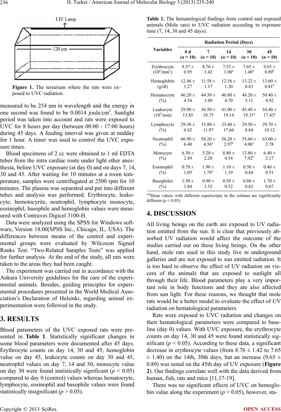 H. Turker / American Journal of Molecular Biology 3 (2013) 235-240 236 Figure 1. The terrarium where the rats were ex- posed to UVC radiation. measured to be 254 nm in wavelength and the energy in one second was found to be 0.0014 joule/cm2. Sunlight period was taken into account and rats were exposed to UVC for 8 hours per day (between 08:00 - 17:00 hours) during 45 days. A feeding interval was given at midday for 1 hour. A timer was used to control the UVC expo- sure times. Blood specimens of 2 cc were obtained to 1 ml EDTA tubes from the intra cardiac route under light ether anes- thesia, before UVC exposure (at day 0) and on days 7, 14, 30 and 45. After waiting for 10 minutes at a room tem- perature, samples were centrifugated at 2500 rpm for 10 minutes. The plasma was separated and put into different tubes and analysis was performed. Erythrocyte, leuko- cyte, hemotocyrite, neutrophil, lymphocyte monocyte, eosinophil, basophile and hemoglobin values were meas- ured with Contraves Digicel 3100-H. Data were analyzed using the SPSS for Windows soft- ware, Version 18.00(SPSS Inc., Chicago, IL, USA). The differences between means of the control and experi- mental groups were evaluated by Wilcoxon Signed Ranks Test. “Two-Related Samples Tests” was applied for further analysis. At the end of the study, all rats were taken to the areas they had been caught. The experiment was carried out in accordance with the Ankara University guidelines for the care of the experi- mental animals. Besides, guiding principles for experi- mental procedures presented in the World Medical Asso- ciation’s Declaration of Helsinki, regarding animal ex- perimentation were followed in the study. 3. RESULTS Blood parameters of the UVC exposed rats were pre- sented in Table 1. Statistically significant changes in some blood parameters were documented after 45 days. Erythrocyte counts on day 14, 30 and 45, hemoglobin value on day 45, leukocyte counts on day 30 and 45, neutrophil values on day 7, 14 and 30, monocyte value on day 30 were found statistically significant (p < 0.05) compared to day 0 (control) values whereas hematocryte, lymphocyte, eosinophil and basophile values were found statistically insignificant (p > 0.05). Ta ble 1. The hematological findings from control and exposed animals (Mole rats) to UVC radiation according to exposure time (7, 14, 30 and 45 days). Radiation Period (Days) Variables 0 d (n = 10)7 (n = 10) 14 (n = 10) 30 (n = 10)45 (n = 10) Erythrocyte (106/mm3) 8.57 ± 0.95 8.76 ± 1.42 7.53 ± 1.00a 7.65 ± 1.40a 9.65 ± 0.89b Hemoglobin (g/dl) 12.46 ± 1.27 11.58 ± 1.17 12.56 ± 1.30 13.22 ± 0.83 13.60 ± 0.81b Hematocryte (%) 46.20 ± 4.54 44.50 ± 3.80 46.80 ± 4.70 48.20 ± 5.11 50.40 ± 4.92 Leukocyte (103/mm) 29.90 ± 13.85 36.50 ± 18.75 41.00 ± 19.14 45.40 ± 19.31a 54.40 ± 17.43a Lymphocyte (%) 29.30 ± 6.42 33.80 ± 11.97 33.40 ± 17.66 29.50 ± 8.84 29.70 ± 10.12 Neutrophil (%) 66.90 ± 6.40 58.20 ± 4.56a 56.20 ± 2.97a 55.60 ± 4.06a 63.00 ± 2.78 Monocyte (%) 6.30 ± 2.49 5.20 ± 2.20 8.80 ± 4.54 13.80 ± 7.92b 6.40 ± 2.17 Eosinophil (%) 0.70 ± 1.05 1.90 ± 1.79a 1.10 ± 1.19 0.50 ± 0.84 0.40 ± 0.51 Basophiles (%) 1.50 ± 1.84 0.90 ± 1.52 0.50 ± 0.52 0.80 ± 0.63 1.70 ± 0.67 a,bMean values with different superscripts in the column are significantly different (p < 0.05). 4. DISCUSSION All living beings on the earth are exposed to UV radia- tion emitted from the sun. It is clear that previously ab- sorbed UV radiation would affect the outcome of the studies carried out on these living beings. On the other hand, mole rats used in this study live in underground galleries and are not exposed to sun emitted radiation. It is too hard to observe the effect of UV radiation on vis- cera of the animals that are exposed to sunlight all through their life. Blood parameters play a very impor- tant role in body functions and they are also affected from sun light. For these reasons, we thought that mole rats would be a better model to evaluate the effect of UV radiation on hematological parameters. Rats were exposed to UVC radiation and changes on their hematological parameters were compared to base- line (day 0) values. With UVC exposure, the erythrocyte counts on day 14, 30 and 45 were found statistically sig- nificant (p < 0.05). According to these data, a significant decrease in erythrocyte values (from 8.76 ± 1.42 to 7.65 ± 1.40) on the 14th, 30th days, but an increase (9.65 ± 0.89) was noted on the 45th day of UV exposure (Figure 2). Our findings correlate well with the data derived from human, fish, rats and mice [11,17-19]. There was no significant effects of UVC on hemoglo- bin value along the experiment (p > 0.05), however, sta- Copyright © 2013 SciRes. OPEN ACCESS 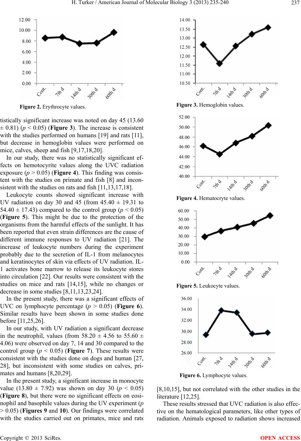 H. Turker / American Journal of Molecular Biology 3 (2013) 235-240 237 Figure 2. Erythrocyte values. tistically significant increase was noted on day 45 (13.60 ± 0.81) (p < 0.05) (Figure 3). The increase is consistent with the studies performed on humans [19] and rats [11], but decrease in hemoglobin values were performed on mice, calves, sheep and fish [9,17,18,20]. In our study, there was no statistically significant ef- fects on hemotocyrite values along the UVC radiation exposure (p > 0.05) (Figure 4). This finding was consis- tent with the studies on primate and fish [8] and incon- sistent with the studies on rats and fish [11,13,17,18]. Leukocyte counts showed significant increase with UV radiation on day 30 and 45 (from 45.40 ± 19.31 to 54.40 ± 17.43) compared to the control group (p < 0.05) (Figure 5). This might be due to the protection of the organisms from the harmful effects of the sunlight. It has been reported that even strain differences are the cause of different immune responses to UV radiation [21]. The increase of leukocyte numbers during the experiment probably due to the secretion of IL-1 from melanocytes and keratinocytes of skin via effects of UV radiation. IL- 1 activates bone marrow to release its leukocyte stores into circulation [22]. Our results were consistent with the studies on mice and rats [14,15], while no changes or decrease in some studies [8,11,13,23,24]. In the present study, there was a significant effects of UVC on lymphocyte percentage (p > 0.05) (Figure 6). Similar results have been shown in some studies done before [11,25,26]. In our study, with UV radiation a significant decrease in the neutrophil, values (from 58.20 ± 4.56 to 55.60 ± 4.06) were observed on day 7, 14 and 30 compared to the control group (p < 0.05) (Figure 7). These results were consistent with the studies done on dogs and human [27, 28], but inconsistent with some studies on calves, pri- mates and humans [8,20,29]. In the present study, a significant increase in monocyte value (13.80 ± 7.92) was shown on day 30 (p < 0.05) (Figure 8), but there were no significant effects on eosi- nophil and basophile values during the UV experiment (p > 0.05) (Figures 9 and 10). Our findings were correlated with the studies carried out on primates, mice and rats Figure 3. Hemoglobin values. Figure 4. Hematocryte values. Figure 5. Leukocyte values. Figure 6. Lymphocyte values. [8,10,15], but not correlated with the other studies in the literature [12,25]. These results stressed that UVC radiation is also effec- tive on the hematological parameters, like other types of radiation. Animals exposed to radiation shows increased Copyright © 2013 SciRes. OPEN ACCESS 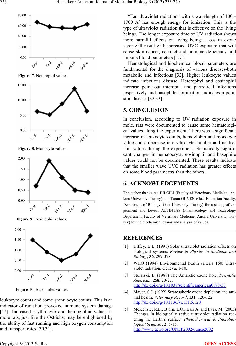 H. Turker / American Journal of Molecular Biology 3 (2013) 235-240 238 “Far ultraviolet radiation” with a wavelength of 100 - 1700 A˚ has enough energy for ionization. This is the type of ultraviolet radiation that is effective on the living beings. The longer exposure time of UV radiation shows more harmful effects on living beings. Loss in ozone layer will result with increased UVC exposure that will cause skin cancer, cataract and immune deficiency and impairs blood parameters [1,7]. Hematological and biochemical blood parameters are fundamental for the diagnosis of various diseases-both metabolic and infectious [32]. Higher leukocyte values indicate infectious disease. Heterophyl and eosinophil increase point out microbial and parasitical infections respectively and basophile domination indicates a para- sitic disease [32,33]. Figure 7. Neutrophil values. Figure 8. Monocyte values. Figure 9. Eosinophil values. Figure 10. Basophiles values. leukocyte counts and some granulocyte counts. This is an indicator of radiation provoked immune system damage [15]. Increased erythrocyte and hemoglobin values in mole rats, just like the Ostrichs, may be enlightened by the ability of fast running and high oxygen consumption and transport rates [30,31]. 5. CONCLUSION In conclusion, according to UV radiation exposure in mole, rats were documented to cause some hematologi- cal values along the experiment. There was a significant increase in leukocyte counts, hemoglobin and monocyte value and a decrease in erythrocyte number and neutro- phil values during the experiment. Statistically signifi- cant changes in hematocryte, eosinophil and basophile values could not be documented. These results indicate that the smaller wave UVC radiation has greater effects on some blood parameters than the others. 6. ACKNOWLEDGEMENTS The author thanks Ali BILGILI (Faculty of Veterinary Medicine, An- kara University, Turkey) and Turan GUVEN (Gazi Education Faculty, Department of Biology, Gazi University, Turkey) for assisting of ex- periment and Levent ALTINTAS (Pharmacology and Toxicology Department, Faculty of Veterinary Medicine, Ankara University, Tur- key) for the biochemical exams and analysis of values. REFERENCES [1] Diffey, B.L. (1991) Solar ultraviolet radiation effects on biological systems. Review in Physics in Medicine and Biology, 36, 299-328. [2] WHO (1994) Environmental health criteria 160: Ultra- violet radiation. Geneva, 1-10. [3] Stolarski, E. (1988) The Antarctic ozone hole. Scientific American, 258, 20-27. http://dx.doi.org/10.1038/scientificamerican0188-30 [4] Mayer, S.J. (1992) Stratospheric ozone depletion and ani- mal health. Veterinary Record, 131, 120-122. http://dx.doi.org/10.1136/vr.131.6.120 [5] McKenzie, R.L., Björn, L.O., Bais A. and Ilyas, M. (2003) Changes in biologically active ultraviolet radiation rea- ching the Earth’s surface. Photochemical & Photobio- logical Sciences, 2, 5-15. http://www.gcrio.org/UNEP2002/6unep2002 Copyright © 2013 SciRes. OPEN ACCESS  H. Turker / American Journal of Molecular Biology 3 (2013) 235-240 239 [6] Schwartz, E. (1988) Connective tissue alterations in the skin of ultraviolet irradiated hairless mice. Journal of In- vestigative Dermatology, 91, 58-161. http://dx.doi.org/10.1111/1523-1747.ep12464405 [7] Zeman, G. (2009) Ultraviolet radiation. http://www.hps.org/hypspublications/articles/uv.html [8] Egami, M.I., Segreto, C., Kerbauy, J. and Juliano, Y. (1991) Effects of whole-body X-irradiation on the peri- pheral blood of primate Cebus apella. Brazilian Journal of Medical and Biological Research, 24, 271-274. [9] Godar, D.E., Thomas, D.P., Miller, S.A. and Lee, W. (1993) Long wavelength UVA radiation induces oxida- tive stress, cytoskeleton damage, and hemolysis. Photo- chemistry and Photobiology, 57, 1018-1026. http://dx.doi.org/10.1111/j.1751-1097.1993.tb02965.x [10] Cotelioglu, U. and Ozcan, M. (1995) The effects of UV-B radiation on some of the blood parameters in rats I. I. The count of leukocyte, the differential leukocyte count, and IgG amount. Med. Bull. Istanbul, 28, 45-49. [11] Ozcan, M. and Çötelioglu, U. (1996) The effects of UV-B radiation on some of the blood parameters in Rats II. I. Erythrocyte number, hemoglobin amount and hemato- cryte value. Med. Bull. Istanbul, 29, 24-27. [12] Duthie, M.S., Kimber, I. and Norval, M. (1999) The ef- fects of ultraviolet radiation on the human immune sys- tem. British Journal of Dermatology, 140, 995-1009. http://dx.doi.org/10.1046/j.1365-2133.1999.02898.x [13] Salo, H.M., Jokinen, E.I., Markkula, S.E., Aaltonen, T.M. and Penttila, H.T. (2000) Comparative effects of UVA and UVB irradiation on the immune system of fish. Pho- tochemistry and Photobiology B: Biology, 56, 154-162. [14] Gidley, D.S., Pecaut, M.J., Miller, G.M., Moyers, M.F. and Nelson, G.A. (2001) Dose and dose rate effects of whole-body gamma-irradiation: II. Hematological varia- bles and cytokines. In Vivo, 15, 209-216. [15] Cetin, E. and Altınsaat Ç. (2006) Effects of ultraviolet radiation on some immunological parameters in rats. American-Eurasian Journal of Agricultural & Environ- mental Sciences, 1, 31-36. [16] Sayed, A.H., Ibrahim, A.T., Mekkawy, I.A. and Mah- moud, U.M. (2007) Effects of ultraviolet radiation on some biochemical and histological parameters of African catfish, Clarias gariepinus (Burchell, 1822). Egyptian Journal of Aquatic Biology and Fisheries, 11, 167-176. [17] Osman, A.G., Koutb, M. and Sayed, Ael-D. (2010) Use of hematological parameters to assess the efficiency of quince (Cydonia oblonga Miller) leaf extract in allevi- ation of the effect of ultraviolet—A radiation on African catfish Clarias gariepinus (Burchell, 1822). Journal of Photochemistry and Photobiology B, 99, 1-8. [18] Verma, P., Sharma, P., Parmar, J., Sharma, P., Agrawal, A. and Goyal, P.K. (2011) Amelioration of radiation-in- duced hematological and biochemical alterations in Swiss albino mice by Panax ginseng extract. Integrative Cancer Therapies, 10, 77-84. http://dx.doi.org/10.1177/1534735410375098 [19] Sukhodub, L.F., Tertyshnyĭ, N.G., Duzhyĭ, I.D. Dand Pli- skachev, V.M. (1991) Ultraviolet irradiation of blood in patients with pulmonary tuberculosis (In Russian). Probl. Tuberk, 7, 65-68. http://www.ncbi.nlm.nih.gov/pubmed?term=%22Duzhy %C4%AD%20ID%22%5BAuthor%5 [20] Broucek, J. and Kovalcik, K. (1988) Effect of artificial ultraviolet light on hematological indices of calves. Pol- nohospodarstvo, 34, 569-577. [21] Loveren, H. van Steerenberg, P.A. and Vos, J.G. (1995) Early detection of immunotoxicity from animal studies to human biomonitoring. Toxicology Letters, 77, 73-80. http://dx.doi.org/10.1016/S0378-4274(97)84688-7 [22] Gahring, L., Baltz, M., Pepysm, B. and Raymond, D. (1984) Effect of ultraviolet radiation on production of epidermal cell thymocyte-activating factor/ interleukin 1 in vivo and in vitro. Proceedings of the National Acade- my of Sciences, 81, 1198-1202. http://dx.doi.org/10.1073/pnas.81.4.1198 [23] Hofer, M., Viklicka, S., Gerasimenko, V.N. and Kaba- chenko, A.N. (1994) Effects of sublethal irradiation with helium ions (300 MeV/nucleon) on basic hematological parameters of mice. Astronauts, 32, 757-760. http://dx.doi.org/10.1016/0094-5765(94)90171-6 [24] Santos, M.S.F., Cavalcanti, M.S.M. and Sampaio, D.A. (2008) Weekly monitoring of the effects of conventional external beam radiation therapy on patients with head and neck, chest, and pelvis cancer by means of blood cells count. Radiologia Brasileira, 41, 1. [25] Bredberg, A. and Forsgren, A. (1984) Effects of in vitro PUVA on human leukocyte function. British Journal of Dermatology, 111, 159-168. http://dx.doi.org/10.1111/j.1365-2133.1984.tb04039.x [26] Jokinen, E.I., Salo, H.M., Markkula, S.E., Aaltonen, T.M. and Immonen, A.K. (2000) Effects of ultraviolet light on immune parameters of the roach. Toxicology Letters, 112- 113, 303-310. http://dx.doi.org/10.1016/S0378-4274(99)00240-4 [27] Nothdurft, W., Fliedner, T.M., Fritz, T.E. and Seed, T.M. (1995) Response of hemopoiesis in dogs to continuous low dose rate total body irradiation. Stem Cells, 13, 261- 267. http://dx.doi.org/10.1002/stem.5530130732 [28] Lundin, A., Michaelsson, G., Venge, P. and Berne, B. (1999) Effects of UVB treatment on neutrophil function in psoriatic patients and healthy subjects. Acta Derm Venereol, 70, 39-45. [29] Lundgren, M.S.F.S., Cavalcanti, M.S.M. and Sampaio, D.A. (2008) Weekly monitoring of the effects of con- ventional external beam radiation therapy on patients with head and neck, chest and pelvis cancer by means of blood cells count. Radiolog ia Brasileira, 41, 9-33. http://dx.doi.org/10.1590/S0100-39842008000100009 [30] Levi, A., Perelman, B., Waner, T., van Grevenbroek, M., van Creveld, C. and Yagil, R. Hematological parameters of the ostrich (Struthio camelus). Avian Pathology, 18, 321-327. http://dx.doi.org/10.1080/03079458908418605 [31] Agaoglu, Z., Yuksek, N., Altug, N. and Kara, A. (2003) Hematological and biochemical parameters in clinical Copyright © 2013 SciRes. OPEN ACCESS  H. Turker / American Journal of Molecular Biology 3 (2013) 235-240 Copyright © 2013 SciRes. 240 OPEN ACCESS healthy Ostriches. Turkish Journal of Veterinary and Ani- mal Sciences, 27, 161-163. [32] Raukur, J. and Simprage, M. (2005) Hematological pa- rameters in the blood of one day old ostriches. Israel Journal of Veterinary Medicine, 60, 112-116. [33] Romdhane, S.B., Romdane, M.N., Mhiri, S., Miled, M.A.B. and Kortas, M. (2000) Les paramètres biochi- miques et hématologiques chez l’autruche (Struthio ca- melus) dans un élevage Tunisien. Revue de Médecine Vé- térinaire, 151, 231-238. |