

Paper Menu >>
Journal Menu >>
 Modern Plastic Surgery, 2013, 3, 130-133 http://dx.doi.org/10.4236/mps.2013.34027 Published Online October 2013 (http://www.scirp.org/journal/mps) Combined Carbon Dioxide Laser Lateral Canthotomy and Femtosecond Laser-Assisted Cataract Surgery* Randal Tanh Hoang Pham1,2 1Aesthetic and Refractive Surgery Medical Center, San Jose, USA; 2Advanced Surgery Medical Center, San Jose, USA. Email: [email protected] Received September 10th, 2013; revised October 6th, 2013; accepted October 13th, 2013 Copyright © 2013 Randal Tanh Hoang Pham. This is an open access article distributed under the Creative Commons Attribution License, which permits unrestricted use, distribution, and reproduction in any medium, provided the original work is properly cited. ABSTRACT Purpose: The purpose of this study was to evaluate a new procedure aimed at lengthening the palpebral fissure to fa- cilitate femtosecond laser-assisted cataract surgery in patients with small eyes. Method: A quick procedure using the CO2 laser was adapted for patients with small eyes undergoing laser-assisted cataract surgery using the Catalys system. The UniPulse CO2 laser (Nidek) was used for laser lateral canthotomy on patients with small palpebral fissures to allow fitting of the Liquid Optic™ Interface eyepiece. Results: Lateral canthotomies were performed on 19 women and 7 men (ages ranged from 45 to 93 years) with lower eyelid lengths equal to or shorter than 32 mm who then underwent femtosecond laser-assisted cataract surgery. A total of 33 eyelids received laser lateral canthotomy with the CO2 laser; only one eyelid had lateral canthotomy with cold-steel tenotomy scissors. Dockings were completed for all 34 eyes with eyelids receiving lateral canthotomies. The 33 eyes with eyelids that received laser canthotomy with the CO2 laser had successful femtosecond laser-assisted cataract surgery. The one eye with the eyelid that received cold-steel canthotomy could not have femtosecond laser-assisted cataract surgery despite successful docking because of pupillary constriction. The findings were statistically significant; Fisher Exact Test showed a p-value of 0.0294. Conclusion: Laser lateral canthotomy with CO2 laser is a safe and effective method to allow docking and completion of femtosecond laser-as- sisted cataract surgery. Keywords: Cataract; Laser; CO2 Laser; Femtosecond Laser; Femtosecond Laser-Assisted Cataract Surgery; Refractive Surgery; Laser Refractive surgery; LASIK; PRK; LASEK; ALK; RLE; EpiLASIK; PRELEX; ICR; Phakic Intraocular Lens Implant; AK; RK; Cataract Surgery San Jose 1. Introduction Femtosecond laser-assisted cataract surgery (FLACS) has recently been available in the United States and around the world. The advantages of this technology in- clude the ability to create a perfectly sized, shaped and centered circular anterior capsular incision, the perfect positioning of the intraocular lens held by a complete edge of the anterior capsule and the ability to break up the cataract, which in turn reduces phacoemulsification time and energy [1,2]. During the procedure, a suction ring is placed directly on the eyeball and the laser is at- tached to the ring using suction; this interface stabilizes the eye relative to the laser. Several factors can affect the ability to create a stable laser-eye interface using the suc- tion ring. In cases where the eye has a short palpebral fissure i.e. short lower eyelid length (LEL), fitting the suction ring to the eyeball may be difficult. In addition, suction can be lost with excessive eye, head and/or body movements [3]. This retrospective study was conducted to identify eyelids that required lateral canthotomy due to a short palpebral fissure, factors that were responsible for femtosecond laser docking failure and to evaluate the safety and efficacy of laser lateral canthotomy (LLC) as an adjunct procedure to femtosecond laser-assisted cata- ract surgery. A review of the literature showed that this study was the first of its kind to compare cold-steel and laser lateral canthotomies when combined with laser- assisted cataract surgery and to address issues related to docking and completion of the femtosecond laser-as- sisted cataract surgery. 2. Materials and Methods Charts were reviewed for all patients who had undergone femtosecond laser-assisted cataract surgery with the *Financial Disclosure: The author has no financial disclosure or con- flict of interest. Copyright © 2013 SciRes. MPS 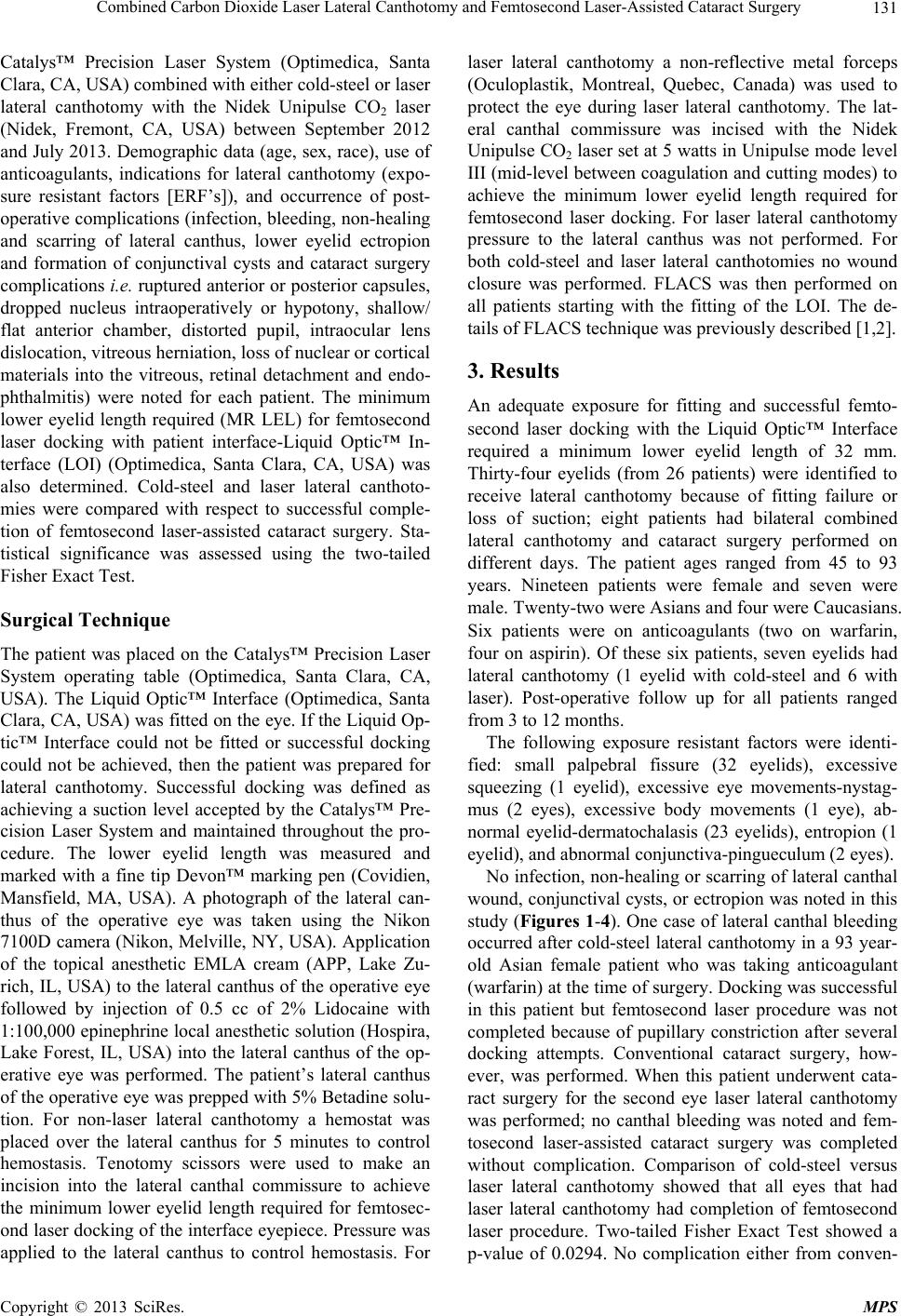 Combined Carbon Dioxide Laser Lateral Canthotomy and Femtosecond Laser-Assisted Cataract Surgery 131 Catalys™ Precision Laser System (Optimedica, Santa Clara, CA, USA) combined with either cold-steel or laser lateral canthotomy with the Nidek Unipulse CO2 laser (Nidek, Fremont, CA, USA) between September 2012 and July 2013. Demographic data (age, sex, race), use of anticoagulants, indications for lateral canthotomy (expo- sure resistant factors [ERF’s]), and occurrence of post- operative complications (infection, bleeding, non-healing and scarring of lateral canthus, lower eyelid ectropion and formation of conjunctival cysts and cataract surgery complications i.e. ruptured anterior or posterior capsules, dropped nucleus intraoperatively or hypotony, shallow/ flat anterior chamber, distorted pupil, intraocular lens dislocation, vitreous herniation, loss of nuclear or cortical materials into the vitreous, retinal detachment and endo- phthalmitis) were noted for each patient. The minimum lower eyelid length required (MR LEL) for femtosecond laser docking with patient interface-Liquid Optic™ In- terface (LOI) (Optimedica, Santa Clara, CA, USA) was also determined. Cold-steel and laser lateral canthoto- mies were compared with respect to successful comple- tion of femtosecond laser-assisted cataract surgery. Sta- tistical significance was assessed using the two-tailed Fisher Exact Test. Surgical Technique The patient was placed on the Catalys™ Precision Laser System operating table (Optimedica, Santa Clara, CA, USA). The Liquid Optic™ Interface (Optimedica, Santa Clara, CA, USA) was fitted on the eye. If the Liquid Op- tic™ Interface could not be fitted or successful docking could not be achieved, then the patient was prepared for lateral canthotomy. Successful docking was defined as achieving a suction level accepted by the Catalys™ Pre- cision Laser System and maintained throughout the pro- cedure. The lower eyelid length was measured and marked with a fine tip Devon™ marking pen (Covidien, Mansfield, MA, USA). A photograph of the lateral can- thus of the operative eye was taken using the Nikon 7100D camera (Nikon, Melville, NY, USA). Application of the topical anesthetic EMLA cream (APP, Lake Zu- rich, IL, USA) to the lateral canthus of the operative eye followed by injection of 0.5 cc of 2% Lidocaine with 1:100,000 epinephrine local anesthetic solution (Hospira, Lake Forest, IL, USA) into the lateral canthus of the op- erative eye was performed. The patient’s lateral canthus of the operative eye was prepped with 5% Betadine solu- tion. For non-laser lateral canthotomy a hemostat was placed over the lateral canthus for 5 minutes to control hemostasis. Tenotomy scissors were used to make an incision into the lateral canthal commissure to achieve the minimum lower eyelid length required for femtosec- ond laser docking of the interface eyepiece. Pressure was applied to the lateral canthus to control hemostasis. For laser lateral canthotomy a non-reflective metal forceps (Oculoplastik, Montreal, Quebec, Canada) was used to protect the eye during laser lateral canthotomy. The lat- eral canthal commissure was incised with the Nidek Unipulse CO2 laser set at 5 watts in Unipulse mode level III (mid-level between coagulation and cutting modes) to achieve the minimum lower eyelid length required for femtosecond laser docking. For laser lateral canthotomy pressure to the lateral canthus was not performed. For both cold-steel and laser lateral canthotomies no wound closure was performed. FLACS was then performed on all patients starting with the fitting of the LOI. The de- tails of FLACS technique was previously described [1,2]. 3. Results An adequate exposure for fitting and successful femto- second laser docking with the Liquid Optic™ Interface required a minimum lower eyelid length of 32 mm. Thirty-four eyelids (from 26 patients) were identified to receive lateral canthotomy because of fitting failure or loss of suction; eight patients had bilateral combined lateral canthotomy and cataract surgery performed on different days. The patient ages ranged from 45 to 93 years. Nineteen patients were female and seven were male. Twenty-two were Asians and four were Caucasians. Six patients were on anticoagulants (two on warfarin, four on aspirin). Of these six patients, seven eyelids had lateral canthotomy (1 eyelid with cold-steel and 6 with laser). Post-operative follow up for all patients ranged from 3 to 12 months. The following exposure resistant factors were identi- fied: small palpebral fissure (32 eyelids), excessive squeezing (1 eyelid), excessive eye movements-nystag- mus (2 eyes), excessive body movements (1 eye), ab- normal eyelid-dermatochalasis (23 eyelids), entropion (1 eyelid), and abnormal conjunctiva-pingueculum (2 eyes). No infection, non-healing or scarring of lateral canthal wound, conjunctival cysts, or ectropion was noted in this study (Figures 1-4). One case of lateral canthal bleeding occurred after cold-steel lateral canthotomy in a 93 year- old Asian female patient who was taking anticoagulant (warfarin) at the time of surgery. Docking was successful in this patient but femtosecond laser procedure was not completed because of pupillary constriction after several docking attempts. Conventional cataract surgery, how- ever, was performed. When this patient underwent cata- ract surgery for the second eye laser lateral canthotomy was performed; no canthal bleeding was noted and fem- tosecond laser-assisted cataract surgery was completed without complication. Comparison of cold-steel versus laser lateral canthotomy showed that all eyes that had laser lateral canthotomy had completion of femtosecond laser procedure. Two-tailed Fisher Exact Test showed a p-value of 0.0294. No complication either from conven- Copyright © 2013 SciRes. MPS 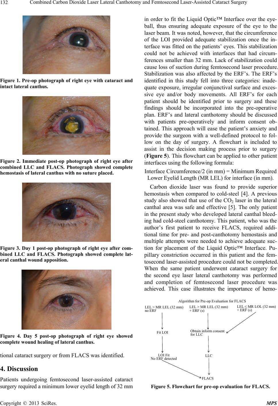 Combined Carbon Dioxide Laser Lateral Canthotomy and Femtosecond Laser-Assisted Cataract Surgery 132 Figure 1. Pre-op photograph of right eye with cataract and intact lateral canthus. Figure 2. Immediate post-op photograph of right eye after combined LLC and FLACS. Photograph showed complete hemostasis of lateral canthus with no suture placed. Figure 3. Day 1 post-op photograph of right eye after com- bined LLC and FLACS. Photograph showed complete lat- eral canthal wound apposition. Figure 4. Day 5 post-op photograph of right eye showed complete wound healing of lateral canthus. tional cataract surgery or from FLACS was identified. 4. Discussion Patients undergoing femtosecond laser-assisted cataract surgery required a minimum lower eyelid length of 32 mm in order to fit the Liquid Optic™ Interface over the eye- ball, thus ensuring adequate exposure of the eye to the laser beam. It was noted, however, that the circumference of the LOI provided adequate stabilization once the in- terface was fitted on the patients’ eyes. This stabilization could not be achieved with interfaces that had circum- ferences smaller than 32 mm. Lack of stabilization could cause loss of suction during femtosecond laser procedure. Stabilization was also affected by the ERF’s. The ERF’s identified in this study fell into three categories: inade- quate exposure, irregular conjunctival surface and exces- sive eye and/or body movements. All ERF’s for each patient should be identified prior to surgery and these findings should be incorporated into the pre-operative plan. ERF’s and lateral canthotomy should be discussed with patients pre-operatively and inform consent ob- tained. This approach will ease the patient’s anxiety and provide the surgeon with a well-defined protocol to fol- low on the day of surgery. A flowchart is included to assist in the decision making process prior to surgery (Figure 5). This flowchart can be applied to other patient interfaces using the following formula: Interface Circumference/2 (in mm) = Minimum Required Lower Eyelid Length (MR LEL) for interface (in mm). Carbon dioxide laser was found to provide superior hemostasis when compared to cold-steel [4]. A previous study also showed that use of the CO2 laser in the lateral canthal area was safe and effective [5]. The only patient in the present study who developed lateral canthal bleed- ing had cold-steel canthotomy. This patient, who was the author’s first patient to receive FLACS, required addi- tional time for pre- and post-canthotomy hemostasis and multiple attempts were needed to achieve adequate suc- tion for placement of the Liquid Optic™ Interface. Pu- pillary constriction occurred in this patient and the fem- tosecond laser-assisted procedure could not be completed. When the same patient underwent cataract surgery for the second eye laser lateral canthotomy was performed and completion of femtosecond laser procedure was achieved. This case illustrates the importance of hemo- Algorithm for Pre-op Evaluation for FLACS LEL > MR LEL (32 mm) no ERF LEL > MR LEL (32 mm) + ERF (s) LEL ≤ MR LOL (32 mm) + ERF ( s ) Fit LOIObtain inform consent for LLC LOI Fit No ERF detectedLLC FLACS Figure 5. Flowchart for pre-op evaluation for FLACS. Copyright © 2013 SciRes. MPS  Combined Carbon Dioxide Laser Lateral Canthotomy and Femtosecond Laser-Assisted Cataract Surgery Copyright © 2013 SciRes. MPS 133 stasis control should lateral canthotomy become indi- cated for femtosecond laser-assisted cataract surgery. This patient was one of the six patients in the study who received anticoagulants and one of two who was on war- farin. None of the patients undergoing CO2 laser lateral canthotomy developed canthal bleeding. This observa- tion reinforces the finding that CO2 laser lateral can- thotomy provides superior hemostasis compared to cold- steel and is therefore indicated in patients who receive anticoagulants [4]. Continued use of anticoagulants is currently considered the standard of care for patients undergoing cataract surgery in many communities in North America, Europe, and Japan [6-8]. CO2 laser lateral canthotomy can be used as an adjunct procedure for conventional cataract surgery and other refractive surgeries such as LASIK, LASEK, PRK, ALK, RLE, EpiLASIK, PRELEX, ICR, phakic intraocular lens implant, AK, RK, etc. In addition, in patients with multi- ple ERF’s, CO2 laser lateral canthotomy can be effective in reducing incidences of suction loss during femtosec- ond laser refractive surgery. This advantage helps achieve successful completion of femtosecond laser-assisted pro- cedures. 5. Conclusion CO2 laser lateral canthotomy is a safe and effective ad- junct procedure for femtosecond laser-assisted cataract surgery. 6. Acknowledgements The author wishes to thank Beverley Murray, PhD for her assistance in the preparation of this manuscript. REFERENCES [1] I. Conrad-Hengerer, et al., “Effects of Femtosecond Laser Fragmentation of the Nucleus with Different Softening Grid Sizes on Effective Phaco Time in Cataract Surgery,” Journal of Cataract & Refractive Surgery, Vol. 38, No. 110, 2012, pp. 1888-1894. http://dx.doi.org/10.1016/j.jcrs.2012.07.023 [2] R. G. Abell, et al., “Catalys Femtosecond Laser-Assisted Cataract Surgery Compared to Conventional Cataract Surgery,” Clinical & Experimental Ophthalmology, Vol. 41, No. 5, 2013, pp. 455-462. http://dx.doi.org/10.1111/ceo.12025 [3] R. Pham, “Lateral Canthotomy for Small Eyes Undergo- ing Laser Refractive Cataract Surgery with Image Guid- ance System,” Presented at the 2013 Annual Symposium of the American Society for Cataract and Refractive Sur- gery, San Francisco. [4] A. M. Goldbaum, et al., “The CO2 Laser in Oculoplastic Surgery,” Survey of Ophthalmology, Vol. 42, No. 3, 1997, pp. 255-267. http://dx.doi.org/10.1016/S0039-6257(97)00097-0 [5] S. S. Baker, et al., “Lateral Canthal Tendon Suspension Using the CO2 Laser: A Modified Technique,” Derma- tologic Surgery, Vol. 21, No. 12, 1995, pp. 1071-1073. http://dx.doi.org/10.1111/j.1524-4725.1995.tb00561.x [6] I. S. Barequet, et al., “Phacoemulsification of Cataract in Patients Receiving Coumadin Therapy: Ocular and He- matologic Risk Assessment,” American Journal of Oph- thalmology, Vol. 144, No. 5, 2007, pp. 719-723. http://dx.doi.org/10.1016/j.ajo.2007.07.029 [7] R. Batra, et al., “Perioperative Management of Antico- agulated Patients Having Cataract Surgery,” Journal of Cataract & Refractive Surgery, Vol. 35, No. 10, 2009, pp. 1815-1820. http://dx.doi.org/10.1016/j.jcrs.2009.05.042 [8] H. Kobayashi, “Evaluation of the Need to Discontinue Antiplatelet and Anticoagulant Medications before Cata- ract Surgery,” Journal of Cataract & Refractive Surgery, Vol. 36, No. 7, 2010, pp. 1115-1119. http://dx.doi.org/10.1016/j.jcrs.2010.01.017 Abbreviations LEL: Lower Eyelid Length MR LEL: Minimum Required Lower Eyelid Length ERF: Exposure Resistant Factor LOI: Liquid Optic™ Interface LLC: Laser Lateral Canthotomy FLACS: Femtosecond Laser-Assisted Cataract Surgery |