

Paper Menu >>
Journal Menu >>
 Journal of Crystallization Process and Technology, 2013, 3, 123-129 http://dx.doi.org/10.4236/jcpt.2013.34020 Published Online October 2013 (http://www.scirp.org/journal/jcpt) Copyright © 2013 SciRes. JCPT 123 Synthesis, Growth, Crystal Structure and Characterization of the o-Toluidinium Picrate Kandasamy Mohana Priyadarshini1, Angannan Chandramohan1*, Thangarak Uma Devi2 1Department of Chemistry, Sri Ramakrishna Mission Vidyalaya College of Arts and Science, Coimbatore, India; 2Department of Physics, Government Arts College for Women, Pudukottai, India. Email: *[email protected] Received July 23rd, 2013; revised July August 23rd, 2013; accepted August 30th, 2013 Copyright © 2013 Kandasamy Mohana Priyadarshini et al. This is an open access article distributed under the Creative Commons Attribu- tion License, which permits unrestricted use, distribution, and reproduction in any medium, provided the original work is properly cited. ABSTRACT A new organic charge transfer molecular complex salt of o-toluidinium picrate (OTP) was synthesised and the single crystals were grown by the slow solvent evaporation solution growth technique using methanol as a solvent at room temperature. Formation of the new crystal has been confirmed by single crystal X-ray diffraction (XRD) and NMR spectroscopic techniques. The crystal structure determined by single crystal X-ray diffraction indicates that both the cation and the anion are interlinked to each other by three types of intermolecular hydrogen bonds, namely N(4)-H(4A)···O(7), N(4)-H(4B)···O(5) and N(4)-H(4C)···O(7). The title compound (OTP) crystallizes in monoclinic crystal system with the centrosymmetric space group P21/c. Fourier transform infrared (FT IR) spectral analysis was used to confirm the presence of various functional groups in the grown crystal. The optical properties were analyzed by the UV-Vis-NIR and fluorescence emission studies. Keywords: Single Crystal; Organic Molecules; Solution Growth; X-Ray Diffraction; Characterization; Nonlinear Optical 1. Introduction The organic materials with aromatic ring, which are of great interest for second and third-order nonlinear optical applications due to their high nonlinearity, high optical damage threshold and their ultrafast, almost purely elec- tronic response. Based on the concepts of the molecular and crystal engineering, the organic molecules offer many possibilities to tailoring the substances with desired properties through optimization of the microscopic hy- perpolarizabilities and the incorporation of the molecules in a crystalline lattice [1-4]. Mulliken suggested that the charge transfer interactions from two aromatic molecules can arise from the transfer of an electron from Lewis base to Lewis acid and these complexes have attracted great attention for nonlinear optical materials. Generally, proton transfer interactions between electron donor and electron acceptor molecules absorb radiation in the visi- ble region leading to the formation of intensely colored charge transfer complexes [5-10]. Picric acid forms crys- talline picrates of various organic molecules through io- nic and hydrogen bonding and π-π interactions and the presence of phenolic OH in the picric acid favors the formation of the salts with various organic bases [11]. The formation of charge transfer complex depending on the nature of the donor-acceptor system and the orientation of anionic and cationic species facilitates the formation of expected N-H·····O hydrogen bonds between amino hydrogen and phenolic oxygen [12]. It has been reported that intramolecular hydrogen bonding interactions are absent in most of the picrate salts [13] and picric acid derivatives are interesting candidates, as the presence of phenolic OH and electron withdrawing nitro groups fa- vors the formation of salts with various organic bases such as N,N-dimethylanilinium picrate [11], 3-Methyl aniliniumpicrate [14], 2-Chloroanilinium picrate [15], Anilinium picrate [16], p-toluidinium picrate [13], 8- hydroxyquinolinium picrate [17], 1,3-Dimethylurea di- methyl ammonium picrate [18], N,N-Dimethyl anilinium picrate [19] have already been reported. The title salt crystallizes in the monoclinic crystal system with cen- trosymmetric space group P21/c, an analogue of p-Tolu- idinium picrate. In the present work, we report the syn- thesis, crystal growth, structural, spectral and optical stu- dies of o-toluidinium picrate single crystal. *Corresponding author. 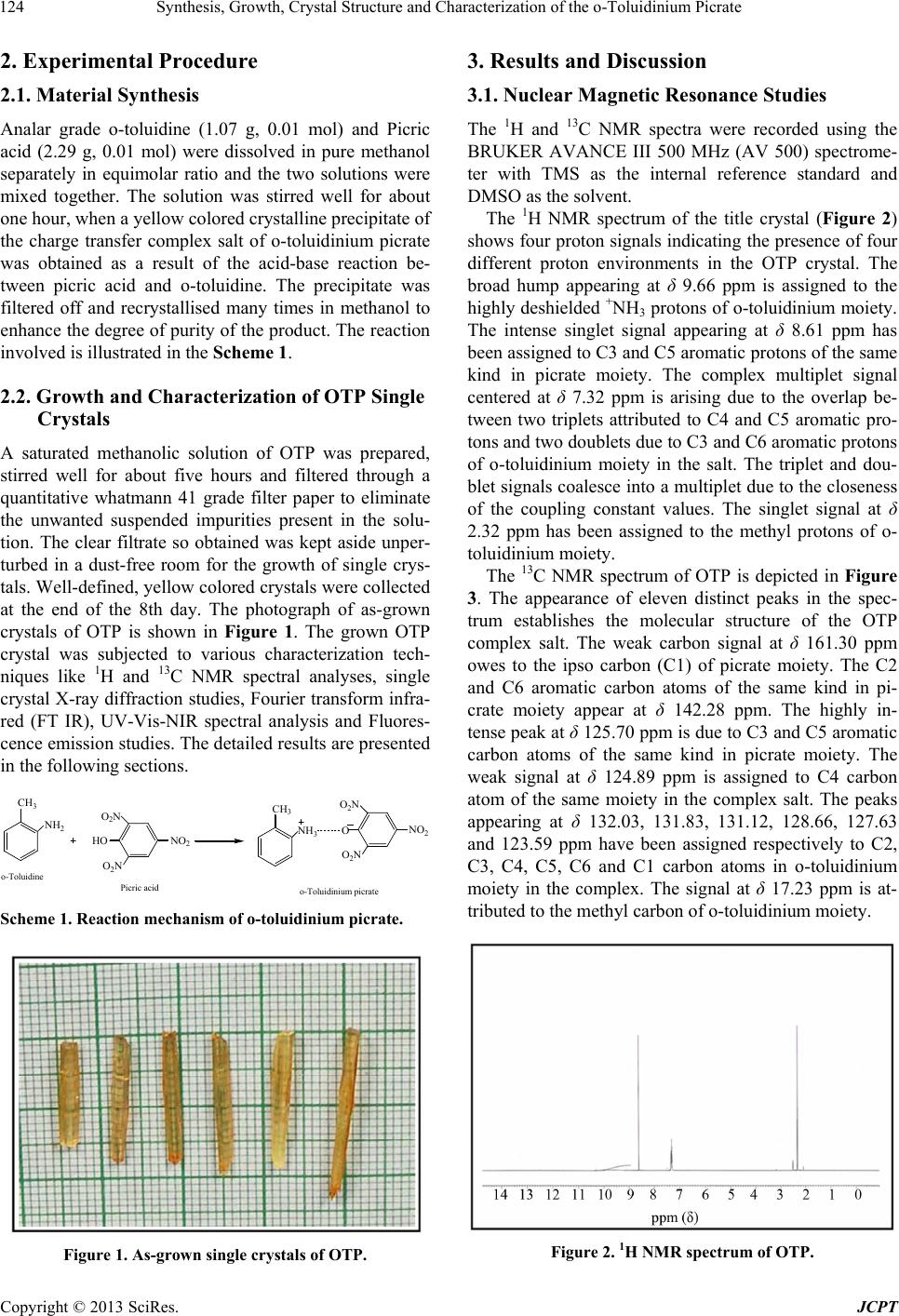 Synthesis, Growth, Crystal Structure and Characterization of the o-Toluidinium Picrate Copyright © 2013 SciRes. JCPT 124 2. Experimental Procedure 2.1. Material Synthesis Analar grade o-toluidine (1.07 g, 0.01 mol) and Picric acid (2.29 g, 0.01 mol) were dissolved in pure methanol separately in equimolar ratio and the two solutions were mixed together. The solution was stirred well for about one hour, when a yellow colored crystalline precipitate of the charge transfer complex salt of o-toluidinium picrate was obtained as a result of the acid-base reaction be- tween picric acid and o-toluidine. The precipitate was filtered off and recrystallised many times in methanol to enhance the degree of purity of the product. The reaction involved is illustrated in the Scheme 1. 2.2. Growth and Characterization of OTP Single Crystals A saturated methanolic solution of OTP was prepared, stirred well for about five hours and filtered through a quantitative whatmann 41 grade filter paper to eliminate the unwanted suspended impurities present in the solu- tion. The clear filtrate so obtained was kept aside unper- turbed in a dust-free room for the growth of single crys- tals. Well-defined, yellow colored crystals were collected at the end of the 8th day. The photograph of as-grown crystals of OTP is shown in Figure 1. The grown OTP crystal was subjected to various characterization tech- niques like 1H and 13C NMR spectral analyses, single crystal X-ray diffraction studies, Fourier transform infra- red (FT IR), UV-Vis-NIR spectral analysis and Fluores- cence emission studies. The detailed results are presented in the following sections. CH 3 NH 2 HO O 2 N O 2 N NO 2 CH 3 NH 3 O O 2 N O 2 N NO 2 o-Toluidine Picric acido-To l u i d i n i um p i crate Scheme 1. Reaction mechanism of o-toluidinium picrate. Figure 1. As-grown single crystals of OTP. 3. Results and Discussion 3.1. Nuclear Magnetic Resonance Studies The 1H and 13C NMR spectra were recorded using the BRUKER AVANCE III 500 MHz (AV 500) spectrome- ter with TMS as the internal reference standard and DMSO as the solvent. The 1H NMR spectrum of the title crystal (Figure 2) shows four proton signals indicating the presence of four different proton environments in the OTP crystal. The broad hump appearing at δ 9.66 ppm is assigned to the highly deshielded +NH3 protons of o-toluidinium moiety. The intense singlet signal appearing at δ 8.61 ppm has been assigned to C3 and C5 aromatic protons of the same kind in picrate moiety. The complex multiplet signal centered at δ 7.32 ppm is arising due to the overlap be- tween two triplets attributed to C4 and C5 aromatic pro- tons and two doublets due to C3 and C6 aromatic protons of o-toluidinium moiety in the salt. The triplet and dou- blet signals coalesce into a multiplet due to the closeness of the coupling constant values. The singlet signal at δ 2.32 ppm has been assigned to the methyl protons of o- toluidinium moiety. The 13C NMR spectrum of OTP is depicted in Figure 3. The appearance of eleven distinct peaks in the spec- trum establishes the molecular structure of the OTP complex salt. The weak carbon signal at δ 161.30 ppm owes to the ipso carbon (C1) of picrate moiety. The C2 and C6 aromatic carbon atoms of the same kind in pi- crate moiety appear at δ 142.28 ppm. The highly in- tense peak at δ 125.70 ppm is due to C3 and C5 aromatic carbon atoms of the same kind in picrate moiety. The weak signal at δ 124.89 ppm is assigned to C4 carbon atom of the same moiety in the complex salt. The peaks appearing at δ 132.03, 131.83, 131.12, 128.66, 127.63 and 123.59 ppm have been assigned respectively to C2, C3, C4, C5, C6 and C1 carbon atoms in o-toluidinium moiety in the complex. The signal at δ 17.23 ppm is at- tributed to the methyl carbon of o-toluidinium moiety. Figure 2. 1H NMR spectrum of OTP.  Synthesis, Growth, Crystal Structure and Characterization of the o-Toluidinium Picrate Copyright © 2013 SciRes. JCPT 125 Figure 3. 13C NMR spectrum of OTP. 3.2. FT-IR Spectroscopy The characteristic vibrational frequencies of the func- tional groups of OTP are identified from the fourier transform infrared (FT-IR) spectrum recorded in the range of 4000 - 400 cm−1 employing Perkin-Elmer FT-IR spectrometer by using the KBr pellet technique. The formation of charge transfer complex during the acid- base interaction of o-toluidine with picric acid is strongly evidenced through the realization of important bands of donor and acceptor in the resultant spectrum of the com- plex salt (Figure 4). The absorption at 3194 cm−1 is due to the +N-H stretching vibration. The absorption band at 3060 cm−1 corresponds to aromatic C-H asymmetric stretching vibration. The broad absorption bands in the region 2940 to 2813 cm−1 are due to the overlapping of C-H asymmetric and symmetric stretching vibration of methyl group and aromatic C-H symmetric stretching vibration. The absorptions at 1535 and 1359 cm−1 con- firm the asymmetric and symmetric stretching vibrations of NO2 group respectively. The C-O stretching vibration is observed at 1192 cm−1. The band at 836 cm−1 is due to C-N stretching vibration. The presence of C=C stretch- ing vibration of aromatic ring is revealed from the ab- sorption bands at 1606, 1570 and 1479 cm−1. The as- signment is in very close agreement with data of the complex salts reported already [7-9]. 3.3. Single Crystal X-Ray Diffraction Studies Single crystal XRD analysis for the grown o-toluidi- nium picrate has been carried out to identify the unit cell parameters and the crystal structure using “ENRAF (BRUKER) NONIUS CAD4” diffractometer with graph- ite monochromated MoKα radiation (λ = 0.71073 Å). The structure was solved by direct methods procedure as implemented in SHELXS 97 [20] program. Cell refine- ment and data reduction were carried out using SAINT program. All the hydrogen atoms were fixed geometri- cally and allowed to ride on their parent atoms. All non-hydrogen atoms were refined using anisotropic dis- placement parameters. The crystal structure analyses of OTP reveals that OTP crystallized in a monoclinic crystal structure with cen- trosymmetric space group P21/c, and the unit cell pa- rameters are a = 11.6475(2) Å, b = 16.4763(5) Å, c = 7.5702(4) Å. Table 1 summarizes the crystal data, inten- sity data collection and refinement details for the OTP single crystals. The selected bond lengths and bond an- gles of OTP charge transfer complex salt are given in Tables 2 and 3 respectively. The protonation on the N1 site of the cation is confirmed from C-N bond distances and C-N-C bond angles. All the bond distances and bond angles of the two molecules in the asymmetric unit are agreed with each other. The crystal structure of OTP consists one molecule each of o-toluidinium cation and picrate anion. The ORTEP of the charge transfer com- plex OTP is clearly shown in the Figure 5 and the atom numbering scheme adopted. A well-defined yellow col- our single crystal of OTP with dimension 0.30 × 0.20 × 0.20 mm was selected for diffraction analysis. A total of 12528 reflections (2541 Unique, R(int) = 0.0239) were collected by using ω/2θ scan mode at 293(2) K in the range of 2.15˚ < θ < 25˚ with the index ranges −13 <= h = 13, −19 <= k <= 19, −7 <= 1 <= 9. The refinement converged to final R-factor of 0.04%. The packing dia- gram (Figure 6) indicates the existence of intermolecular hydrogen bonds in the three dimensional network be- tween the constituent molecules. The structure is based on asymmetric part of o-Toluidinium picrate which con- tains 2-methyl anilinium cation and picrate anion con- nected by three intermolecular N-H····O hydrogen bonds with a donor-acceptor distance of 2.7697(19), 2.853(2) and 2.762(2) Å. The corresponding data for the H-bonds are listed in Table 4. In the charge transfer complex of OTP, the carbon skeleton of the anionic picrate species Figure 4. FT-IR spectrum of OTP.  Synthesis, Growth, Crystal Structure and Characterization of the o-Toluidinium Picrate Copyright © 2013 SciRes. JCPT 126 Table 1. Crystallographic data for OTP. Empirical formula C13H12N4O7 Formula weight 336.27 Temperature 293(2) k Wavelength 0.71073 Å Crystal system, space group Monoclinic, P21/c Unit cell dimensions a = 11.6475(2) Å, α = 90˚ b = 16.4763(5) Å, β = 94.1980(10)˚ c = 7.5702(4) Å, γ = 90˚ Volume 1448.88(9) Å3 Z, Calculated density 4, 1.542 Mg/m3 Absorption coefficient 0.128 mm−1 F(000) 696 Crystal size 0.30 × 0.20 × 0.20 mm Theta range for data collection 2.15 to 25.00 deg Limiting indices −13 <= h <= 13, −19 <= k <= 19, −7 <= 1 <= 9 Reflections collected/unique 12528/2541 [R(int) = 0.0239] Completeness to theta = 25.00 99.9% Absorption correction Semi-empirical from equivalents Max. and min. transmission 0.9924 and 0.9245 Refinement method Full-matrix least-squares on F2 Data/restraints/parameters 2541/0/220 Goodness-of-fit on F2 1.046 Final R indices [I > 2 sigma(I)] R1 = 0.0402, wR2 = 0.1084 R indices (all data) R1 = 0.0468, wR2 = 0.1152 Extinction coefficient 0.0042(11) Largest diff. peak and hole 0.382 and −0.264 e.Å−3 Figure 5. ORTEP diagram of OTP. Table 2. Selected bond lengths in OTP (Å). C(1)-C(6) 1.374(2) C(1)-C(2) 1.432(2) C(1)-N(1) 1.461(2) C(2)-O(7) 1.270(2) C(2)-C(3) 1.429(2) C(3)-C(4) 1.365(2) C(3)-N(2) 1.462(2) C(4)-C(5) 1.386(3) C(5)-C(6) 1.380(3) C(5)-N(3) 1.447(2) C(7)-C(12) 1.380(2) C(7)-C(8) 1.386(2) C(7)-N(4) 1.470(2) C(8)-C(9) 1.391(3) C(8)-C(13) 1.503(3) C(9)-C(10) 1.376(3) C(10)-C(11) 1.375(3) C(11)-C(12) 1.380(3) C(13)-H(13A) 0.9600 N(1)-O(1) 1.216(2) N(1)-O(2) 1.221(2) N(2)-O(4) 1.209(2) N(2)-O(3) 1.213(2) N(3)-O(6) 1.215(2) N(3)-O(5) 1.228(2) N(4)-H(4A) 0.8900 Figure 6. Packing arrangement of molecules showing in- termolecular N-H···O hydrogen bonding interactions and π-π interactions viewed down in the c-axis.  Synthesis, Growth, Crystal Structure and Characterization of the o-Toluidinium Picrate Copyright © 2013 SciRes. JCPT 127 Table 3. Selected bond angles in OTP (˚). C(6)-C(1)-C(2) 124.17(16) C(6)-C(1)-N(1) 116.84(16) C(2)-C(1)-N(1) 118.88(15) O(7)-C(2)-C(3) 122.26(15) O(7)-C(2)-C(1) 125.48(16) C(3)-C(2)-C(1) 112.14(14) C(4)-C(3)-C(2) 125.28(15) C(4)-C(3)-N(2) 117.71(15) C(2)-C(3)-N(2) 116.99(14) C(3)-C(4)-C(5) 118.03(16) C(6)-C(5)-C(4) 121.50(16) C(6)-C(5)-N(3) 119.09(16) C(4)-C(5)-N(3) 119.37(16) C(1)-C(6)-C(5) 118.77(16) C(12)-C(7)-C(8) 122.89(16) C(12)-C(7)-N(4) 118.12(15) C(8)-C(7)-N(4) 118.98(15) C(7)-C(8)-C(9) 116.38(17) C(7)-C(8)-C(13) 122.73(16) C(9)-C(8)-C(13) 120.89(17) C(10)-C(9)-C(8) 121.68(18) C(11)-C(10)-C(9) 120.34(18) C(10)-C(11)-C(12) 119.74(18) C(11)-C(12)-C(7) 118.96(17) C(8)-C(13)-H(13A) 109.5 H(13A)-C(13)-H(13B) 109.5 O(1)-N(1)-O(2) 123.16(18) O(1)-N(1)-C(1) 118.20(17) O(2)-N(1)-C(1) 118.65(16) O(4)-N(2)-O(3) 123.06(17) O(4)-N(2)-C(3) 118.66(16) O(3)-N(2)-C(3) 118.24(16) O(6)-N(3)-O(5) 123.45(16) O(6)-N(3)-C(5) 119.62(17) O(5)-N(3)-C(5) 116.93(17) Table 4. Hydrogen bond parameters in OTP. D-H···A d(D-H) d(H···A) d(D···A) <(DHA) N(4)-H(4A)···O(7)#1 0.89 1.89 2.7697(19) 172.0 N(4)-H(4B)···O(5)#2 0.89 2.14 2.853(2) 136.9 N(4)-H(4C)···O(7) 0.89 1.88 2.762(2) 172.5 #1 x, −y + 1/2, z + 1/2; #2 –x + 1, −y, −z + 1. and cationic o-Toluidinium species is non-planar and is shown in the torsion angle of N(1)-C(1)-C(2)-C(3), N(2)-C(3)-C(4)-C(5), N(3)-C(5)-C(6)-C(1), N(4)-C(7)- C(8)-C(13), C(13)-C(8)-C(9)-C(10), C(10)-C(11)-C(12)- C(7) which are 179.28(16)˚, −178.89(16)˚, 178.76(16)˚, −0.8(3)˚, 178.8(2)˚, −0.6(3)˚ respectively. 3.4. UV-Vis-NIR Transmission Studies The optical transmission spectrum of OTP crystals was recorded in the region 200 - 1100 nm employing a Shi- madzu UV-1061 UV-Vis spectrophotometer in solution using DMSO as the solvent. The recorded transmission spectrum of OTP is shown in Figure 7. The lower cutoff wavelength of the OTP crystal was around 488 nm. The attained percentage of transmittance was around 97 for OTP complex in the region between 500 and 1100 nm. Hence, this crystal can be used for the suitable optical applications due to its wide transparency range in the part of visible region above 488 nm and in the entire near infrared region. 3.5. Fluorescence Emission Studies Fluorescence may be expected generally in molecules that are aromatic or contain multiple conjugated double bonds with a high degree of resonance stability [21].The fluorescence emission spectrum of OTP was recorded using HORIBA JASCO V-670 FLUOROLOG 3 spectro- fluorometer. The fluorescence emission spectrum was recorded in the range from 500 to 900 nm and depicted in Figure 8. Two peaks at 555 and 607 nm observed in the emission spectrum indicates that OTP crystal has a green- orange fluorescence emission. 4. Conclusion The organic molecular charge transfer complex salt OTP Figure 7. Optical transmission spectrum of OTP. 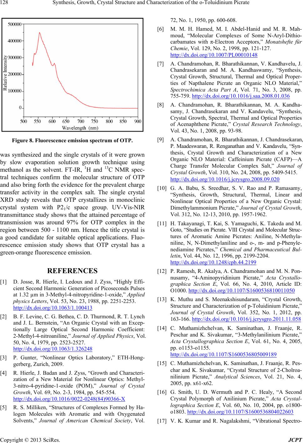 Synthesis, Growth, Crystal Structure and Characterization of the o-Toluidinium Picrate Copyright © 2013 SciRes. JCPT 128 Figure 8. Fluorescence emission spectrum of OTP. was synthesized and the single crystals of it were grown by slow evaporation solution growth technique using methanol as the solvent. FT-IR, 1H and 13C NMR spec- tral techniques confirm the molecular structure of OTP and also bring forth the evidence for the prevalent charge transfer activity in the complex salt. The single crystal XRD study reveals that OTP crystallizes in monoclinic crystal system with P21/c space group. UV-Vis-NIR transmittance study shows that the attained percentage of transmission was around 97% for OTP complex in the region between 500 - 1100 nm. Hence the title crystal is a good candidate for suitable optical applications. Fluo- rescence emission study shows that OTP crystal has a green-orange fluorescence emission. REFERENCES [1] D. Josse, R. Hierle, I. Ledoux and J. Zyss, “Highly Effi- cient Second Harmonic Generation of Picoseconds Pulses at 1.32 µm in 3-Methyl-4-nitropyridine-1-oxide,” Applied physics Letters, Vol. 53, No. 23, 1988, pp. 2251-2253. http://dx.doi.org/10.1063/1.100413 [2] B. F. Levine, C. G. Bethea, C. D. Thurmond, R. T. Lynch and J. L. Bernstein, “An Organic Crystal with an Excep- tionally Large Optical Second Harmonic Coefficient: 2-Methyl-4-nitroaniline,” Journal of Applied Physics, Vol. 50, No. 4, 1979, pp. 2523-2527. http://dx.doi.org/10.1063/1.326248 [3] P. Gunter, “Nonlinear Optics Laboratory,” ETH-Hong- gerberg, Zurich, 2009. [4] R. Hierle, J. Badan and J. Zyss, “Growth and Characteri- zation of a New Material for Nonlinear Optics: Methyl- 3-nitro-4-pyridine-1-oxide (POM),” Journal of Crystal Growth, Vol. 69, No. 2-3, 1984, pp. 545-554. http://dx.doi.org/10.1016/0022-0248(84)90366-X [5] R. S. Milliken, “Structures of Complexes Formed by Ha- logen Molecules with Aromatic and with Oxygenated Solvents,” Journal of American Chemical Society, Vol. 72, No. 1, 1950, pp. 600-608. [6] M. M. H. Hamed, M. I. Abdel-Hanid and M. R. Mah- moud, “Molecular Complexes of Some N-Aryl-Dithio- carbamates with π-Electron Acceptors,” Monatshefte für Chemie, Vol. 129, No. 2, 1998, pp. 121-127. http://dx.doi.org/10.1007/PL00010148 [7] A. Chandramohan, R. Bharathikannan, V. Kandhavelu, J. Chandrasekaran and M. A. Kandhaswamy, “Synthesis, Crystal Growth, Structural, Thermal and Optical Proper- ties of Napthalene Picrate an Organic NLO Material,” Spectrochimica Acta Part A, Vol. 71, No. 3, 2008, pp. 755-759. http://dx.doi.org/10.1016/j.saa.2008.01.036 [8] A. Chandramohan, R. Bharathikannan, M. A. Kandha- samy, J. Chandrasekaran and V. Kandavelu, “Synthesis, Crystal Growth, Spectral, Thermal and Optical Properties of Acenaphthene Picrate,” Crystal Research Technology, Vol. 43, No. 1, 2008, pp. 93-98. [9] A. Chandramohan, R. Bharathikannan, J. Chandrasekaran, P. Maadeswaran, R. Renganathan and V. Kandavelu, “Syn- thesis, Crystal Growth and Characterization of a New Organic NLO Material: Caffeinium Picrate (CAFP)—A Charge Transfer Molecular Complex Salt,” Journal of Crystal Growth, Vol. 310, No. 24, 2008, pp. 5409-5415. http://dx.doi.org/10.1016/j.jcrysgro.2008.09.020 [10] G. A. Babu, S. Sreedhar, S. V. Rao and P. Ramasamy, “Synthesis, Growth, Structural, Thermal, Linear and Nonlinear Optical Properties of a New Organic Crystal: Dimethylammonium Picrate,” Journal of Crystal Growth, Vol. 312, No. 12-13, 2010, pp. 1957-1962. [11] H. Takayanagi, T. Kai, S. Yamaguchi, K. Takeda and M. Goto, “Studies on Picrate. VIII Crystal and Molecular Struc- tures of Aromatic Amine Picrates: Aniline, N-Methyla- niline, N, N-Dimethylaniline and o-, m- and p-Phenyle- nediamine Picrates,” Chemical and Pharmaceutical Bul- letin, Vol. 44, No. 12, 1996, pp. 2199-2204. http://dx.doi.org/10.1248/cpb.44.2199 [12] P. Ramesh, R. Akalya, A. Chandramohan and M. N. Pon- nusamy, “4-Aminopyridinium Picrate,” Acta Crystallo- graphica Section E, Vol. 66, No. 4, 2010, Article ID: O1000. http://dx.doi.org/10.1107/S1600536810011050 [13] K. Muthu and S. Meenakshisundaram, “Crystal Growth, Structure and Characterization of p-Toluidinium Picrate,” Journal of Crystal Growth, Vol. 352, No. 1, 2012, pp. 163-166. http://dx.doi.org/10.1016/j.jcrysgro.2011.11.058 [14] C. Muthamizhchelvan, K. Saminathan, J. Fraanje, R. Peschar and K. Sivakumar, “3-Methylanilinium Picrate,” Acta Crystallographica Section E, Vol. 61, No. 4, 2005, pp. o1153-o1155. http://dx.doi.org/10.1107/S1600536805009189 [15] C. Muthamizhchelvan, K. Saminathan, J. Fraanje, R. Pes- char and K. Sivakumar, “Crystal Structure of 2-Cholroa- nilinium Picrate,” Analytical Sciences, Vol. 21, No. 4, 2005, pp. x61-x62. [16] G. Smith, U. D. Wermuth and P. C. Healy, “A Second Crystal Polymorph of Anilinium Picrate,” Acta Crystal- lographica Section E, Vol. 60, No. 10, 2004, pp. o1800- o1803. http://dx.doi.org/10.1107/S1600536804022603 [17] V. K. Kumar and R. Nagalakshmi, “Vibrational Spectro- 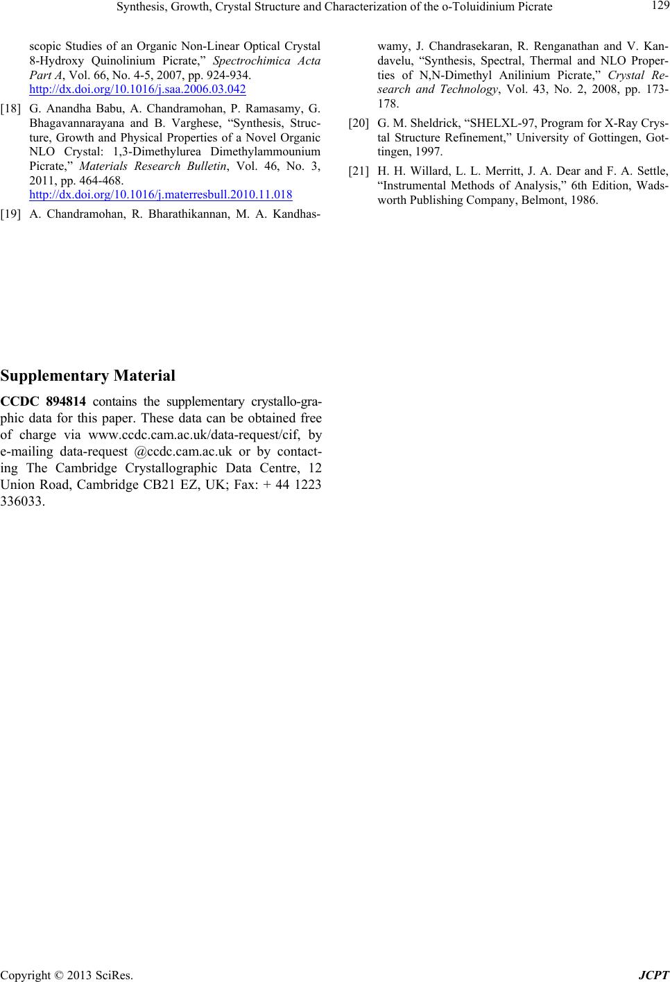 Synthesis, Growth, Crystal Structure and Characterization of the o-Toluidinium Picrate Copyright © 2013 SciRes. JCPT 129 scopic Studies of an Organic Non-Linear Optical Crystal 8-Hydroxy Quinolinium Picrate,” Spectrochimica Acta Part A, Vol. 66, No. 4-5, 2007, pp. 924-934. http://dx.doi.org/10.1016/j.saa.2006.03.042 [18] G. Anandha Babu, A. Chandramohan, P. Ramasamy, G. Bhagavannarayana and B. Varghese, “Synthesis, Struc- ture, Growth and Physical Properties of a Novel Organic NLO Crystal: 1,3-Dimethylurea Dimethylammounium Picrate,” Materials Research Bulletin, Vol. 46, No. 3, 2011, pp. 464-468. http://dx.doi.org/10.1016/j.materresbull.2010.11.018 [19] A. Chandramohan, R. Bharathikannan, M. A. Kandhas- wamy, J. Chandrasekaran, R. Renganathan and V. Kan- davelu, “Synthesis, Spectral, Thermal and NLO Proper- ties of N,N-Dimethyl Anilinium Picrate,” Crystal Re- search and Technology, Vol. 43, No. 2, 2008, pp. 173- 178. [20] G. M. Sheldrick, “SHELXL-97, Program for X-Ray Crys- tal Structure Refinement,” University of Gottingen, Got- tingen, 1997. [21] H. H. Willard, L. L. Merritt, J. A. Dear and F. A. Settle, “Instrumental Methods of Analysis,” 6th Edition, Wads- worth Publishing Company, Belmont, 1986. Supplementary Material CCDC 894814 contains the supplementary crystallo-gra- phic data for this paper. These data can be obtained free of charge via www.ccdc.cam.ac.uk/data-request/cif, by e-mailing data-request @ccdc.cam.ac.uk or by contact- ing The Cambridge Crystallographic Data Centre, 12 Union Road, Cambridge CB21 EZ, UK; Fax: + 44 1223 336033. |