

Paper Menu >>
Journal Menu >>
 Journal of Signal and Information Processing, 2013, 4, 66-71 doi:10.4236 /jsip.2013.43B012 Published Online August 2013 (http://www.scirp.org/journal/jsip) Copyright © 2013 SciRes. JSIP Fuzzy C Mean Thresholding based Level Set for Automated Segmentation of Skin Lesions Ammara Masood, Adel Ali Al-Jumaily Scho ol of Electrical, Mechanical and Mechatronic Engineer ing, Universi ty of Techn ology, Sydney, Austral ia. Email: ammara.masoo [email protected]. edu.au, Adel.Al-[email protected] Received April, 2013. ABSTRACT Accurate se gmentation is an importa nt and challe nging task in any comp uter vision s ystem. It also plays a vital role in computerized analysis of skin lesion images. This paper presents a new segmentation method that combines the advan- tages of fuzzy C mean algorithm, thresholding and level set method. 3-class Fuzzy C mean thresholding is applied to initialize level set automatically and also for estimating controlling parameters for level set evolution. Parameters for performance evaluation are presented and segmentation results are compared with some other state-of-the-art segmenta- tion methods. Increased true detection rate and reduced false positive and false negative errors confirm the effectiveness of proposed method for skin cancer detection. Keywords: Skin Cancer; Segmentation; Diagnosis; Fuzzy; T hresholding; Level Se ts 1. Introduction The incidence of skin cancer is rapidly increasing thro ugh-out the world [1]. An estimated 76,250 new cas- es of invasive melanoma were diagnosed in the US in 2012, with an estimated 9,180 resulting in death [2]. Australia has one of the highest rates of skin cancer in the world. Over 1890 Australians die from skin cancer each year [3]. Melanoma is capable of deep invasion. The most dangerous characteristic of melanoma is that it can spread widely over the body via the lymphatic ves- sels and blood vessels. Thus, early diagnosis of mela- noma is a key factor for prognosis of the disease. Analysis of skin lesion images taken using dermo- scopic imaging technique [4] is a commonly used me- thod for diagnosis of skin cancer but, this method re- quires great deal of experience [5]. Due to the lack of reproducibility and subjectivity of human interpretation, the development of computerized image analysis tech- niques is of paramount importance. Segmentation is one of the most important and difficult task in computerized image analysis process. The accuracy of the subsequent steps highly depends on the success of image segmenta- tion technique. Segmentation o f ski n lesi ons is d iffic ult because of the great variety of lesion shapes, sizes, and colors along with different types and textures of human skin. Other difficulties that make it a challenging task include, low contrast between the lesion and the surrounding skin, smooth transitio n between the lesion and the skin, reflec- tions d ue to wrong i llu mina tio n and art ifacts suc h as s ki n texture, air bubbles and hair. Image segmentation algorithms available in literature can be broadly classified into two categories: 1) discon- tinuity based segmentatio n 2) Similar ity based seg menta- tion, which c a n further be classified into thresholding [6], clustering [7], and region based approach [8]. Compara- tive analysis of these approaches in the area of skin le- sion segmentation can be found in literature [9-11]. Active contour is a popular approach used to estimate boundaries in medical images. Two types of algorithms lie under this category: 1) parametric active contours [12] which adapt a deformable curve until it fits the object boundary. 2) Geometric active contours based on level set theory. Some of the active contour models need user interventio n for initialization. Thus auto matic approaches like gradient vector flow algorithm [13, 14] based on anisotropic diffusion [15] and robust algorithms like adaptive snakes and shape probability association model [16] are ta king important plac e in liter a ture. Level set (LS) methods, as one of the automatic proc- ess approach have shown effective results for medical image segmentation. However, intensive computational requirements and regulation of controlling parameters make it a complex and time consuming method. To overcome such shortcomings, fuzzy clustering has been used to facilitate the LS segmentation for automatic segmentation of ultrasound, computed tomography and  Fuzzy C Mean Thresholding based Level Set for Automated Segmentation of Skin Lesions Copyright © 2013 SciRes. JSIP 67 magnetic resonance imaging [17-19]. Fuzzy C mean (FCM) based thresholding [20] and LS algorithm are different computational models that have been applied individuall y for segme ntation of der moscopic images [21, 22]. Similarly, some work is done on using clustering for initial segmentation followed by further edge preserving and refining steps for border tracing of skin lesions [23]. We analyzed the advantages and limitations of these methods for skin lesion segmentation. The observations showed that FCM works well for making rough cluster- ing of pixels which can further be used to get better threshold values for image segmentation. In addition to this, if the thresholded image is used as initial estimate for level set evolution, it can result in more accurate segmentation as compared to standard level set method [24,25] and region based active contours [26,27]. We, hereby presented a technique, based on combining these methods to achieve higher accuracy in segmentation of skin lesion images . This paper introduces 3-class FCM based thresholding to be used for ini tializi n g the LS evolu tion a nd re gulati ng the controlling parameters. Performance evaluation is done by comparing the diagnosis results of this method with three other segmentation methods i.e. FCM cluster- ing [28], region based active contours [26, 29] and adap- tive thresholding [11]. Results show that the proposed approach performs reasonably well for the segmentation of ski n lesion i mages. The pap er is or ganized as follows: Section 2 provides details of the proposed segmentation method. Section 3 discusses experimental results and Section 4 provides performance evaluation. Concluding remarks ar e given in Sect ion 5. 2. Methodology This section describes our technique for segmentation of skin cancer images. The main parts of our proposed al- gorithm are: 2.1. Image Pre-Processing For skin lesion images there are certain extraneous arti- facts such as skin texture, air bubbles, dermoscopic gel, presence of ruler markings and hair that make border de- tection a bit difficult. In order to reduce the effect of these artifacts on segmentation results, the images need to be pre-processed with a smoothing filter. We found that the best segmentation results were obtained using median filter with 7x7 mask to smooth the images before seg- mentation. Thus, the first step of the overall process is to get a filtered image. 2.2. Fuzzy C-Mean Thresholding FCM clustering is used to partition N objects into C classes. In our method, N is equal to the number of pixels in the image i.e. N=Nx x Ny and C=3 for 3-class FCM clustering. The FCM algorithm uses iterative optimization of an objective function based on a weighted similarity measure between the pixels in the image and each of the c-clus ter cen ters . A local extre mism of the objecti ve func- tion indicates an op timal clustering o f the input d ata. The objective function that is minimized is given by (1) (1) where and & where . ‖*‖ is a norm expressing the simi- larity between any measured data value and the cluster centre; m∈ [1, ∞] is a weighting exponent and can be any real number greater than 1. Calculations suggest that best choice of m is in the in- terval [1.5, 2.5], so m=2 is used here as it is widely ac- cepted as a good choice of fuzzification parameter. The fuzzy c-partition of given data set is the fuzzy partition matrix U= [ with i =1 , 2 …. C a nd j =1, 2, 3…N , where indicate the degree of membership of jth pixel to ith cluster. The membership functions are subject to satisfy the following conditions. for j=1,2,3,….N ; for i= 1,2….C; The aim of FCM algorith m is to find an optimal fuzz y c-partition by evolving the fuzzy partition matrix U= [ iteratively and computing the cluster centres. In order to achieve this, the algorithm tries to minimize the objective function Q (1) b y iteratively updating the clus- ter centres and the membership functions using the fol- lowing equations. (2) (3) After performing FCM clustering, finally each pixel is assigned to the cluster for which its membership value is maximum. Based on the intensity distribution obtained using histogram of the image, the threshold value is calculated by taking mean of maximum of cluster 1 and minimum of cluster 2 or maximum of cluster 2 and minimum of cluster 3. This method of threshold selection takes into acco unt the intensity distrib ution i n the ima ge. This choice helps in obtaining opti mum threshold values for different images obtained under different conditions. The overall FCM thresholding algorithm is presented in Figure 1. The output of this stage is a binary image (Bi) which has been used in the followed steps. 2.3. Fuzzy C-mean Thresholding based Level Set Segmentation Segmentation of images by means of active contours is a well-established approach. Active contours [12] are used  Fuzzy C Mean Thresholding based Level Set for Automated Segmentation of Skin Lesions Copyright © 2013 SciRes. JSIP 68 to detect objects in a given image using techniques of curve evolution. The basic idea is to evolve a curve, sub- ject to constraints from a given image, for detecting ob- jects in that image. Figure 1. Algorith m for FCM thresholding. In pr o b lems o f curve evo l uti o n, le ve l set methods have been extensively used. LS methods are established on dynamic implicit interfaces and partial differential equa- tions (PDEs). In traditional LS formulation [25], the contours denoted by C, are represented by the zero level set C(t)= {(x,y)| (t,x,y)=0)of a level set function (t, x,y). T he evolving eq uation of the level set functio n can b e written in the following general form (4) (4) which is called levels set equation. The function F is the speed function that represents the comprehensive forces, including the internal force from the interface geometry and the external force from image gradient or/and artifi- cial mome ntums . In order to stop the level set e volutio n near the opti ma l solution, the advancing force has to be regularized by an edge indication function g. The edge indication function used here is given by (5) (5) where is the filtered image. T raditional LS method is computationall y intens ive and has certain limi tations like need of re-initialization of level set function to signed distance function for stable curve evolution [26]. There- fore, in this paper fast leve l set for mulation has been used which was proposed by Li. et al. [24]. This method is computatio nally more efficient and can be implemented by using simple finite difference scheme. In order to segment the skin lesion image the overall iterative proc- ess for levels se t e volution is given by (6). (6) where is the term for attracting towards the variational boundary and is the penalty term that forces to approach the genuine signed distance function automati- cally. This fast level set formulation proposed in [24] pro- vides a benefit of flexible initialization where roughly obtained region from thresholding can be used to con- struct initial level set function. Taking advantage of this facility binary image (Bi) obtained from FCM threshold- ing algorithm, discussed in the previous section is used here for automatic initializatio n of the level set fu nction. The initial level se t function is given as [18] . (7) where is the regulator for dirac function [25] defined as follows: (8) Controlling parameters are also adaptively determined fro m the bina ry i mage (Bi) fo r re gular izi ng the LS evo lu- tion process automatically. The weighting coefficient of penalty term is taken as the ratio of area of on pixels in the binary image (Bi) to its perimeter pixels. The time step is take n a s 0 .2 / so that ( remains s maller than 0.25 which is necessary to ensure stable evolution as found in [24]. is the coefficient of con- tour length for smoothness regulation and its value is taken here as 0.1/. The val ue of can be increased for accelerating the evolution process but it leads to smoother contours. Thus, care must be taken especially for skin lesion images, where over smoothened images may lose significant details about boundary of lesion, which is an important feature for correct diagnosis. The balloon force which determines the advancing direction  Fuzzy C Mean Thresholding based Level Set for Automated Segmentation of Skin Lesions Copyright © 2013 SciRes. JSIP 69 and speed of the evolvi ng curve is given as ( 9) (9) where is the modulating argument which i s t aken her e as 0.5 through experimental analysis. Fig ure 2 shows a systematic diagram of the proposed method. Each block represents different steps of the algorithm. Figure 2. Flowchart of proposed algorithm. 3. Experimental Results Segmentation results obtained using the proposed algo- rithm, FCM clustering, Adaptive Thresholding (AT) and Region based Active Contours (RBACs) are presented in Figures 3-7 for some of the skin lesion images. While presenting results, we tried to present images having dif- ferent common problems of dermoscopic images which can badly affect the segmentation process. Figure 3 shows an image of dysphasic nevi ha ving poor illumina- tion and uneven boundary of lesion. Fig ure 4 represents melanoma lesio n with uneven border and hair on it. Fig- ure 5 is melanoma lesion with smooth transaction be- tween lesion and skin. Figure 6 is a melanoma lesion present on a skin with spots and redness effect. Figure 7 is a benign lesion surrounded by a lot of hair. FCM clustering and AT provide rough segmentation but cannot track the boundary exactly. Region based ac- tive contours provided results comparable to proposed method in some cases but this method cannot track the border in the presence of many hair (see Figure 7(f)) or spotty skin (see Figure 6(f)). Similarly, it missed the exact boundary in the case where there is a smooth transaction between lesion and skin (see Figure 5(f)). Figure 3. Se g menta ti o n res ul t s of dy s pla s ti c ne vi ( a) or ig i nal image (b) Ground truth by expert (c) Proposed method (d) FCM clustering (e) AT (f) RBACs. Figure 4. Segmentation results of melanoma (a) original image (b) Ground truth by expert (c) Proposed method (d) FCM clustering (e) AT (f) RBACs. Figure 5. Segmentation results of melanoma (a) original image (b) Ground truth by expert (c) Proposed method (d) FCM clustering (e) AT (f) RBACs. Figure 6. Segmentation results of melanoma (a) original image (b) Ground truth by expert (c) Proposed method (d) FCM clustering (e) AT (f) RBACs. Figure 7. Segmentation results of benign lesion (a) original image (b) Ground truth by expert (c) Proposed method (d) FCM clustering (e) AT (f) RBACs. Analysis of segmentation results showed accuracy of proposed method even in the presence of all these artifacts. 4. Evaluation of Segmentation Resu l t s The o bj ec tive eva lua ti o n o f se g me ntatio n al go ri t hms on a 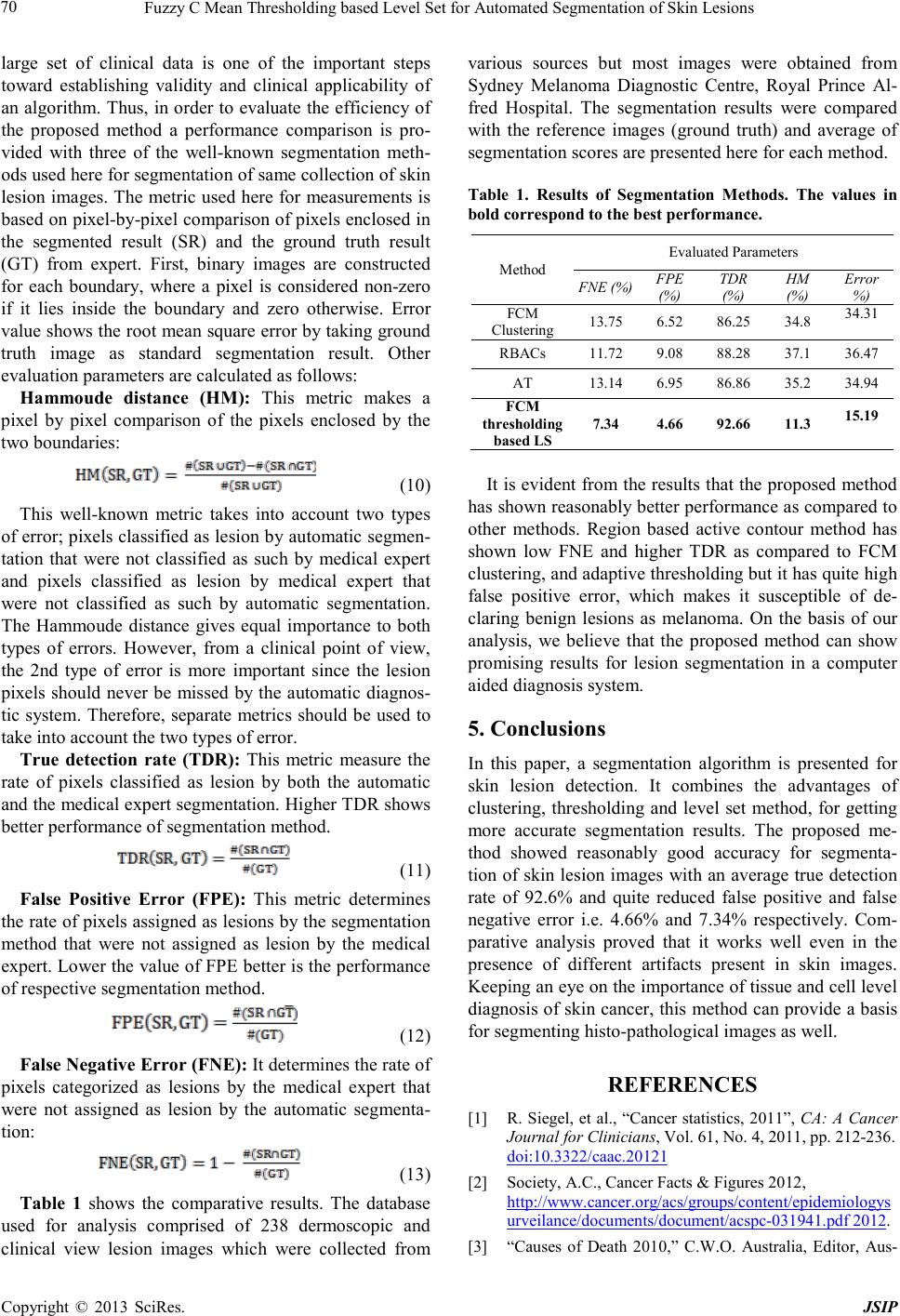 Fuzzy C Mean Thresholding based Level Set for Automated Segmentation of Skin Lesions Copyright © 2013 SciRes. JSIP 70 large set of clinical data is one of the important steps toward establishing validity and clinical applicability of an al gorith m. T hus, in or der to evaluate the ef ficienc y of the proposed method a performance comparison is pro- vided with three of the well-known segmentation meth- ods used here for segmentation of same collection of skin lesion images. The metric used here for measurements is based on pixel-by-pixel comparison of pixels enclosed in the segmented result (SR) and the ground truth result (GT) from expert. First, binary images are constructed for each boundary, where a pixel is considered non-zero if it lies inside the boundary and zero otherwise. Error value s hows t he ro ot mean sq uare e rror b y taking gr ound truth image as standard segmentation result. Other evaluation pa ra meters are calculated as follows: Hammoude distance (HM): This metric makes a pixel by pixel comparison of the pixels enclosed by the two boundaries: (10) This well-known metric takes into account two types of error; p ixels classified a s lesion by au tomatic seg men- tation that were not classified as such by medical expert and pixels classified as lesion by medical expert that were not classified as such by automatic segmentation. The Hammoude distance gives equal importance to both types of errors. However, from a clinical point of view, the 2nd type of error is more important since the lesion pixels should never be missed by the automatic diagnos- tic system. Therefore, separate metrics should be used to take into account the two types of error. True detection rate (TDR): This metric measure the rate of pixels classified as lesion by both the automatic and the medical expert segmentation. Higher TD R sho ws better performance of segmentation method. (11) False Positive Error (FPE): This metric determines the rate of pixels assigned as lesions by the segmentation method that were not assigned as lesion by the medical expert. Lower the value of FPE better is the performance of r espe c tive segmentatio n method. (12) False Negative Error (FNE): It determines the rate of pixels categorized as lesions by the medical expert that were not assigned as lesion by the automatic segmenta- tion: (13) Table 1 shows the comparative results. The database used for analysis comprised of 238 dermoscopic and clinical view lesion images which were collected from various sources but most images were obtained from Sydney Melanoma Diagnostic Centre, Royal Prince Al- fred Hospital. The segmentation results were compared with the reference images (ground truth) and average of segmentation scores are presented here for each method. Table 1. Results of Segmentation Methods. The values in bold correspond to t he b est performance. Method Evaluated Parameters FNE (%) FPE (%) TDR (%) HM (%) Error %) FCM Clustering 13.75 6.52 86.25 34.8 34.31 RBACs 11.72 9.08 88.28 37.1 36.47 AT 13.14 6.95 86.86 35.2 34.94 FCM thresholding based LS 7.34 4.66 92.66 11.3 15.19 It is evident from the results that the proposed method has shown reasonably better performance as compared to other methods. Region based active contour method has shown low FNE and higher TDR as compared to FCM cluste ri n g, and ada ptive thre sho lding but it has quite hig h false positive error, which makes it susceptible of de- claring benign lesions as melanoma. On the basis of our analysis, we believe that the proposed method can show promising results for lesion segmentation in a computer aided d ia gnosis system. 5. Conclusions In this paper, a segmentation algorithm is presented for skin lesion detection. It combines the advantages of clustering, thresholding and level set method, for getting more accurate segmentation results. The proposed me- thod showed reasonably good accuracy for segmenta- tion of skin lesion images with an average true detection rate of 92.6% and quite reduced false positive and false negative error i.e. 4.66% and 7.34% respectively. Com- parative analysis proved that it works well even in the presence of different artifacts present in skin images. Keeping an eye on the importance of tissue and cell level diagnosis of skin cancer, this method can provide a basis for segmenti ng histo -patho logica l images as well. REFERENCES [1] R. Siegel, et al., “Cancer statistics, 2011”, CA: A Cancer Journal for Clinicians, Vol. 61, No. 4, 2011, pp. 21 2-236. doi:10.3322/caac.20121 [2] Society, A.C., Cancer F acts & Figur es 2012, http://www.cancer.org/acs/groups/content/epidemiologys urveilance/documents/document/acspc-031941.pdf 2012. [3] “Causes of Death 2010,” C.W.O. Australia, Editor, Aus- 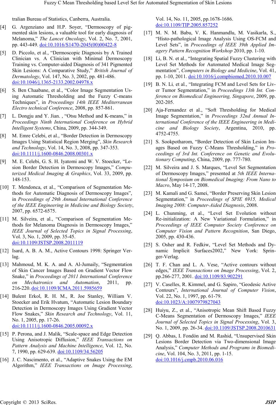 Fuzzy C Mean Thresholding based Level Set for Automated Segmentation of Skin Lesions Copyright © 2013 SciRes. JSIP 71 tralian Bureau of Statistics, Canberra, Austr alia. [4] G. Argenziano and H.P. Soyer, “Dermoscopy of pig- mented skin lesions, a valuable tool for early diagnosis of Melanoma,” The Lancet Oncology, Vol. 2, No. 7, 2001, pp. 443-449. doi:10.1016/S1470-2045(00)00422-8 [5] D. Piccolo, et al., “Dermoscopic Diagnosis by A Trained Clinician vs. A Clinician with Minimal Dermoscopy Trainin g vs. Computer-aided Diagnosis of 341 Pigmented Skin Lesions: A Comparative Study,” British Journal of Dermatology, Vol. 147, No. 3, 2002, pp. 481-486. doi:10.1046/j.1365-2133.2002. 04978.x [6] S. Ben Chaabane, et al., “Color Image Segmentation Us- ing Automatic Thresholding and the Fuzzy C-means Techniques”, in Proceedings 14th IEEE Mediterranean Electro techn ical Conference, 2008, pp. 857 -861. [7] L. Dongju and Y. Jian . , “Otsu Me thod and K-means,” in Proceedings Ninth International Conference on Hybrid Intelligent Systems, China , 2009, pp. 344-349. [8] M. Emre Celebi, et al., “Bo rder Detection in Dermoscopy Images Using Statistical Region Merging”, Skin Resea rch and Technology, V ol. 14, No. 3, 2008, pp. 347-353. doi:10.1111/j.1600-0846.2008.00301.x [9] M. E. Celebi, G. S. H. Iyatomi and W. V. Stoecker, “Le- sion Border Detection in Dermoscopy Images,” Co mp u- terized Medical Imaging & Graphics, Vol. 33, 2009, pp. 148-153. [10] T. Mendonca, et al., “Comparison of Segmentation Me- thods for Automatic Diagnosis of Dermoscopy Images”, in Proceedings of 29th Annual International Conference of the IEEE Engineering in Medicine and Biology Society, 2007, pp . 6572-6575. [11] M. Silveira, et al., “Comparison of Segmentation Me- thods for Melanoma Diagnosis in Dermoscopy Images,” IEEE Journal of Selected Topics in Signal Processing, Vol. 3, No. 1, 2009, pp. 35-45. doi:10.1109/JS TSP.2008.20111 19 [12] Isard, A. B. A. M., Active Contours 1998: Springer Ver- lag. [13] Mahmoud, M. K. A. and A. Al-Jumaily, “Segmentation of Skin Cancer Images Based on Gradient Vector Flow Snake,” in Proceedings of 2011 International Conference on Mechatronics and Automation, 2011, pp. 216-220. doi:10.1109/IC MA.2011.59 85659 [14] Bulent Erkol, R. H. M., R. Joe Stanley, William V. Stoecker and Erik Hvatum, “Automatic Lesion Boundary Detection in Dermoscopy Images Using Gradient Vector Flow Sn akes,” Skin Research and Technology, Vol. 11, No. 1, 2005, pp. 17-26. doi:10.1111/j.1600-0846.2005.00092.x [15] P. Perona, an d J. Mali k, “Scale-sp ace and Edge Detection Using Anisotropic Diffusion,” IEEE Transactions on Pattern Analysis and Machine Intelligence, Vol. 12, No. 7, 1990, pp. 629-639. doi:10.1109/34.56205 [16] J. C. Nascimento , et al., “Adaptive Snakes Using the EM Algorithm,” IEEE Transactions on Image Processing, Vol. 14, N o. 11, 2005, pp.1678-1686. doi:10.1109/TIP.2005.85 7252 [17] M. N. M. Babu, V. K. Hanmandlu, M. Vasikarla, S., “Histo-pathological Image Analysis Using OS-FCM and Level Sets”, in Proceedings of IEEE 39th Applied Im- agery Pattern Recognition Workshop 201 0, pp. 1-10. [18] Li, B . N. et al., “Integrating Spatial Fuzzy Clustering with Level Set Methods for Automated Medical Image Seg- mentati o n”, Computers in Biology and Medicine, Vol. 41, pp. 1-10, 2011. doi:10.1016/j.compbiomed.2010.10.007 [19] B. N. Li , et al., “Integrati ng FCM and Level Sets for Liv- er Tumor Segmentation,” in Proceedings 13th Int. Con- ference on Biomedical Engineering, Singapore, 2009 , pp. 202-205. [20] Aja-Fernandez et al., “Soft Thresholding for Medical Image Segmentation,” in Proceedings 32nd Annual In- ternational Conference of the IEEE Engineering in Medi- cine and Biology Society, Argentina, 2010, pp. 4752-4755. [21] S. Sookpotharom, “Border Detection of Skin Lesion Im- ages Based on Fuzzy C-Means Thresholding,” in Pro- ceedings of 3rd Int. Conference on Genetic and Evolu- tionary Computing, Chi na , 2009, pp. 777-780. [22] M. Silveira and J. S. Marques, “Level Set Segmentation of Dermoscopy Images,” presented at 5th IEEE Interna- tional Symposium on Biomedical Imaging: From Nano to Macro, May 14-1 7, 2008. [23] M. Kamali and G. Samei, “Border Preserving Skin Lesion Segmentation,” in Proceedings of SPIE 6915, Medical Imagi ng 2008: Comp ut e r -A ided D iagnosis , 20 08. [24] L. Chunming, et al., “Level Set Evolution without Re-initialization: A New Variational Formulation,” in Proceedings of IEEE Computer Society Conference on Computer Vision and Pattern Recognition, San Diego, 2005, pp . 430-436. [25] S. Osher and R. Fedkiw, “Level Set Methods and Dy- namic Implicit Surfaces2002,” New York: Sprin- ger-Ver l ag. [26] T. F. Chan and L. A. Vese, “Active contours without edges,” IEEE Transactions on Image Processing, Vol. 2, pp.266-277, 2001. doi:10.1109/83.902291 [27] V. Caselles, R. Kimmel, and G. Sapiro , “Geodesi c Active Contours”, International Journal of Computer Vision, Vol. 22, No. 1, 1997, pp. 61-79. doi:10.1023/A:1007979827043 [28] Huiyu, Z., et al., “Anisotropic Mean Shift Based Fuzzy C-Means Segmentation of Dermoscopy Images,” IEEE Journal of Selected Topics in Signal Processing, Vol. 3, No. 1, 2009 , pp. 26-34. doi:10.1109/JSTSP.2008.2010631 [29] Q. Abbas, I. Fondón and M. Rashid, “Unsupervised Skin Lesions Border Detection via Two-dimensional Image Analysis,” Computer Methods and Programs in Biomedi- cine, Vol. 104, No. 3, 2011, pp. 1-15. doi:10.1016/j.cmpb.2010.06.016 |