

Paper Menu >>
Journal Menu >>
 Advances in Bioscience and Biotechnology, 2013, 4, 941-944 ABB http://dx.doi.org/10.4236/abb.2013.410124 Published Online October 2013 (http://www.scirp.org/journal/abb/) Characterization of purified β-glucosidase produced from Trichoderma viride through bio-processing of orange peel waste Muhammad Irshad1*, Zahid Anwar1, Muhammad Ramzan2, Zahed Mahmood2, Haq Nawaz3 1Department of Biochemistry, NSMC, University of Gujrat, Gujrat, Pakistan 2Department of Chemistry and Biochemistry, University of Agriculture, Faisalabad, Pakistan 3Institute of Animal Nutrition & Feed Technology, University of Agriculture, Faisalabad, Pakistan Email: *[email protected] Received 22 June 2013; revised 23 July 2013; accepted 12 August 2013 Copyright © 2013 Muhammad Irshad et al. This is an open access article distributed under the Creative Commons Attribution Li- cense, which permits unrestricted use, distribution, and reproduction in any medium, provided the original work is properly cited. ABSTRACT In the present study, solid state fermentation was car- ried out using orange peel waste to produce β-gluco- sidase from Trichoderma viride. A locally isolated fungal strain T. viride was cultured in the solid state medium of orange peel (50% w/w moisture) under optimized fermentation conditions and maximum act- ivity of 515 ± 12.4 U/mL was recorded after 4th day of incubation at pH 5.5 and 30˚C. Indigenously pro- duced β-glucosidase was subjected to the ammonium sulfate precipitation and Sephadex-G-100 gel filtra- tion chromatography. In comparison to the crude ex- tract β-glucosidase was 5.1-fold purified with specific activity of 758 U/mg. The enzyme was shown to have a relative molecular weight of 62 kDa as evidenced by sodium dodecyl sulphate polyacrylamide gel electro- phoresis. The purified β-glucosidase displayed 6 and 60˚C as an optimum pH and temperature respect- ively. Keywords: Orange Peel Waste; β-Glucosidase; T. viride; Purification; SDS-PAGE; Characterization 1. INTRODUCTION The major components of plant cell walls are cellulose, hemicellulose and lignin and among all of them, cellu- lose is about 35% to 50% which is the most common and most abundant component of all plant matter [1]. Among many of the developing countries, it’s a routine practice that such agricultural wastes have not been fully dis- carded, which has become a major source of pollution. A large variety of micro-organisms including Trichoderma, Aspergillus, Penicillium, and Fusarium have the ability to produce enzymes like cellulases, and under mild fer- mentation environment to hydrolyze insoluble polysac- charides to soluble sugars [1,2]. Trichoderma is one of the most efficient cellulases producer organisms which is being studied for the production of cellulose degrading enzymes. Cellulose degrading enzymes system is a com- plex of three major enzymes that can be divided into three main types: 1) Endoglucanase, 2) Exoglucanases, and 3) β-glucosidase [1,3-5]. From the last few years, cellulase is being used in many of the industrial applications, especially in the field of cotton processing; paper recycling, and animal feed additives animal feed industry, agriculture as well as in the field of research and development [4,5]. One of the potential applications of cellulase is the production of fuel ethanol from lignocellulosic biomass. The most pro- mising technology for the conversion of the lignocellu- losic biomass to fuel ethanol is based on the enzymatic breakdown of cellulose using cellulase enzymes [6,7]. Pakistan is an agricultural land that produced abundant magnitude of agricultural wastes that can be utilized for the production of useful industrial enzymes. Enzymatic hydrolysis of such wastes is one of the attractive solu- tions of this problematic issue that also provides an en- vironmentally friendly means of depolymerizing cellu- lose and other carbohydrates at high yields. With respect to the factors affecting culture conditions, productivity and properties of enzymes (cellulase complex), it was considered of significance to purify and characterize this enzyme through kinetic studies to explore such factors. Keeping in mind the broad range of industrial applica- tions of cellulases, this study was performed to purify and characterize the β-glucosidase from T. viridi to pre- sent its potential application for industrial application. *Corresponding author. OPEN ACCESS  M. Irshad et al. / Advances in Bioscience and Biotechnology 4 (2013) 941-944 942 The present study was also focused on providing a po- tential solution for the management of large magnitude of solid wastes. 2. MATERIALS AND METHODS 2.1. Agro-Industrial Substrate Agro-industrial waste orange peel was obtained from local fruit market, Gujrat, Pakistan. The collected sub- strate crushed into pieces, oven dried and finally ground to fine particle size before to use. 2.2. Fungal Culture and Inoculum Development The pure culture of T. viride was obtained from the De- partment of Biochemistry University of Gujrat, Pakistan. A homogeneous inoculum of T. viride was developed in an Erlenmeyer flask containing 30 mL of Potato Dex- trose broth at 30˚C ± 1˚C for 5 days after sterilizing the potato dextrose broth at 15 lbs/inch2 pressure and 121˚C for 15 min, and incubated under stationary conditions for the development of fungal spore suspension. 2.3. Pretreatment of Agro-Industrial Waste 10 g of moisture free orange peel was pretreated with 2% HCl by adopting thermal treatment methodology as de- scribed earlier [1]. After pretreatment the slurry of sub- strate was filtered through four layers of muslin cloth, residue were washed 4 to 5 times with distilled water to remove extra acidity and used for production of β-glu- cosidase under optimum fermentation conditions. 2.4. Solid-State Fermentation Strategy Basel salt media was used to moist the pretreated orange peel in an Erlenmeyer flask for β-glucosidase production. The initial pH value of the medium was adjusted to 5 before sterilization at 121˚C and 15.0 lbs/inch2 pressure for 15 min. The autoclaved medium was inoculated with 5 mL of freshly prepared fungal inoculum and incubated at 30˚C ± 1˚C for stipulated fermentation time period. 2.5. Extraction of β-Glucosidase β-glucosidase was extracted from the fermented biomass by adding citrate buffer 0.05 M of pH 4.8 in 1:10 ratio and the flasks were shaken at 120 rpm for 30 min. The contents were filtered through muslin cloth and filtrates were centrifuged at 10,000 g for 10 min. After that su- pernatants were carefully collected and used to determine enzyme activity and for purification purposes. 2.6. Determination of Activity & Protein Contents β-glucosidase activity was determined by the method of Gielkens [8] while, the protein contents of the crude and purified enzyme extracts were determined by following the method of Bradford [9], with Bovine serum albumin as standard. 2.7. Purification and SDS-PAGE of β-Glucosidase Crude extract of β-glucosidase obtained from T. viridi was centrifuged (10,000 g) for 15 min followed by the ammonium sulfate fractionation as described by Iqbal et al. [1]. Total proteins and activity of partially purified β- glucosidase were determined before and after dialysis of ammonium sulfate precipitation. β-glucosidase was ly- ophilized and subjected to gel filtration chromatography using Sephadex-G-100 column [10]. The flow rate was maintained at 0.5 mL·min−1 and up to 20 active fractions were collected. To determine the molecular weight of purified β-glucosidase SDS-PAGE was performed on a 5% stacking and a 12% resolving gel according to the method of Laemmli [11]. 2.8. Characterization of Purified β-Glucosidase Characterization of purified β-glucosidase was performed to investigate the effect of pH and temperature. β-gluco- sidase was incubated in buffers of different pH (2 - 10), followed by standard assay protocol. To determine the thermal features β-glucosidase was incubated under dif- ferent temperatures ranging from 25˚C to 70˚C for 1 h time period followed by normal assay protocol as previ- ously described. 3. RESULTS AND DISCUSSION 3.1. Production and Purification of β-Glucosidase A locally isolated fungal strain T. viride was cultured under optimized fermentation conditions in the solid state medium of orange peel (50% w/w moisture) and maximum activity of 515 ± 12.4 U/mL was recorded after 4th day of incubation at pH 5.5 and 30˚C. T. viridi showed high levels of β-glucosidase production under SSF as well as growth rate of cells. The eco-friendly pro- cedure has been adopted to utilize low cost substrates to induce enzymes production by T. viridi. The separated cell free supernatant as crude enzyme solution containing β-glucosidase with activity of 103,000 U/200 mL and specific activity of 149 U/mg was subjected to partial purification by ammonium sulfate precipitation. The cru- de enzyme was maximally precipitated at 85% satura- tion with specific activity of 208 U/mg and 1.4 fold puri- fication. The optimally active fraction was loaded on Sephadex G-100 column, and after gel filtration the en- zyme was purified up to 5.1 fold with specific activity of Copyright © 2013 SciRes. OPEN ACCESS 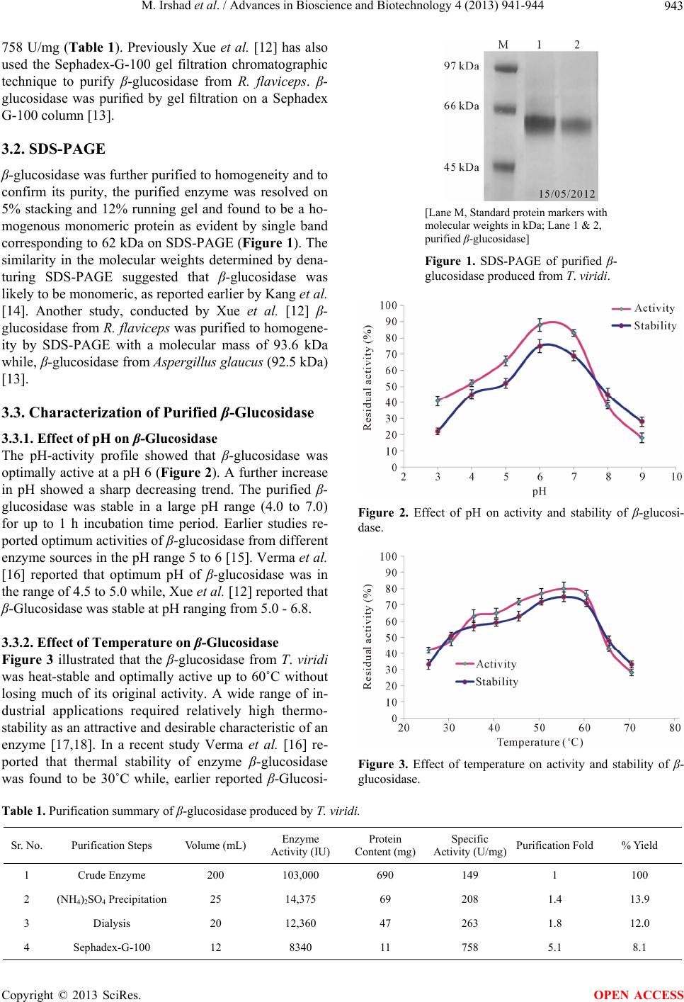 M. Irshad et al. / Advances in Bioscience and Biotechnology 4 (2013) 941-944 Copyright © 2013 SciRes. 943 758 U/mg (Table 1). Previously Xue et al. [12] has also used the Sephadex-G-100 gel filtration chromatographic technique to purify β-glucosidase from R. flaviceps. β- glucosidase was purified by gel filtration on a Sephadex G-100 column [13]. 3.2. SDS-PAGE β-glucosidase was further purified to homogeneity and to confirm its purity, the purified enzyme was resolved on 5% stacking and 12% running gel and found to be a ho- mogenous monomeric protein as evident by single band corresponding to 62 kDa on SDS-PAGE (Figure 1). The similarity in the molecular weights determined by dena- turing SDS-PAGE suggested that β-glucosidase was likely to be monomeric, as reported earlier by Kang et al. [14]. Another study, conducted by Xue et al. [12] β- glucosidase from R. flaviceps was purified to homogene- ity by SDS-PAGE with a molecular mass of 93.6 kDa while, β-glucosidase from Aspergillus glaucus (92.5 kDa) [13]. [Lane M, Standard protein markers with molecular weights in kDa; Lane 1 & 2, purified β-glucosidase] Figure 1. SDS-PAGE of purified β- glucosidase produced from T. viridi. 3.3. Characterization of Purified β-Glucosidase 3.3.1. Effect of pH on β-Glucosidase The pH-activity profile showed that β-glucosidase was optimally active at a pH 6 (Figure 2). A further increase in pH showed a sharp decreasing trend. The purified β- glucosidase was stable in a large pH range (4.0 to 7.0) for up to 1 h incubation time period. Earlier studies re- ported optimum activities of β-glucosidase from different enzyme sources in the pH range 5 to 6 [15]. Verma et al. [16] reported that optimum pH of β-glucosidase was in the range of 4.5 to 5.0 while, Xue et al. [12] reported that β-Glucosidase was stable at pH ranging from 5.0 - 6.8. Figure 2. Effect of pH on activity and stability of β-glucosi- dase. 3.3.2. Effect of Temperature on β-Glucosidase Figure 3 illustrated that the β-glucosidase from T. viridi was heat-stable and optimally active up to 60˚C without losing much of its original activity. A wide range of in- dustrial applications required relatively high thermo- stability as an attractive and desirable characteristic of an enzyme [17,18]. In a recent study Verma et al. [16] re- ported that thermal stability of enzyme β-glucosidase was found to be 30˚C while, earlier reported β-Glucosi- Figure 3. Effect of temperature on activity and stability of β- glucosidase. Table 1. Purification summary of β-glucosidase produced by T. viridi. Sr. No. Purification Steps Volume (mL) Enzyme Activity (IU) Protein Content (mg) Specific Activity (U/mg)Purification Fold % Yield 1 Crude Enzyme 200 103,000 690 149 1 100 2 (NH4)2SO4 Precipitation 25 14,375 69 208 1.4 13.9 3 Dialysis 20 12,360 47 263 1.8 12.0 4 Sephadex-G-100 12 8340 11 758 5.1 8.1 OPEN ACCESS  M. Irshad et al. / Advances in Bioscience and Biotechnology 4 (2013) 941-944 944 dase was stable above 30˚C and below 45˚C. In compari- son the earlier reported the present β-glucosidase from T. viridi was reasonably more stable and active for up to one hour incubation at 60˚C that suggests its potential for industrial applicability. 4. CONCLUSIONS 1) Bio-utilization and conversion of agro are based on waste materials into useful products. 2) T. viridi produces high titers of β-glucosidase dur- ing solid state bio-processing of an agro-industrial or- ange peel waste material. 3) An extra thermo-stability feature of an indigenous T. viride β-glucosidase suggests its potential for industrial applicability and striking prospect for application of this enzyme. 5. ACKNOWLEDGEMENTS The authors are grateful to the Department of Biochemistry and Mo- lecular Biology, University of Gujrat, Pakistan for providing financial support and laboratory facilities. REFERENCES [1] Iqbal, H.M.N., Ahmed, I., Zia, M.A. and Irfan, M. (2011) Purification and characterization of the kinetic parameters of cellulase produced from wheat straw by Trichoderma viride under SSF and its detergent compatibility. Advan- ces in Bioscience and Biotechnology, 2, 149-156. [2] Ilyas, U., Ahmed, S., Majeed, A. and Nadeem, M. (2012) Biohydrolysis of Saccharum spontaneum for cellulase production by Aspergillus terreus. African Journal of Biotechnology, 11, 4914-4920. [3] Yin, L.J., Lin, H.H. and Xiao, Z.R. (2010) Purification and characterization of a cellulase from Bacillus subtilis YJ1. Journal of Marine Science and Technology, 18, 466- 471. [4] Iqbal, H.M.N., Kamal, S., Ahmed, I. and Naveed, M.T. (2012) Enhanced bio-catalytic and tolerance properties of an indigenous cellulase through xerogel immobilization. Advances in Bioscience and Biotechnology, 3, 308-313. http://dx.doi.org/10.4236/abb.2012.34044 [5] Yano, S., Ozaki, H., Matsuo, S., Ito, M., Wakayama, M. and Takagi, K. (2012) Production, purification and char- acterization of D-aspartate oxidase from the fungus Tri- choderma harzianum SKW-36. Advances in Bioscience and Biotechnology, 3, 7-13. http://dx.doi.org/10.4236/abb.2012.31002 [6] Iqbal, H.M.N., Asgher, M., Ahmed, I. and Hussain, S. (2010) Media optimization for hyper-production of car- boxymethyl cellulase using proximally analyzed agro- industrial residue with Trichoderma harzianum under SSF. International Journal for Agro Veterinary and Med- ical Sciences, 4, 47-55. [7] Iqbal, H.M.N., Kyazze, G. and Keshavarz, T. (2013) Advances in the valorization of lignocellulosic materials by biotechnology: An overview. BioResources, 8, 3157- 3176. [8] Gielkens, M.M.C., Dekkers, E., Visser, J. and De-Graaff, L.H. (1999) Two cellobiohydrolase-encoding genes from Aspergillus niger require D-xylose and the xylanolytic transcriptional activator XlnR for their expression. Ap- plied and Environmental Microbiology, 65, 4340-4545. [9] Bradford, M.M. (1976) A rapid and sensitive method for the quantitation of microgram quantities of protein utiliz- ing the principle of protein-dye binding. Analytical Bio- chemistry, 72, 248-254. http://dx.doi.org/10.1016/0003-2697(76)90527-3 [10] Ahmed, I., Zia, M.A., Iftikhar, T. and Iqbal, H.M.N. (2011) Characterization and detergent compatibility of purified protease produced from Aspergillus niger by utilizing agro wastes. BioResources, 6, 4505-4522. [11] Laemmli, U.K. (1970) Cleavage of structural proteins during the assembly of the head of bacteriophage T4. Nature, 227, 680-685. http://dx.doi.org/10.1038/227680a0 [12] Xue, Y.P., Jin, L.Q., Liu, Z.Q., Zhang, J.F. and Zheng, Y.G. (2008) Purification and characterization of betaglu- cosidase from Reticulitermes flaviceps and its inhibition by valienamine and validamine. African Journal of Bio- technology, 7, 4595-601. [13] Ma, S.J., Leng, B., Xu, X.Q., Zhu, X.Z., Shi, Y. and Tao, Y.M. (2011) Purification and characterization of b-1,4- glucosidase from Aspergillus glaucus. African Journal of Biotechnology, 10, 19607-19614. [14] Kang, S.K., Cho, K.K., Ahn, J.K., Bok, J.D., Kang, S.H. and Woo, J.H. (2005) Three forms of thermo-stable lac- tose-hydrolase from Thermus sp. IB-21: Cloning, expres- sion, and enzyme characterization. Journal of Biotech- nology, 116, 337-346. http://dx.doi.org/10.1016/j.jbiotec.2004.07.019 [15] Dharmawardhana, D.P., Ellis, B.E. and Carlson, J.E. (1999) cDNA cloning and heterologous expression of co- niferin β-glucosidase. Plant Molecular Biology, 40, 365- 372. http://dx.doi.org/10.1023/A:1006226931512 [16] Verma, O.P. (2011) Isolation, purification and charac- terization of ß-glucosidase from Rauvolfia serpentine. Journal of Chemical Engineering & Process Technology, 2, 119. http://dx.doi.org/10.4172/2157-7048.1000119 [17] Asgher, M. and Iqbal, H.M.N. (2011) Characterization of a novel manganese peroxidase purified from solid state culture of Trametes versicolor IBL-04. BioResources, 6, 4302-4315. [18] Iqbal, H.M.N., Asgher, M. and Bhatti, H.N. (2011b) Op- timization of physical and nutritional factors for synthesis of lignin degrading enzymes by a novel strain of Tram- etes versicolor. BioResources, 6, 1273-1287. Copyright © 2013 SciRes. OPEN ACCESS |