

Paper Menu >>
Journal Menu >>
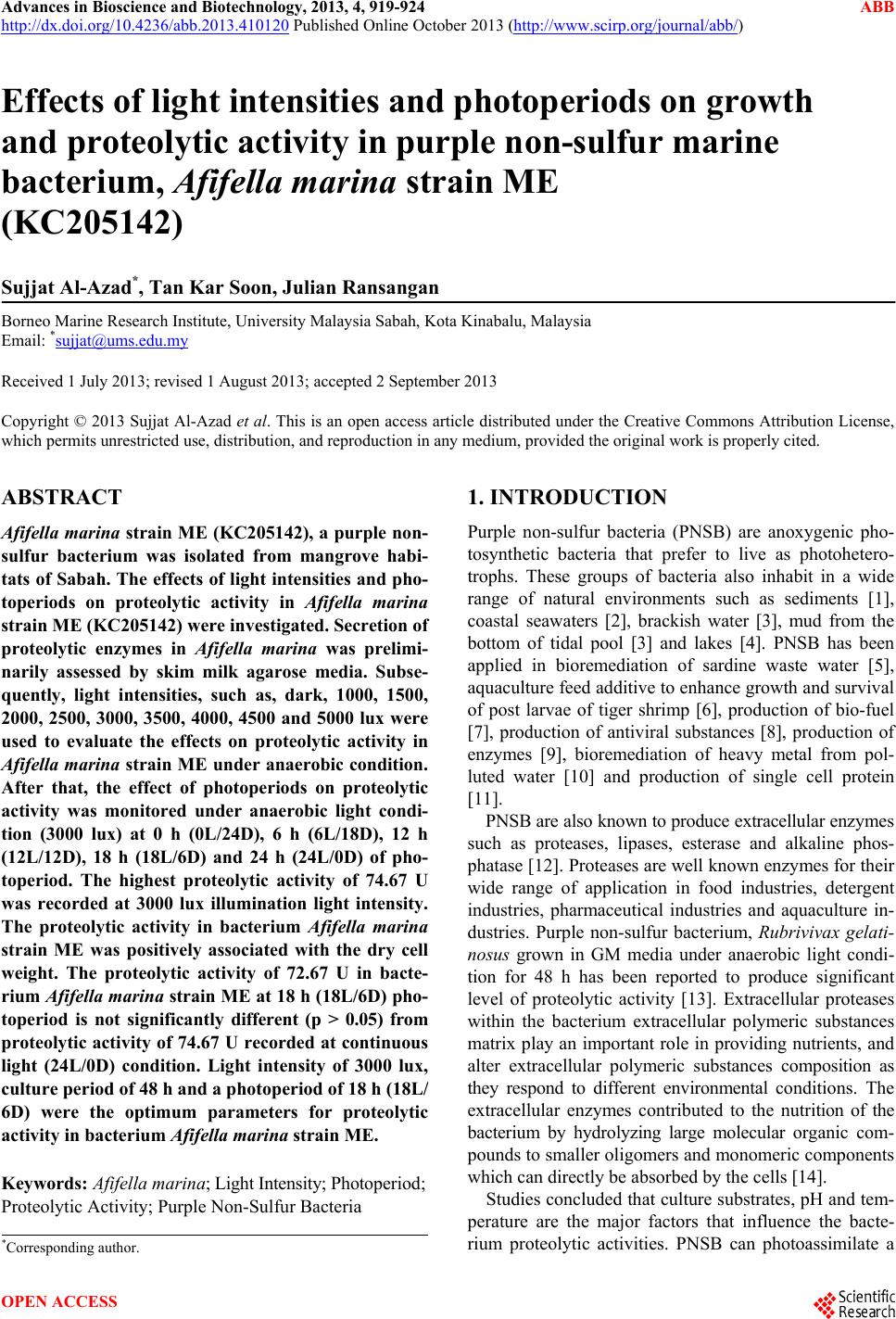 Advances in Bioscience and Biotechnology, 2013, 4, 919-924 ABB http://dx.doi.org/10.4236/abb.2013.410120 Published Online October 2013 (http://www.scirp.org/journal/abb/) Effects of light intensities and photoperiods on growth and proteolytic activity in purple non-sulfur marine bacterium, Afifella marina strain ME (KC205142) Sujjat Al-Azad*, Tan Kar Soon, Julian Ransangan Borneo Marine Research Institute, University Malaysia Sabah, Kota Kinabalu, Malaysia Email: *[email protected] Received 1 July 2013; revised 1 August 2013; accepted 2 September 2013 Copyright © 2013 Sujjat Al-Azad et al. This is an open access article distributed under the Creative Commons Attribution License, which permits unrestricted use, distribution, and reproduction in any medium, provided the original work is properly cited. ABSTRACT Afifella marina st rain ME (KC205142), a purple non- sulfur bacterium was isolated from mangrove habi- tats of Sabah. The effects of light intensities and pho- toperiods on proteolytic activity in Afifella marina strain ME (KC205142) were investigated. Secretion of proteolytic enzymes in Afifella marina was prelimi- narily assessed by skim milk agarose media. Subse- quently, light intensities, such as, dark, 1000, 1500, 2000, 2500, 3000, 3500, 4000, 4500 and 5000 lux were used to evaluate the effects on proteolytic activity in Afifella marina strain ME under anaerobic condition. After that, the effect of photoperiods on proteolytic activity was monitored under anaerobic light condi- tion (3000 lux) at 0 h (0L/24D), 6 h (6L/18D), 12 h (12L/12D), 18 h (18L/6D) and 24 h (24L/0D) of pho- toperiod. The highest proteolytic activity of 74.67 U was recorded at 3000 lux illumination light intensity. The proteolytic activity in bacterium Afifella marina strain ME was positively associated with the dry cell weight. The proteolytic activity of 72.67 U in bacte- rium Afifella marina strain ME at 18 h (18L/6D) pho- toperiod is not significantly different (p > 0.05) from proteolytic activity of 74.67 U recorded at continuous light (24L/0D) condition. Light intensity of 3000 lux, culture period of 48 h and a photoperiod of 18 h (18L/ 6D) were the optimum parameters for proteolytic activity in bacterium Afifella marina strain ME. Keywords: Afifella marina; Light Intensity; Photoperiod; Proteolytic Activity; Purple Non-Sulfur Bacteria 1. INTRODUCTION Purple non-sulfur bacteria (PNSB) are anoxygenic pho- tosynthetic bacteria that prefer to live as photohetero- trophs. These groups of bacteria also inhabit in a wide range of natural environments such as sediments [1], coastal seawaters [2], brackish water [3], mud from the bottom of tidal pool [3] and lakes [4]. PNSB has been applied in bioremediation of sardine waste water [5], aquaculture feed additive to enhance growth and survival of post larvae of tiger shrimp [6], production of bio-fuel [7], production of antiviral substances [8], production of enzymes [9], bioremediation of heavy metal from pol- luted water [10] and production of single cell protein [11]. PNSB are also known to produce extracellular enzymes such as proteases, lipases, esterase and alkaline phos- phatase [12]. Proteases are well known enzymes for their wide range of application in food industries, detergent industries, pharmaceutical industries and aquaculture in- dustries. Purple non-sulfur bacterium, Rubrivivax gelati- nosus grown in GM media under anaerobic light condi- tion for 48 h has been reported to produce significant level of proteolytic activity [13]. Extracellular proteases within the bacterium extracellular polymeric substances matrix play an important role in providing nutrients, and alter extracellular polymeric substances composition as they respond to different environmental conditions. The extracellular enzymes contributed to the nutrition of the bacterium by hydrolyzing large molecular organic com- pounds to smaller oligomers and monomeric components which can directly be absorbed by the cells [14]. Studies concluded that culture substrates, pH and tem- perature are the major factors that influence the bacte- rium proteolytic activities. PNSB can photoassimilate a *Corresponding author. OPEN ACCESS  S. Al-Azad et al. / Advances in Bioscience and Biotechnology 4 (2013) 919-924 920 wide variety of organic compounds such as acetate, py- ruvate, dicarboxylic acids, fatty acids and sugar [15]. However, high level of complex organic polymers in- duces the proteolytic activities, while simple compounds inhibit the bacterium proteolytic activities [16]. Low temperature always results in slow biological activity while high temperature around 40˚C has been reported to inhibit the growth of PNSB [17]. A temperature of 30˚C was reported to be the optimum temperature for prote- lytic activities in bacteria such as Bacillus sp. [18]. The maximal protease production was reported in basal me- dium, where pH 8 was documented to be the optimum pH value for proteolytic activity in Bacillus sp. [19]. Light intensity is an important factor that controls the growth of PNSB. The optimum light intensity for the growth of PNSB is in the range of 2500 lux to 3000 lux. The highest dry cell weight of 5.6 g/l of Rhodocyclus gelatinosus is obtained when cultured in glutamate- malate (GMM) media at 3000 lux of illumination light intensity [17]. Azad et al. [6] recorded the highest dry cell weight of 4.8 g/l in Rhodovulum sulfidopilum that was grown in GMM media under anaerobic condition at 2500 lux light intensity and a temperature of 30˚C ± 2˚C. Protease production in Rubrivivax gelatinosus was re- ported to be light inducible, therefore, it is likely that the light intensity also influences the proteolytic activity in PNSB. However, limited information is known about the effects of light intensities and photoperiods on the prote- olytic activities in PNSB. Therefore, the objectives of this study were to assess the effects of light intensities and photoperiods on the proteolytic activities in Afifella marina strain ME. 2. MATERIALS AND METHODS 2.1. Bacterium and Culture Afifella marina, strain ME (KC205142) was obtained from the culture collection of Borneo Marine Research Institute, University Malaysia Sabah, Malaysia. This strain was isolated from exposed mangrove mud sample of KingFisher, Sabah, Malaysia. A single colony of Afifella marina strain ME was inoculated in Mc. Corney bottle containing 25 ml of 112 media and incubated an- aerobically under 2500 lux illumination light intensity at a temperature of 30˚C ± 2˚C for 7 days. The composition of 112 media (g/l) was: 10.0 g of yeast-extract, 0.5 g of magnesium sulphate, 30 g of sodium chloride and 1.0 g of dipotassium hydrogen phosphate. 2.2. Inoculum Preparation One milliliter of the best grown culture was taken from previously culture bottle and transferred into a new Mc. Corney bottle containing 24 ml of freshly prepared ster- ile 112 media. The bottles were then incubated the same way as stated previously. A 48 h culture was used as the inoculum in subsequent experiments. 2.3. Preliminary Test for Proteolytic Activity in Afifella marina Strain ME Afifella marina strain ME was tested for the production of proteolytic enzyme by incubating the cells on solidi- fied 112 media supplemented with 10% skim milk. A 48 h culture plate was opened and observed the present of clear zone around the bacteria colony. 2.4. Proteolytic Activity Characterization A 48 h inoculum with 10% (v/v) was inoculated into triplicate bottles containing sterilised 112 media and in- cubated anaerobically at a temperature of 30˚C ± 2˚C under 2500 lux of light intensity for 120 h. Three bottles from each level of light intensity were selected randomly after 0 h, 24 h, 48 h, 96 h, and 120 hours of incubation to analyse the dry cell weight (g/l) and proteolytic activity (U). After 0 h, 24 h, 48 h, 72 h, 96 h and 120 h, three bottles of culture were taken randomly for analysis of the dry cell weight (g/l) and proteolytic activity (U). 2.5. Proteolytic Activity in Afifella marina Strain ME with Levels of Light Intensities Proteolytic activities in bacterium Afifella marina strain ME was monitored with various light intensities. Tripli- cate bottles of 112 media were inoculated with 10% (v/v) 48 h inoculum and incubated anaerobically at 30˚C ± 2˚C under dark, 1000, 1500, 2000, 2500 (control), 3000, 3500, 4000, 4500 and 5000 lux illumination of light in- tensities. 2.6. Proteolytic Activity in Afifella marina Strain ME with Levels of Photoperiod Triplicate bottles of 112 media were inoculated with 10% (v/v) 48 h of inoculum and incubated anaerobically at 30˚C ± 2˚C with 3000 lux light intensity under photo- periods of 0 h light (0L/24D), 6 h light (6L/18D), 12 h light (12L/12D), 18 h light (18L/6D) and 24 h light (24L/0D). The light intensity of 3000 lux was selected base on previous experiment, which showed the highest proteolytic activity in Afifella ma rina strain ME. The dry cell weight (g/l) and proteolytic activity (U) of Afifella marina strain ME after 0 h, 24 h, 48 h, 96 h, and 120 h of incubation were analysed. 2.7. Analytical Parameters Dry Cell Weight The bacterium cells were collected by centrifuging 10 ml of culture at 4400 rpm for 20 minutes at 4˚C. The proc- ess was repeated 3 times, after washing and re-suspended Copyright © 2013 SciRes. OPEN ACCESS  S. Al-Azad et al. / Advances in Bioscience and Biotechnology 4 (2013) 919-924 921 the cells in distilled water. Subsequently, the dry cell weight is obtained by drying the cell pallet in oven at a temperature of 105˚C until constant weight achieved. The dry cell weight was calculated as the formula given by Sawada et al. [20]: Drycell weightg/l Final weightInitial weight1000 v where: Initial weight = plastic centrifuge tube weight (g) Final weight = plastic centrifuge tube weight (g) + dry cell weight (g) v = Volume of sample (ml) 2.8. Proteolytic Activity Proteolytic activity in bacterium Afifella marina strain ME was assayed using azocasein as substrates according to method described by Winkler and Stuckman [21]. In this assay, digestion of azoproteins with proteolytic en- zymes at 38˚C results in the formation of coloured com- ponents soluble in trichloroacetic acid. This coloured compound has maximum absorption at a wavelength of 440 nm. The proteolytic activity is expressed as enzy- matic unit (U), where one unit of proteolytic activity is defined as the amount of enzyme that produces an in- crease in absorbance of 0.01 under assay conditions. The absorbance was measured using 100 VIS spectropho- tometer (Busk Scientific, USA). 2.9. Statistical Analysis Statistical analyses were performed using the SPSS Windows Statistical Package (version 19.0). Tests were judged with significant at p < 0.05 level. All variables were tested for normality and homogeneity of variances. Data which satisfy the assumptions of normality and homogeneity, parametric tests (one-way ANOVA, Inde- pendent t-test, and Pearson correlation test) were per- formed. 3. RESULTS AND DISCUSSION 3.1. Proteolytic Enzymes Production in Afifella marina Strain ME A clear zone was observed when Afifella marina strain ME was grown on skim milk plate under anaerobic light condition for 48 h (Figure 1). Similar observations were reported in other studies where purple non-sulfur bacteria such as Rhodopseudomonas palustris grown in Malate basal medium at 28˚C [22] and Rubrivivax gelatinosus KDDS1 grown in Glutamate-malate (GM) medium un- der anaerobic light conditions at light intensity of 3000 lux and a temperature of 35˚C for 48 h [13] exhibited proteolytic activity. The result of this study shows that Afifella marina strain ME do secrete of extracellular pro- tease under anaerobic light conditions. 3.2. Growth and Proteolytic Activity Profile of Afifella marina Strain ME Afifella marina strain ME incubated under anaerobic light condition at light intensity of 2500 lux and at a tem- perature of 30˚C had shown a sigmoid growth curve. The growth began with a negligible lag phase, followed by an exponential phase until 72 h of incubation then entered a deceleration phase until 120 h of incubation (Figure 2). The highest dry cell weight of 4.34 g/l was achieved at 72 h culture. The optimum culturing period was similar to other studies. Rhodomicrobium vannielii grown in GM media produced the highest dry cell weight of 1.1 g/l at 72 h culture under anaerobic light condition (2000 lux) [23]. Rhodovulum sulfidophilum grown in sardine proc- essing effluence recorded highest dry cell weight of 2.8 g/l at 72 h culture under anaerobic light condition (2500 Bacterial colony Clear zone Skim milk with agar Figure 1. Zone clearance by Afifella marina strain ME grown on skim milk plate under anaerobic light condition (2500 lux) and at a temperature of 30˚C ± 2˚C for 48 h. Figure 2. Dry cell weight (g/l) and proteolytic activities (U) of Afifella marina strain ME in 112 media incubated under an- aerobic light condition (2.5 k lux illumination, control) for the period of 120 hours at a temperature of 30˚C ± 2˚C. Copyright © 2013 SciRes. OPEN ACCESS  S. Al-Azad et al. / Advances in Bioscience and Biotechnology 4 (2013) 919-924 922 lux) [5]. The decrease in dry cell weight after 72 h was believed to be due to the cells had entered stationary phase or were dead [23]. This suggested the optimum dry cell weight production of Afifella marina strain ME is at 72 h culture. Interestingly, the proteolytic activity corresponding with growth where the proteolytic activity of Afifella marina strain ME was increased drastically to 54.67 U within 48 h of incubation. Subsequently, the level prote- olytic activity of Afifella marina remains constant at 54.7 U until 120 h of incubation. The maximum proteolytic activity of 54.7 U was recorded at 48 h of incubation in this study. The optimum incubation time of 48 h re- corded in Afifella marina strain ME was in agreement with Shivanand and Jayaraman [24], where 48 h incuba- tion was the optimum incubation time for Bacillus aqui- maris strain VITP4 too. Bacteria were reported to syn- thesis extracellular enzymes actively within 24 h of in- cubation. However, the quantitative enzymes activities might increase until 72 h of incubation [18]. This study shows that the proteolytic activities of Afifella marina strain ME optimum at 48 h culture. The maximum proteolytic activity of 57.4 U at 48 h culture in Afifella marina strain ME was doubled the proteolytic activities (25 U) of Rubrivivax gelatinosus that was grown in GM media under anaerobic light con- dition (3000 lux) at a temperature of 35˚C [13]. This suggests that Afifella marina strain ME is a good prote- olytic enzymes producer among PNSB. Compared to other microbial proteolytic producers, the proteolytic activity in Afifella marina strain ME, the purple non- sulfur bacteria was much lower than that the proteolytic activity (350 to 410 U) in Bacillus sp. [25]. Maximum proteolytic activity of Bacillus aq uimaris strain VITP4 of 630 U/ml was recorded when cultured in zobell broth at 37˚C for 48 h of incubation [24]. It is not surprising be- cause Bacillus spp. is a well known proteolytic enzymes producer. It is reasonable to believe that higher prote- olytic activity in Bacillus aquimaris strain VITP4 than Afifella marina strain ME is due to Bacillus spp. are naturally biosynthesis higher level of proteolytic en- zymes than PNSB. However, the proteolytic avtivities in Afifella marina strain ME (57.4 U) is comparable to pro- teolytic activity in probiotic bacteria strain TP3B that isolated from tilapia (51.0 U) [26]. This indicates the Afifella marina strain ME could be a potential probiotic bacteria. 3.3. Effect of Light Intensities on Dry Cell Weight and Proteolytic Activity in Afifella marina Strain ME Dry cell weight and proteolytic activity of Afifella ma- rina strain ME were strongly affected by light intensity. Increase in light intensities from dark to 3000 lux was associated with increase in dry cell weight from 1.28 to 4.97 g/l and proteolytic activity from 16.67 ± 2.31 U to 74.67 ± 2.31 U (Figure 3). This observation was com- parable to other study where the proteolytic activity of Bacillus aquimaris strain VITP4 grown in zobell broth at 37˚C also nearly corresponded with the growth and it was maximal at stationary phase [24]. The maximum dry cell weight of 4.97 g/l and prote- olytic activity of 74.67 ± 2.31 U in Afifella marina strain ME were recorded at 3000 lux intensity in Afifella ma- rina strain ME under anaerobic conditions at 48h culture. These values were significantly higher (p < 0.05) than that the dry cell weight and proteolytic activities re- corded at other light intensities. Rhodocyclus gelatinosus grown in GM media also yield the maximum dry cell weight of 5.6 g/l under anaerobic light condition at light intensity of 3000 lux and a temperature of 30˚C ± 2˚C [17]. PNSB prefers to grow as photoheterotrophs in the presence of light and dissolved organic matter [3]. There- fore, increase in dry cell weight is associated with in- crease in light intensity. On the other hand, proteolytic activity in purple non-sulfur bacterium, Rubrivivax ge- latinosus was reported to be light inducible and light dependent [13]. The light inducible proteinase complex is associated with thylakoid membranes. The light in- ducible serine type proteinase is released under light stress condition [27]. So, higher proteolytic activity of Afifella marina strain ME in higher light intensity sug- gests that it may be also light dependent. However, the dry cell weight and proteolytic activity were then decreased to 3.2 g/l and 50.00 ± 2.00 U, re- spectively at 4000 lux of light intensity. Rhodomicrobium bvanielii grown in GM media under anaerobic light con- dition at 4000 lux also results in low growth of 1.1 g/l [23]. This simply indicates that high light intensity of ≥4000 lux inhibit the growth of cell due to the heat generated in the medium. The lower the proteolytic activity at high light intensity of ≥4000 lux can be explained by the low dry cell weight. The cells were Figure 3. Effect of light intensities on maximum dry cell weight (g/l) and proteolytic activities (U) in Afifella marina strain ME in 112 media incubated under anaerobic light condi- tions at a temperature of 30˚C ± 2˚C. Copyright © 2013 SciRes. OPEN ACCESS  S. Al-Azad et al. / Advances in Bioscience and Biotechnology 4 (2013) 919-924 923 dead due to high temperature in the culture then resulting in proteolytic activity also decreased. This study showed a strong positive correlation (R = 0.839, p < 0.01) between proteolytic activity and yield in dry cell weight. One of the functions of extracellular enzymes in heterotrophic microorganisms is contributed to the nutrition by hydrolyzing large molecular organic compounds before being absorbed by cells [14]. The higher proteolytic activity at higher dry cell weight sug- gests that Afifella marina might secrete proteolytic en- zyme to hydrolyze high molecular weight compound outside the cell, prior to the uptake of growth. However, further studies are essential to validate this assumption. 3.4. Effect of Photoperiod on Dry Cell Weight and Extracellular Proteolytic Activity in Afifella marina Strain ME Dry cell weight and proteolytic activity in Afi fella marina strain ME was influenced by photoperiod where increase in photoperiod associated with increase in proteolytic activity (Figure 4). The highest dry cell weight of 4.97 g/l and proteolytic activity of 74.7 ± 2.31 U were re- corded at continuous light condition (24L/0D). These values were significantly higher (p < 0.05) than that dry cell weight and proteolytic activity at dark (0L/24D), 6 h light (6L/18D) and 12 h light (12L/12D). It is not sur- prising because continuous light condition favours the growth of photosynthetic bacterial cells [28]. However, there was no significance difference (p > 0.05) in dry cell weight and proteolytic activity between continuous light condition (24L/0D) (74.7 ± 2.31 U) and 18 h (18L/6D) light condition (72.7 ± 4.6 U). This indi- cates that reducing in photoperiod still works without significantly reducing the growth and production of me- tabolites is possible. Eroglu et al. [29] reported Rhodo- bacter sphaeroides grown in olive mill wastewater under anaerobic light condition (200 W/m2) at a temperature of 32˚C showed no significant difference in dry cell weight Figure 4. Effect of photoperiod on proteolytic activities (U) in Afifella marina strain ME in 112 media incubated under an- aerobic light condition (3000 lux) at 48 h culture and a tem- perature of 30˚C ± 2˚C. between 12 h light (12L/12D) of 0.5 g/l and continuous light condition (24L/0D) of 0.55 g/l. Moreover, Liqin et al. [30] also reported 18 h photoperiod (18L/6D) was the optimum photoperiods for extracellular polymeric sub- stances (105 g/l) produced by photosynthetic bacteria at a temperature of 30˚C and light intensity of 80 µE/m2s. Thus, present study suggests that the continuous light culture condition (24L/0D) can be replaced by 18 h of photoperiods (18L/6D) to yield the similar proteolytic activity in Afifella marina strain ME. 4. CONCLUSION Afifella marina strain ME (KC205142) produces pro- tease under anaerobic light conditions at temperature of 30˚C ± 2˚C. The proteolytic activity was positively correlated with the dry cell weight. Optimum proteolytic activity was recorded at 48 h of incubation. The optimum light intensity and photoperiods of proteolytic activity in Afifella marina strain ME are at 3000 lux and 18 h (18L/ 6D) light, respectively. 5. ACKNOWLEDGEMENTS This study was supported by the project FRG0274-SG-2/2010, a grant from Ministry of Higher Education Malaysia to University Malaysia Sabah. REFERENCES [1] Srinivas, T.N.R., Anil Kumar, P., Sasikala, C., Ramana, C.V. and Imhoff, J.F. (2007) Rhodobacterium from tidal waters, and emended description of the genus Rhodo- bacter. International Journal of Systematic and Evolu- tionary Microbiology, 57, 1984-1987. http://dx.doi.org/10.1099/ijs.0.65077-0 [2] Imhoff, J.F. and Hiraishi, A. (2005) Genus I. Rhodobium, In: Garrity, G.M., Brenner, D.J., Krieg, N.R. and Staley, J.T., Eds., Bergey’s Manual of Systematic Bacteriology, Vol. 2, Part C: The Alpha-, Beta-, Delta- and Epsilonpro- Teobacteria, Springer, NewYork, 571-574. [3] Hiraishi, A., Urata, K. and Satoh, T. (1995) A new genus of marine budding phototrophic bacteria, Rhodobium gen. nov., which includes Rhodobium orientis sp. nov. and Rhodobium marinum comb. nov. International Journal of Systematic Bacteriology, 45, 226-234. http://dx.doi.org/10.1099/00207713-45-2-226 [4] Imhoff, J.F. and Truper, H.G. (1991) A hand book on the biology of bacteria: Ecophysiology, isolation, identifica- tion, and application. Springer Verlas, New York. [5] Azad, S.A., Vikineswary, S., Chong, V.C. and Ramach- andran, K.B. (2003) Rhodovulum sulfidophilum in the treatment and utilization of sardine processing wastewater. Letters in Applied Microbiology, 38, 13-18. http://dx.doi.org/10.1046/j.1472-765X.2003.01435.x [6] Azad, S.A., Chong, V.C. and Vikineswary, S. (2002) Pho- totrophic bacteria as feed suplliment for rearing Penaeus Copyright © 2013 SciRes. OPEN ACCESS  S. Al-Azad et al. / Advances in Bioscience and Biotechnology 4 (2013) 919-924 Copyright © 2013 SciRes. 924 OPEN ACCESS monodon larvae. Journal of the World Aquaculture So- ciety, 33, 991-994. http://dx.doi.org/10.1111/j.1749-7345.2002.tb00490.x [7] Tsygankov, A.A., Fedorov, A.S., Talipova, L.V., Laurinavichene, T.V., Miyake, J. and Gogotov, I.N. (1998) Use of immobilized phototrophic microorganisms for wastewater treatment and simultaneous production of hydrogen. Applied Biochemistry and Microbiology, 34, 362-366. [8] Hirotani, H., Ohigashi, H., Kobayashi, M., Koshinizu, K. and Takashi, E. (1991) Inactivation of T5 phage by cis- vaccenic acid and antivirus substances from Rhodopseu- domonas capsulate, and by unsaturated fatty acids and related alcohols. FEMS Microbiology Letters, 77, 13-18. http://dx.doi.org/10.1111/j.1574-6968.1991.tb04314.x [9] Brandl, H., Cross, R.A., Lenz, R.W., Lloyd, R. and Fuller, R.C. (1991) The accumulation of poly (3-hydroxyalka- noates) in Rhodobacter sphaeroides. Archieves of Micro- biology, 155, 337-340. [10] Vatsala, T.M. (1987) Uptake of metal ions by photo- synthetic bacterium. Current Science, 56, 1225-1226. [11] Noparatnaraporn, N. and Nagai, S. (1986) Selection of Rhodobacter sphaeroides P47 as a useful source of single cell protein. Journal of General and Applied Microbio- logy, 1, 351-359. http://dx.doi.org/10.2323/jgam.32.351 [12] Tielen, P., Rosenau, F., Wilhelm, S., Jaeger, K.E., Flem- ming, H.C. and Wingender, J. (2010) Extracellular en- zymes affect biofilm formation of mucoid Pseudomonas aeruginosa. Microbiology, 156, 2239-2252. http://dx.doi.org/10.1099/mic.0.037036-0 [13] Oda, K., Tanskull, S., Oyama, H. and Noparatnaraporn, N. (2004) Purification and characterization of alkaline serine proteinase from photosynthetic bacterium, Rubrivivax gelatinosus KDDS1. Bioscience, Biotechnology and Bio- chemistry, 68, 650-655. http://dx.doi.org/10.1271/bbb.68.650 [14] Prakash, B., Veeregowda, B.M. and Krishnappa, G. (2003) Biofilms: A survival strategy of bacteria. Current Science 85, 1299-1307. [15] Hanada, S.Y., Kawasw, A., Hiraishi, S., Takaichi, K., Matsuura, K., Shimada, and Nagashima, K.V.P. (1997) Porphyrobacter terpidarius sp. nov., a moderately ther- mophilic aerobic photosynthetic bacterium isolated from a hot spring. International Journal of Systematic and Evolutionary Microbiology, 47, 408-413. [16] Chrost, R.J. (1991) Environmental control of the syn- thesis and activity of aquatic microbial ectoenzymes. In: Chrost, R.J., Ed., Microbial Enzymes in Aquatic Environ- ments. Springer, Berlin, Heidelberg, New York, 29-59. [17] Prasertsan, P., Choorit, W. and Suwanno, S. (1993) Opti- misation for growth of Rhodocyclus gelatinosus in sea- food processing effluents. World Journal of Microbiology and Biotechnology, 9, 593-596. http://dx.doi.org/10.1007/BF00386302 [18] Willerding, A.L., de Oliveira, L.A., Moreira, F.W., Ger- mano, M.G. and Chagas, A.F. (2011) Lipase activity among bacteria isolated from amazonian soils. Enzyme Research, 2011, Article ID: 720194. http://dx.doi.org/10.4061/2011/720194 [19] Jain, D., Pancha, I., Mishra, S.K., Shrivastav, A. and Mishra, S. (2011) Purification and characterization of haloalkaline thermoactive, solvent stable and SDS-in- duced protease from Bacillus sp.: A potential additive for laundry detergents. Bioresource Technology, 115, 228- 236. http://dx.doi.org/10.1016/j.biortech.2011.10.081 [20] Sawada, H., Parr, R.C. and Roger, P.L. (1977) Pho- tosynthetic bacteria in waste water treatment. Journal of Fermentation Technology, 55, 326-336. [21] Winkler, U.K. and Stuckman, M. (1978) Glycogen, hy- aluronate, and some other polysaccharides greatly en- hance the formation of exolipase by serratia marcescens. Journal of Bacteriology, 138, 663-670. [22] Wang, Y. (2011) Use of probiotics Bacillus coagulans, Rhodopseudomonas palustris and Lactobacillus acido- philus as growth promoters in grass carp (Ctenopharyn- godon idella) fingerlings. Aquaculture Nutrition, 17, 372- 78. http://dx.doi.org/10.1111/j.1365-2095.2010.00771.x [23] Ainon, H., Tan, C.J. and Vikineswary, S. (2006) Bio- logical characterization of Rhodomicrobium vannielii isolated from a hot spring at Gadek, Malacca, Malaysia. Malaysian Journal of Microbiology, 2, 15-21. [24] Shivanand, P. and Jayaraman, G. (2009) Production of extracellular protease from halotolerant bacterium, Ba- cillus aquimaris strain VITP4 isolated from Kumta coast. Process Biochemistry, 44, 1088-1094. http://dx.doi.org/10.1016/j.procbio.2009.05.010 [25] Boominadhan, U., Rajakumar, R., Sivakumaar, P.K.V. and Joe, M.M. (2009) Optimization of protease enzyme production using Bacillus sp. isolated from different wastes. Botany Research International, 2, 83-87. [26] Bairagi, A., Ghosh, K.S., Sen, S.K. and Ray, A.K. (2002) Enzyme producing bacterial flora isolated from fish di- gestive tracts. Aquaculture International, 10, 109-121. http://dx.doi.org/10.1023/A:1021355406412 [27] Lensch, M., Herrmann, R.G., and Sokolenko, A. (2001) Identification and characterization of SppA, a novel light inducible chloroplast proteinase complex associated with thylakoid membranes. Journal of Biology and Chemistry, 276, 33645-33651. http://dx.doi.org/10.1074/jbc.M100506200 [28] Singh, S. and Das, S. (2011) Screening, production, opti- mization and characterization of cyanobacterial poly- saccharide. World Journal of Microbiology and Biotech- nology, 27, 1971-1980. http://dx.doi.org/10.1007/s11274-011-0657-y [29] Eroglu, E., Gunduz, U., Yucel, M. and Eroglu, I. (2010) Photosynthetic bacterial growth and productivity under continuous illumination or diurnal cycles with olive mill wastewater as feedstock. International Journal of Hydro- gen Energy, 35, 5293-5300. http://dx.doi.org/10.1016/j.ijhydene.2010.03.063 [30] Liqin, S., Changhai, W. and Lei, S. (2008) Effects of light regime on extracellular polysaccharide production by Porphyridium cruentum cultured in flat plate photobio- reactors. The 2nd International Conference on Bioinfor- matics and B iom edi ca l Eng in eer in g, ICBBE 2008, Shanghai, 16-18 May 2008, 1488-1491. |