

Paper Menu >>
Journal Menu >>
 Open Journal of Gastroenterology, 2013, 3, 289-294 OJGas http://dx.doi.org/10.4236/ojgas.2013.36049 Published Online October 2013 (http://www.scirp.org/journal/ojgas/) Use of the urinary trypsinogen-2 dipstick test in early diagnosis of pancreatitis after endoscopic retrograde cholangiopancreatography (ERCP) Hasan El-Garem1, Enas Hamdy2, Sherif Hamdy1, Mohammad El-Sayed1*, Aish a Elsharkawy1, Azmi Mohammed Saleh1 1Department of Endemic Medicine and Hepatology, Faculty of Medicine, Cairo University, Cairo, Egypt 2Department of Clinical Pathology, Faculty of Medicine, Cairo University, Cairo, Egypt Email: *[email protected] Received 20 August 2013; revised 25 September 2013; accepted 6 October 2013 Copyright © 2013 Hasan El-Garem et al. This is an open access article distributed under the Creative Commons Attribution License, which permits unrestricted use, distribution, and reproduction in any medium, provided the original work is properly cited. ABSTRACT Background: Acute pancreatitis is one of the most se- rious complications of ERCP. Early diagnosis of post ERCP pancreatitis helps physicians to provide inten- sive care and possible medical treatment as early as possible. Trypsinogen-2 in urine is a good diagnostic and prognostic marker of acute pancreatitis. Objec- tives: To evaluate the diagnostic value of urinary trypsinogen-2 dipstick test for early diagnosis of post ERCP pancreatitis. Methods: A total of 37 patients with obstructive jaundice were tested with the uri- nary trypsinogen-2 dipstick test and serum levels of amylase and lipase before ERCP and 6 hours after ERCP. Results: Post ERCP pancreatitis was diag- nosed in 6 (16%) of 37 patients. The sensitivity, speci- ficity, positive predictive value and negative predic- tive value of urinary trypsinogen-2 dipstick test at 6 hours after ERCP were 100%, 97%, 86%, 100% re- spectively. At the cutoff level (130 U/L) for lipase, the positive predictive value and negative predictive va- lue all were (100%), however, the positive predictive value and negative predictive value for amylase levels at cutoff (122 U/L) were 60%, 100% respectively. Se- rum lipase level was the best test for diagnosing post ERCP pancreatitis followed by the urinary trypsino- gen-2 dipstick test. Conclusions: The urinary trypsi- nogen-2 dipstick test can be used as a rapid and easy test for early diagnosis of post ERCP pancreatitis with high sensitivity and specificity. Keywords: ERCP; Pancreatitis; Urinary Trypsinogen-2 Dipstick Test 1. INTRODUCTION Since its first reported application in 1968, endoscopic retrograde cholangiopancreatography (ERCP) has be- come a valuable procedure for examination and treat- ment of pancreaticobiliary diseases [1]. One of the most serious complications of (ERCP) is acute pancreatitis. The reported incidence varies from 1.3% to 24.4% [2]. Measurement of serum amylase and lipase levels after the procedure may have a possible role for early recog- nition of post-ERCP pancreatitis [3]. Asymptomatic ele- vation in serum amylase and lipase activities after ERCP is common, occurring in approximately 25% to 75% of all patients, owing to the lack of specificity of pancreatic enzymes [4]. Reports indicate that serum and urinary trypsinogen-2 concentrations increase in patients with acute pancreatitis and that trypsinogen-2 levels can ser- ve as a more sensitive diagnostic marker for pancreatitis relative to amylase or lipase [1,5]. A rapid test strip has been developed for the detection of trypsinogen-2 in urine (The urinary trypsinogen-2 dipstick test—UT2DST- actim pancreatitis) which is based on the immunochro- matography principle and shows a good sensitivity and specificity in diagnosing acute pancreatitis [6]. The aim of this study was to evaluate the diagnostic value of urinary trypsinogen-2 dipstick test for early diagnosis of post-ERCP pancreatitis. 2. PATIENTS AND METHODS Thirty seven adult patients with obstructive jaundice were referred to Kasr El-Aini Endoscopy Unit to perform ERCP. 2.1. Inclusion Criteria 1) Adult patients above 18 years. *Corresponding author. OPEN ACCESS  H. El-Garem et al. / Open Journal of Gastroenterology 3 (2013) 289-294 290 2) Patients having biliary obstruction as diagnosed by elevated serum bilirubin and abdominal imaging show- ing biliary obstruction. 2.2. Exclusion Criteria 1) Pancreatitis within 60 days of ERCP. 2) History of drug that may lead to acute pancreatitis. 3) Age less than 18 years. 4) Pregnant patients. 5) Known renal disease. 6) Known acute pancreatitis. 7) History of pancreatic/biliary surgery. After an informed consent by the patients the selected patients were subjected to: Full clinical assessment (history taking and clinical examination), laboratory investigations including (com- plete blood count (CBC), Bilirubin (total and direct), (ALT), (AST), alkaline phosphatase (ALP), Prothrom- bin time and concentration (PT & PC), urea, creatinine, serum amylase, serum lipase, urinary trypsinogen-2 dip- stick test (UT2DST). 3. SPECIMEN COLLECTION Two samples of 5 ml fasting venous blood were collec- ted one before & the other 6 hours after ERCP for each patient, the samples were left to clot and then centrifuged at 3000 rpm for 5 minutes. The serum was then separated & stored at −20˚C for measuring amylase and lipase. Two Urine samples were taken one before & the other 6hours after ERCP for trypsinogen-2 determination by UT2-DST. 3.1. Principle of UT2DST The Actim© pancreatitis dipstick (MedixBiochemica, Kau- niainen, Finland) strip, an immunochromatographic test, was used for urine trypsinogen-2 determination (de- tection limit 50 µg/L). The tip of the strip was im- mersed into a urine-containing vial and was held for 20 seconds before being completely taken out of the vial. The strip was then kept at room temperature for 5 mi- nutes to observe whether urine reacted with blue latex particles covered by monoclonal antitrypsinogen-2 anti- bodies. Excess (>50 µg/L) urinary trypsinogen-2 caused the occurrence of 2 blue stripes (one for trypsinogen-2 & the other as control), while only one stripe (referred to as the control stripe) was observed when urinary trypsino- gen-2 concentration was within the normal range. The appearance of the control stripe confirmed the accuracy of the assay, while no blue stripes on the test strip sug- gested an erroneous test, in which case the test should be repeated. Hyperamylasemia was defined as an increase of serum amylase of greater than the upper limit of normal (82 U/L). Hyperlipasemia was defined as an increase of serum lipase of greater than the upper limit of normal (60 U/L). Acute pancreatitis was diagnosed by abdominal pain persistent for at least 24 hours, associated with serum amylase and lipase equal to or greater than three times the upper normal limit [4]. Abdominal ultrasonography was done for all patients with special emphasis on the pancreas and biliary system to establish the presence of biliary obstruction and to diagnose its cause. ERCP: The examination was done by using Olympus duodenoscope video system Lucera CV 260SC or using the Pentax lateral view endoscope ED-3440T and ED- 3485T. The contrast material used was urograffin. The can- nulation of the ductal system was done under X-Ray screening to show the cause, the level and site of obstruc- tion. Precut sphincterotomy was performed if cannula- tion was difficult All patients were monitored at least for six hours after the procedure to detect symptoms and signs of complications (e.g. tachycardia, hypotension, fever, vomiting and abdominal pain). Measurement of serum amylase and lipase was done as mentioned by sampling of blood at six hours post-ERCP. Urine sample was taken 6 hours post ERCP for Trypsinogen-2 deter- mination by UT2DST. Patients were then either hospi- talized or followed up. 3.2. Statistical Analysis Data were statistically described in terms of mean and standard deviation (SD), frequencies (number of cases) and percentages when appropriate. Comparison of quan- titative variables between the study groups was done using t-student test for parametric data or Mann Whit- ney U test for non-parametric data in independent sam ples. For comparing categorical data, Fischer’s exact test was performed when appropriate. A probability value (P value) less than 0.05 was considered statistically signi- ficant. Kappa was used for the agreement. All statistical calculations were done using SPSS (Statistical Package for the Social Science; SPSS Inc., Chicago, IL, USA) version 15 for Microsoft Windows. 4. RESULTS The study included 37 patients referred to the gastroin- testinal endoscopy unit, Kasr El Aini hospital to perform ERCP. The age of the patients ranged between 22 and 77 years with a mean of 50 ± 1 years. Those patients were 23 males (62%) and 14 females (38%). From the 37 pa- tients, 6 of them developed pancreatitis post ERCP (16.2%) (3 males and 3 females) while 31 of them did not (83.8%) as shown in Figure 1. Copyright © 2013 SciRes. OPEN ACCESS 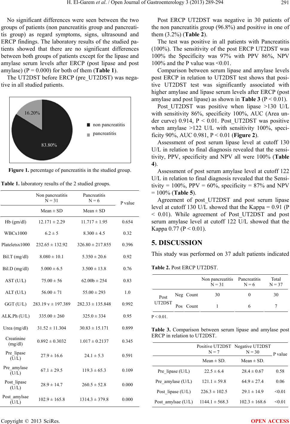 H. El-Garem et al. / Open Journal of Gastroenterology 3 (2013) 289-294 291 No significant differences were seen between the two groups of patients (non pancreatitis group and pancreati- tis group) as regard symptoms, signs, ultrasound and ERCP findings. The laboratory results of the studied pa- tients showed that there are no significant differences between both groups of patients except for the lipase and amylase serum levels after ERCP (post lipase and post amylase) (P = 0.000) for both of them (Table 1). The UT2DST before ERCP (pre_UT2DST) was nega- tive in all studied patients. non pancreatitis pancreatitis 16.20% 83.80% Figure 1. percentage of pancreatitis in the studied group. Table 1. laboratory results of the 2 studied groups. Non pancreatitis N = 31 Pancreatitis N = 6 Mean ± SD Mean ± SD P value Hb (gm/dl) 12.171 ± 2.29 11.717 ± 1.95 0.654 WBCx1000 6.2 ± 5 8.300 ± 4.5 0.32 Plateletsx1000 232.65 ± 132.92 326.80 ± 217.855 0.396 Bil.T (mg/dl) 8.080 ± 10.1 5.350 ± 20.6 0.92 Bil.D (mg/dl) 5.000 ± 6.5 3.500 ± 13.8 0.76 AST (U/L) 75.00 ± 56 62.00b ± 254 0.83 ALT (U/L) 56.00 ± 71 55.00 ± 293 1.0 GGT (U/L) 283.19 v ± 197.389 282.33 ± 135.848 0.992 ALK.Ph (U/L) 335.00 ± 260 325.0 ± 334 0.95 Urea (mg/dl) 31.52 ± 11.304 30.83 ± 15.171 0.899 Creatinine (mg/dl) 0.892 ± 0.3032 1.017 ± 0.2137 0.345 Pre_lipase (U/L) 27.9 ± 16.6 24.1 ± 5.3 0.591 Pre_amylase (U/L) 67.1 ± 29.5 119.3 ± 65.3 0.109 Post_lipase (U/L) 28.9 ± 14.7 260.5 ± 52.8 0.000 Post_amylsae (U/L) 102.9 ± 165.8 1314.3 ± 379.8 0.000 Post ERCP UT2DST was negative in 30 patients of the non pancreatitis group (96.8%) and positive in one of them (3.2%) (Table 2). The test was positive in all patients with Pancreatitis (100%). The sensitivity of the post ERCP UT2DST was 100% the Specificity was 97% with PPV 86%, NPV 100% and the P value was <0.01. Comparison between serum lipase and amylase levels post ERCP in relation to UT2DST test shows that posi- tive UT2DST test was significantly associated with higher amylase and lipase serum levels after ERCP (post amylase and post lipase) as shown in Table 3 (P < 0.01). Post_UT2DST was positive when lipase >130 U/L with sensitivity 86%, specificity 100%, AUC (Area un- der curve) 0.914, P < 0.01. Post_UT2DST was positive when amylase >122 U/L with sensitivity 100%, speci- ficity 90%, AUC 0.981, P < 0.01 (Figur e 2). Assessment of post serum lipase level at cutoff 130 U/L in relation to final diagnosis revealed that the sensi- tivity, PPV, specificity and NPV all were 100% (Table 4). Assessment of post serum amylase level at cutoff 122 U/L in relation to final diagnosis revealed that the Sensi- tivity = 100%, PPV = 60%, specificity = 87% and NPV = 100% (Table 5). Agreement of post_UT2DST and post serum lipase level at cutoff 130 U/L showed that the Kappa = 0.91 (P < 0.01). While agreement of Post_UT2DST and post serum amylase level at cutoff 122 U/L showed that the Kappa 0.77 (P < 0.01). 5. DISCUSSION This study was performed on 37 adult patients indicated Table 2. Post ERCP UT2DST. Non pancreatitis N = 31 Pancreatitis N = 6 Total N = 37 NegCount30 0 30 Post UT2DST PosCount1 6 7 P < 0.01. Table 3. Comparison between serum lipase and amylase post ERCP in relation to UT2DST. Positive UT2DST N = 7 Negative UT2DST N = 30 Mean ± SD. Mean ± SD. P value Pre_lipase (U/L) 22.5 ± 6.4 28.4 ± 0.67 0.58 Pre_amylase (U/L)121.1 ± 59.8 64.9 ± 27.4 0.06 Post_lipase (U/L)226.3 ± 102.5 29.1 ± 14.9 <0.01 Post_amylsae (U/L)1144.1 ± 568.3 102.3 ± 168.6 <0.01 Copyright © 2013 SciRes. OPEN ACCESS 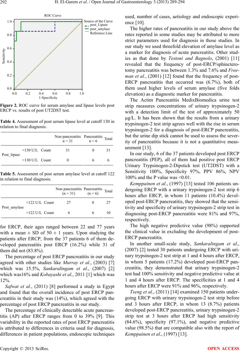 H. El-Garem et al. / Open Journal of Gastroenterology 3 (2013) 289-294 292 Source of the Curve p ost_Lipase p ost_am y lase Reference Line 1.0 0.8 0.6 0.4 0.2 0.0 Sensitivity 0.0 0.2 0.4 0.6 0.8 1.0 1-Specificit y ROC Curve Figure 2. ROC curve for serum amylase and lipase levels post ERCP vs. results of post UT2DST test. Table 4. Assessment of post serum lipase level at cutoff 130 in relation to final diagnosis. Non pancreatitis n = 31 Pancreatitis n = 6 Total <130 U/L Count 31 0 31 Post_lipase >130 U/L Count 0 6 6 Table 5. Assessment of post serum amylase level at cutoff 122 in relation to final diagnosis. Non pancreatitis (n = 31) Pancreatitis (n = 6) Tot al <122 U/L Count 27 0 27 Post_amylase >122 U/L Count 4 6 10 for ERCP, their ages ranged between 22 and 77 years with a mean ± SD of 50 ± 1 years. Upon studying the patients after ERCP, from the 37 patients 6 of them de- veloped pancreatitis post ERCP (16.2%) while 31 of them did not (83.8%). The percentage of post ERCP pancreatitis in our study agreed with other studies like Murray et al., (2003) [7] which was 15.5%, Sankaralingam et al., (2007) [2] which was16% and Kobayashi et al., 2011 [1] which was 12%. Safwat et al., (2011) [8] performed a study in Egypt and found that the overall incidence of post ERCP pan- creatitis in their study was (14%), which agreed with the percentage of post ERCP pancreatitis in our study. The percentage of clinically detectable acute pancreas- titis (AP) after ERCP ranges from 0 to 39% [9]. This variability in the reported rates of post ERCP pancreatitis is attributed to differences in criteria used for diagnosis, differences in patient populations, endoscopic techniques used, number of cases, aetiology and endoscopic experi- ence [10]. The higher rates of pancreatitis in our study above the rates reported in some studies may be attributed to more strict parameters used for diagnosis in those studies. In our study we used threefold elevation of amylase level as a marker for diagnosis of acute pancreatitis. Other stud- ies as that done by Testoni and Bagnolo, (2001) [11] revealed that the frequency of post-ERCP/sphinctero- tomy pancreatitis was between 1.3% and 7.6% and Free- man et al., (2001) [12] found that the frequency of post- ERCP pancreatitis that occurred was (6.7%), both of them used higher levels of serum amylase (five folds elevation) as a diagnostic marker for pancreatitis. The Actim Pancreatitis MedixBiomedica urine test strip measures concentrations of urinary trypsinogen-2 with a detection limit of the test of approximately 50 μg/L. It has been shown that the results from a urinary trypsinogen-2 test strip agrees well with the rise in serum trypsinogen-2 for a diagnosis of post-ERCP pancreatitis, but the urine dip stick cannot be used to assess the sever- ity of pancreatitis because it is not a quantitative meas- urement [13]. In our study, 6 of the 37 patients developed post ERCP pancreatitis (PEP), all of them had positive post ERCP Urinary Trypsinogen-2-Dipstick test (UT2DST) with a Sensitivity 100%, Specificity 97%, PPV 86%, NPV 100% and the P value was <0.01. Kemppainen et al., (1997) [13] tested 106 patients un- dergoing ERCP with a urinary trypsinogen-2 test strip 6 hours after ERCP, in whom 11 patients (10.4%) devel- oped post-ERCP pancreatitis, they showed that the sensi- tivity and specificity of urinary trypsinogen-2 strip test in diagnosing post-ERCP pancreatitis were 81% and 97%, respectively. The high negative predictive value (98%) supported the clinical value in excluding the development of post- ERCP pancreatitis. In another small-scale study, Sankaralingam et al., (2007) [2] tested 30 patients undergoing ERCP with uri- nary trypsinogen-2 test strip at 1 and 4 hours after ERCP, in whom 5 patients (17.2%) developed post-ERCP pan- creatitis, they demonstrated that urinary trypsinogen-2 test had 100% sensitivity and negative predictive value at 1 and 4 hours after ERCP. The specificities at 1 and 4 hours after ERCP were 91% and 96%, respectively. Tseng et al., (2011) [14] examined 150 patients under- going ERCP with urinary trypsinogen-2 test strip before and 3 hours after ERCP, in whom 13 (8.7%) patients developed post-ERCP pancreatitis, urinary trypsinogen-2 strip test at 3 hours after ERCP had high sensitivity (84.6%), specificity (97.1%), and negative predictive value (98.5%) that are compatible also with the report of Kemppainen et al., (1997) [13]. Copyright © 2013 SciRes. OPEN ACCESS 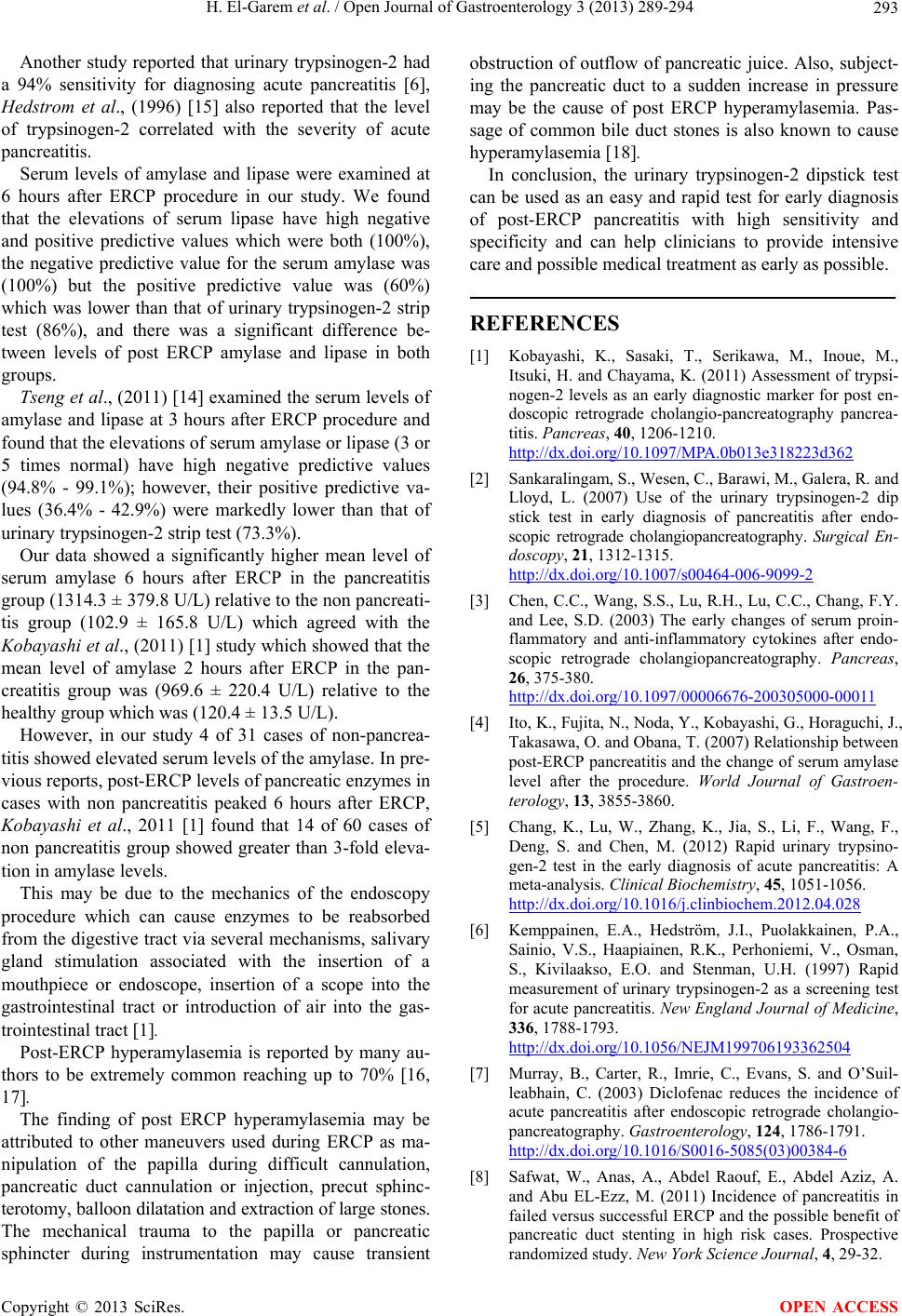 H. El-Garem et al. / Open Journal of Gastroenterology 3 (2013) 289-294 293 Another study reported that urinary trypsinogen-2 had a 94% sensitivity for diagnosing acute pancreatitis [6], Hedstrom et al., (1996) [15] also reported that the level of trypsinogen-2 correlated with the severity of acute pancreatitis. Serum levels of amylase and lipase were examined at 6 hours after ERCP procedure in our study. We found that the elevations of serum lipase have high negative and positive predictive values which were both (100%), the negative predictive value for the serum amylase was (100%) but the positive predictive value was (60%) which was lower than that of urinary trypsinogen-2 strip test (86%), and there was a significant difference be- tween levels of post ERCP amylase and lipase in both groups. Tseng et al., (2011) [14] examined the serum levels of amylase and lipase at 3 hours after ERCP procedure and found that the elevations of serum amylase or lipase (3 or 5 times normal) have high negative predictive values (94.8% - 99.1%); however, their positive predictive va- lues (36.4% - 42.9%) were markedly lower than that of urinary trypsinogen-2 strip test (73.3%). Our data showed a significantly higher mean level of serum amylase 6 hours after ERCP in the pancreatitis group (1314.3 ± 379.8 U/L) relative to the non pancreati- tis group (102.9 ± 165.8 U/L) which agreed with the Kobayashi et al., (2011) [1] study which showed that the mean level of amylase 2 hours after ERCP in the pan- creatitis group was (969.6 ± 220.4 U/L) relative to the healthy group which was (120.4 ± 13.5 U/L). However, in our study 4 of 31 cases of non-pancrea- titis showed elevated serum levels of the amylase. In pre- vious reports, post-ERCP levels of pancreatic enzymes in cases with non pancreatitis peaked 6 hours after ERCP, Kobayashi et al., 2011 [1] found that 14 of 60 cases of non pancreatitis group showed greater than 3-fold eleva- tion in amylase levels. This may be due to the mechanics of the endoscopy procedure which can cause enzymes to be reabsorbed from the digestive tract via several mechanisms, salivary gland stimulation associated with the insertion of a mouthpiece or endoscope, insertion of a scope into the gastrointestinal tract or introduction of air into the gas- trointestinal tract [1]. Post-ERCP hyperamylasemia is reported by many au- thors to be extremely common reaching up to 70% [16, 17]. The finding of post ERCP hyperamylasemia may be attributed to other maneuvers used during ERCP as ma- nipulation of the papilla during difficult cannulation, pancreatic duct cannulation or injection, precut sphinc- terotomy, balloon dilatation and extraction of large stones. The mechanical trauma to the papilla or pancreatic sphincter during instrumentation may cause transient obstruction of outflow of pancreatic juice. Also, subject- ing the pancreatic duct to a sudden increase in pressure may be the cause of post ERCP hyperamylasemia. Pas- sage of common bile duct stones is also known to cause hyperamylasemia [18]. In conclusion, the urinary trypsinogen-2 dipstick test can be used as an easy and rapid test for early diagnosis of post-ERCP pancreatitis with high sensitivity and specificity and can help clinicians to provide intensive care and possible medical treatment as early as possible. REFERENCES [1] Kobayashi, K., Sasaki, T., Serikawa, M., Inoue, M., Itsuki, H. and Chayama, K. (2011) Assessment of trypsi- nogen-2 levels as an early diagnostic marker for post en- doscopic retrograde cholangio-pancreatography pancrea- titis. Pancreas, 40, 1206-1210. http://dx.doi.org/10.1097/MPA.0b013e318223d362 [2] Sankaralingam, S., Wesen, C., Barawi, M., Galera, R. and Lloyd, L. (2007) Use of the urinary trypsinogen-2 dip stick test in early diagnosis of pancreatitis after endo- scopic retrograde cholangiopancreatography. Surgical En- doscopy, 21, 1312-1315. http://dx.doi.org/10.1007/s00464-006-9099-2 [3] Chen, C.C., Wang, S.S., Lu, R.H., Lu, C.C., Chang, F.Y. and Lee, S.D. (2003) The early changes of serum proin- flammatory and anti-inflammatory cytokines after endo- scopic retrograde cholangiopancreatography. Pancreas, 26, 375-380. http://dx.doi.org/10.1097/00006676-200305000-00011 [4] Ito, K., Fujita, N., Noda, Y., Kobayashi, G., Horaguchi, J., Takasawa, O. and Obana, T. (2007) Relationship between post-ERCP pancreatitis and the change of serum amylase level after the procedure. World Journal of Gastroen- terology, 13, 3855-3860. [5] Chang, K., Lu, W., Zhang, K., Jia, S., Li, F., Wang, F., Deng, S. and Chen, M. (2012) Rapid urinary trypsino- gen-2 test in the early diagnosis of acute pancreatitis: A meta-analysis. Clinical Biochemistry, 45, 1051-1056. http://dx.doi.org/10.1016/j.clinbiochem.2012.04.028 [6] Kemppainen, E.A., Hedström, J.I., Puolakkainen, P.A., Sainio, V.S., Haapiainen, R.K., Perhoniemi, V., Osman, S., Kivilaakso, E.O. and Stenman, U.H. (1997) Rapid measurement of urinary trypsinogen-2 as a screening test for acute pancreatitis. New England Journal of Medicine, 336, 1788-1793. http://dx.doi.org/10.1056/NEJM199706193362504 [7] Murray, B., Carter, R., Imrie, C., Evans, S. and O’Suil- leabhain, C. (2003) Diclofenac reduces the incidence of acute pancreatitis after endoscopic retrograde cholangio- pancreatography. Gastroenterology, 124, 1786-1791. http://dx.doi.org/10.1016/S0016-5085(03)00384-6 [8] Safwat, W., Anas, A., Abdel Raouf, E., Abdel Aziz, A. and Abu EL-Ezz, M. (2011) Incidence of pancreatitis in failed versus successful ERCP and the possible benefit of pancreatic duct stenting in high risk cases. Prospective randomized study. New York Science Journal, 4, 29-32. Copyright © 2013 SciRes. OPEN ACCESS  H. El-Garem et al. / Open Journal of Gastroenterology 3 (2013) 289-294 Copyright © 2013 SciRes. OPEN ACCESS 294 [9] Sherman, S. and Lehman, G.A. (1991) ERCP- and endo- scopic sphincterotomy-induced pancreatitis. Pancreas, 6, 350-367. http://dx.doi.org/10.1097/00006676-199105000-00013 [10] Andriulli, A., Loperfido, S., Napolitano, G., Niro, G., Valvano, M.R., Spirito, F., Pilotto, A. and Forlano, R. (2007) Incidence rate of post-ERCP complications: A systematic survey of prospective studies. American Jour- nal of Gastroenterology, 102, 1781-1788. http://d x.doi.org/10.1111/j. 1572-0241.2007.01279.x [11] Testoni, P.A. and Bagnolo, F. (2001) Pain at 24 hours associated with amylase levels greater than 5 times the upper normal limit as the most reliable indicator of post- ERCP pancreatitis. Gastrointestinal Endoscopy, 53, 33- 39. http://dx.doi.org/10.1067/mge.2001.111390 [12] Freeman, M.L., DiSario, J.A., Nelson, D.B., Fennerty, M.B., Lee, J.G., Bjorkman, D.J., Overby, C.S., Aas, J., Ryan, M.E., Bochna, G.S., Shaw, M.J., Snady, H.W., Erickson, R.V., Moore, J.P. and Roel, J.P. (2001) Risk factors for post-ERCP pancreatitis, a prospective, multi- center study. Gastrointestinal Endoscopy, 54, 425-434. http://dx.doi.org/10.1067/mge.2001.117550 [13] Kemppainen, E., Hedström, J., Puolakkainen, P., Halt- tunen, J., Sainio, V., Haapiainen, R. and Stenman, U.H. (1997) Urinary trypsinogen-2 test strip in detecting ERCP- induced pancreatitis. Endoscopy, 29, 247-251. http://dx.doi.org/10.1055/s-2007-1004184 [14] Tseng, C.W., Chen, C.C., Lin, S.Z., Chang, F.Y., Lin, H.C. and Lee, S.D. (2011) Rapid urinary trypsinogen-2 test strip in the diagnosis of pancreatitis after endoscopic retrograde cholangiopancreatography. Pancreas, 40, 1211- 1214. http://dx.doi.org/10.1097/MPA.0b013e31821fcdcf [15] Hedström, J., Korvuo, A., Kenkimäki, P., Tikanoja, S., Haapiainen, R., Kivilaakso, E. and Stenman, U.H. (1996) Urinary trypsinogen-2 test strip for acute pancreatitis. Lancet, 347, 729-730. http://dx.doi.org/10.1016/S0140-6736(96)90078-1 [16] Chen, Y.K. and Pleskow, D.K. (2007) SpyGlass single- operator peroralcholangiopancreatoscopy system for the diagnosis and therapy of bile-duct disorders: A clinical feasibility study (with video). Gastrointestinal Endoscopy, 65, 832-841. http://dx.doi.org/10.1016/j.gie.2007.01.025 [17] Jaik, N.P., Hoey, B.A. and Stawicki, S.P. (2008) Evolv- ing role of endoscopic retrograde cholangiopancreato- graphy in management of extrahepatic hepatic ductal in- juries due to blunt trauma: Diagnostic and treatment al- gorithms. HPB Surgery, 2008, Article ID: 259141. [18] Freeman, M.L. (2002) Adverse outcomes of ERCP. Gas- trointestina l E ndoscopy 56, S273-S282. http://dx.doi.org/10.1016/S0016-5107(02)70025-4 |