

Paper Menu >>
Journal Menu >>
 Open Journal of Anesthesiology, 2013, 3, 353-355 http://dx.doi.org/10.4236/ojanes.2013.38075 Published Online October 2013 (http://www.scirp.org/journal/ojanes) 353 Successful Anesthetic Management for Surgical Repair of Atrio-Esophageal Fistula Following Radiofrequency Ablation for Atrial Fibrillation Atsushi Yasuda, Paul H. Alfille, Lisa T. Wollman-Kliman Department of Anesthesia, Critical Care and Pain Medicine, Massachusetts General Hospital, Boston, USA. Email: [email protected] Received July 17th, 2013; revised August 15th, 2013; accepted August 26th, 2013 Copyright © 2013 Atsushi Yasuda et al. This is an open access article distributed under the Creative Commons Attribution License, which permits unrestricted use, distribution, and reproduction in any medium, provided the original work is properly cited. ABSTRACT Introduction: Atrio-esophageal fistula is a rar e but often fatal complication of radiofre quency ablation for atrial fibril- lation. Here we report a successful case in anesthetic management of surgical repair of atrio-esophageal fistula. Case Report: The patient was a 56-year-old man status post radiofrequency ablation for atrial fibrillation one month before presenting with fever and symptoms and signs of cerebral emboli. He was diagnosed as having atrio-esophageal fistula, which required emergent surgical repair. In the operating room, rapid sequence induction was performed with avoid- ance of positive pressure ventilation before securing airway. Double lumen tube was used for lung isolation for left tho- racotomy. Upon exploration, a small fistula was identified. Both atrial and esophageal defects were ligated and an in- tercostal muscle flap was placed. The patient’s heart rhythm was atrial flutter/atrial fibrillation with marginal hemody- namics during the procedure, but cardioversion was delayed until the fistula was repaired and no remaining air, blood clot or gastric content in the heart was confirmed by epicardial ultrasound. The patient tolerated the surgery and was transferred to ICU, intubated and ventilated. He recovered from surgery and was transferred to a reha bilitation hospital with residual expressive aphasia. Conclusion: We had a successful case in anesthetic management for surgical repair of atrio-esophageal fistula by preventing massive bleeding as well as multiple air embolization through the fistula. Keywords: Atrial Fibrillation; Radiofrequency Ablation; Complication; Atrio-Esophageal Fistula; Anesthetic Management 1. Introduction Atrial fibrillation is the most common clinically relevant heart rhythm disorder. Radiofrequency ablation is widely performed as a treatment for recurrent, drug-resistant atrial fibrillation. Atrio-esophageal fistula is a rare but often fatal complication of radiofrequency ablation for atrial fibrillation. There are no reports on anesthetic management for surgical repair of atrio-esophageal fis- tula. Here we report a successful case in anesthetic man- agement of 56-year-old man who underwent emergent surgical repair of atrio-esophageal fistula. 2. Case Report The patient was a 56-year-old man (85 kgw) with history of chronic deep venous thromboses (DVT) and factor V Leiden deficiency on warfarin therapy and recurrent par- oxysmal atrial fibrillation. He underwent successful ra- diofrequency ablation/pulmonary vein isolation for atrial fibrillation at an outside institution. The initial post-pro- cedure course was uneventful and he maintained normal sinus rhythm. One month later he developed symptoms of progressive malaise, high fevers, and intermittent speech difficulty, which required hospitalization to the outside hospital. Blood cultures drawn at presentation later grew streptococcus viridans. Transesophageal echo- cardiogram was negative for endocarditis but revealed a small patent foramen ovale. Lower extremity non-inva- sive sonography showed bilateral DVTs. MRI of the brain showed multiple subacute embolic foci. He was transferred to our tertiary hospital 3 days after the initial admission. On arrival to our institution, CT scan of the chest was performed, showing air pockets in the left atrial append- age, left ventricle, and left superior pulmonary vein (Fig- ure 1). A gastrografin swallow test showed extravasation Copyright © 2013 SciRes. OJAnes 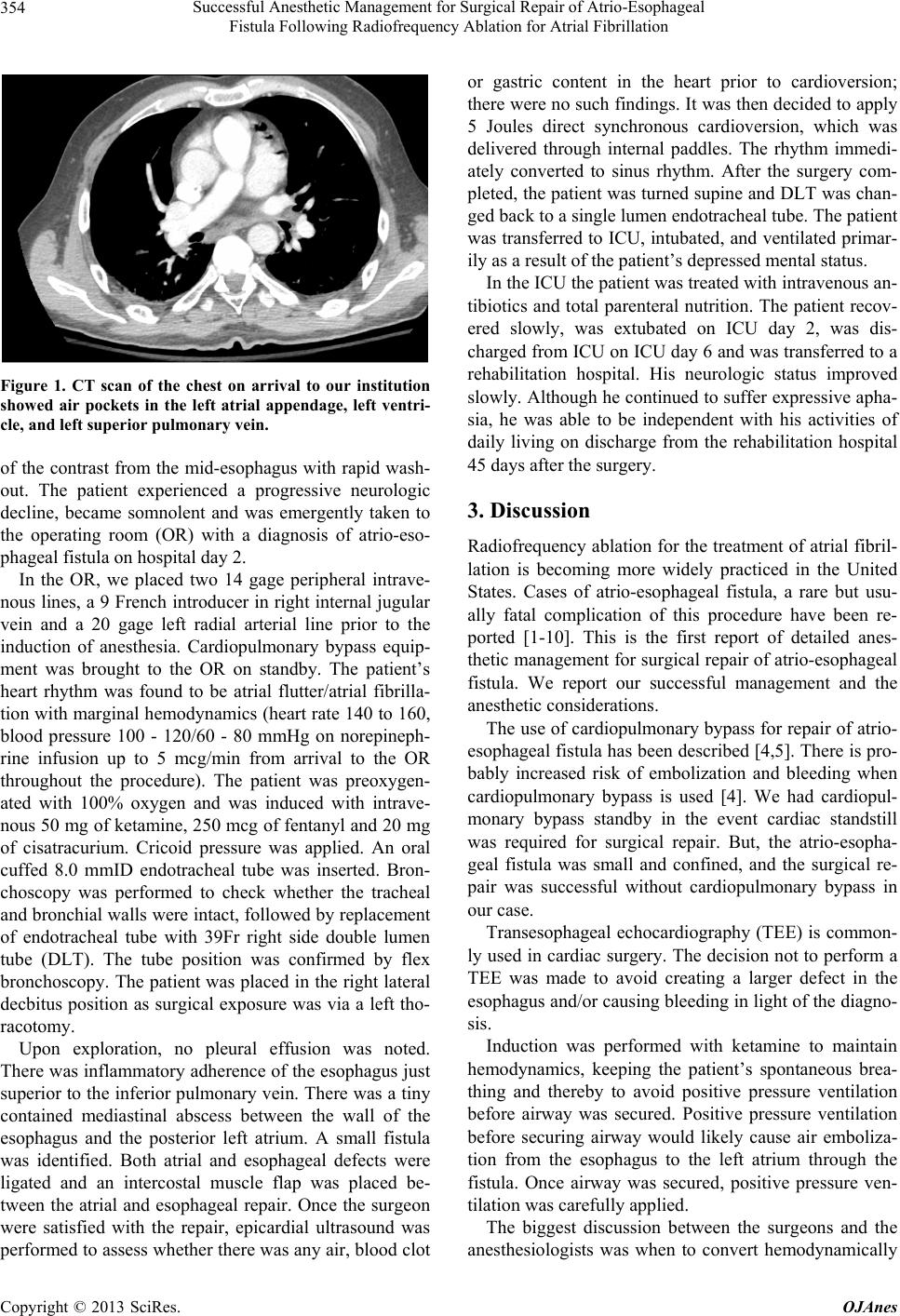 Successful Anesthetic Management for Surgical Repair of Atrio-Esophageal Fistula Following Radiofrequency Ablation for Atrial Fibrillation 354 Figure 1. CT scan of the chest on arrival to our institution showed air pockets in the left atrial appendage, left ventri- cle, and left super i or pulmonary vein. of the contrast from the mid-esophagus with rapid wash- out. The patient experienced a progressive neurologic decline, became somnolent and was emergently taken to the operating room (OR) with a diagnosis of atrio-eso- phageal fistula on hospital day 2. In the OR, we placed two 14 gage peripheral intrave- nous lines, a 9 French in troducer in right internal jugular vein and a 20 gage left radial arterial line prior to the induction of anesthesia. Cardiopulmonary bypass equip- ment was brought to the OR on standby. The patient’s heart rhythm was found to be atrial flutter/atrial fibrilla- tion with marginal hemodynamics (heart rate 140 to 160, blood pressure 100 - 120/60 - 80 mmHg on norepineph- rine infusion up to 5 mcg/min from arrival to the OR throughout the procedure). The patient was preoxygen- ated with 100% oxygen and was induced with intrave- nous 50 mg of ketamine, 250 mcg of fentanyl and 20 mg of cisatracurium. Cricoid pressure was applied. An oral cuffed 8.0 mmID endotracheal tube was inserted. Bron- choscopy was performed to check whether the tracheal and bronchial walls were intact, followed by replacement of endotracheal tube with 39Fr right side double lumen tube (DLT). The tube position was confirmed by flex bronchoscopy. The patient was placed in the right lateral decbitus position as surgical exposure was via a left tho- racotomy. Upon exploration, no pleural effusion was noted. There w as inflammato ry adherenc e of the esophagu s just superior to the inferior pulmonary vein. There was a tin y contained mediastinal abscess between the wall of the esophagus and the posterior left atrium. A small fistula was identified. Both atrial and esophageal defects were ligated and an intercostal muscle flap was placed be- tween the atrial and esophageal repair. Once the surgeon were satisfied with the repair, epicardial ultrasound was performed to assess whether there was any air, blood clot or gastric content in the heart prior to cardioversion; there were no such findings. It was then decided to apply 5 Joules direct synchronous cardioversion, which was delivered through internal paddles. The rhythm immedi- ately converted to sinus rhythm. After the surgery com- pleted, the patient was turned supine and DLT was chan- ged back to a single lumen endotracheal tube. The patient was transferred to ICU, intubated, and ventilated pr imar- ily as a result of the patient’s depressed mental status. In the ICU the patient was treated with intravenous an- tibiotics and total parenteral nutrition. The patient recov- ered slowly, was extubated on ICU day 2, was dis- charged from ICU on ICU day 6 and was tran sferred to a rehabilitation hospital. His neurologic status improved slowly. Although he continued to suffer expressive apha- sia, he was able to be independent with his activities of daily living on discharge from the rehabilitation hospital 45 days after the surgery. 3. Discussion Radiofrequency ablation for the treatment of atrial fibril- lation is becoming more widely practiced in the United States. Cases of atrio-esophageal fistula, a rare but usu- ally fatal complication of this procedure have been re- ported [1-10]. This is the first report of detailed anes- thetic management for surgical repair of atrio-esophageal fistula. We report our successful management and the anesthetic consideration s . The use of cardiopulmonary bypass for repair of atrio- esophageal fistula has been described [4,5]. There is pro- bably increased risk of embolization and bleeding when cardiopulmonary bypass is used [4]. We had cardiopul- monary bypass standby in the event cardiac standstill was required for surgical repair. But, the atrio-esopha- geal fistula was small and confined, and the surgical re- pair was successful without cardiopulmonary bypass in our case. Transesophageal echocardiography (TEE) is common- ly used in cardiac surgery. The decision not to perform a TEE was made to avoid creating a larger defect in the esophagus and/or causing bleeding in light of the diagno- sis. Induction was performed with ketamine to maintain hemodynamics, keeping the patient’s spontaneous brea- thing and thereby to avoid positive pressure ventilation before airway was secured. Positive pressure ventilation before securing airway would likely cause air emboliza- tion from the esophagus to the left atrium through the fistula. Once airway was secured, positive pressure ven- tilation was carefully applied. The biggest discussion between the surgeons and the anesthesiologists was when to convert hemodynamically Copyright © 2013 SciRes. OJAnes  Successful Anesthetic Management for Surgical Repair of Atrio-Esophageal Fistula Following Radiofrequency Ablation for Atrial Fibrillation Copyright © 2013 SciRes. OJAnes 355 unstable atrial fibrillation to sinus rhythm. The patient became hemodynamically unstable shortly after arrival to the OR. According to ACLS protocol, hemodynamically unstable atrial fibrillation should be cardioverted. But in this specific case, atrial fibrillation kept atrial pressure low enough to avoid hemorrhage from the atria to the chest or esophagus. Also converting to sinus rhythm in- creases the likelihood of embolization of air, blood clot, gastric contents in the left atrium into the brain or other organs. And also the patient had had atrial fibrillatio n for at least several days and the patient had very high risk for thrombus formation due to factor V Leiden deficiency and DVT. We did not know whether there was thrombus or air in the left atria or how big it would be if thrombus was present at this point. Cardioversion likely could have ended up causing massive bleeding or embolization. Hemodynamics was managed instead by infusing pheny- lephrine and norepinephrine to maintain a systolic blood pressure around 100 mmHg. Only once air in the atria was suctioned, and no other air, blood clots or gastric contents were detected by direct epicardial ultrasound and the fistula was surgically repaired, caridoversion was performed by directly applying paddles to the heart. The patient was kept intubated and transferred to the ICU due to the hemodyn amic instab ility and n eurolog ical deterioration shown preoperatively which could impair airway protection postoperatively. The patient was treat- ed for sepsis with broad spectrum of antibiotics initially to cover the likely bacterial species inh abiting esophagus. The role of esophageal stent was considered intraope- ratively. There was a successful case of esophageal stent for atrio-esophageal fistula, using esophagoscopy [7], even though most of the literature did recommend avoid- ance of esophagoscopy [4-6,9]. But more data is needed to determine which cases are indicated for esophageal stent and esophagoscopy. The key to managing these patients’ safety is the pre- vention of massive bleeding and multiple air emboliza- tion during the surgical repair and then later the postop- erative management of sepsis and multiple organ failure, which are the main causes of death from this complica- tion. 4. Conclusion We had a successful case in anesthetic management for surgical repair of atrio-esophageal fistula with avoidance of positive pressure ventilation before securing airway and careful control of hemodynamics by delaying car- dioversion to prevent massive bleeding as well as multi- ple air embolization through the fistula. REFERENCES [1] A. M. Gillinov, G. Pettersson and T. W. Rice, “Esophag- eal Injury during Radiofrequency Ablation for Atrial Fib- rillation,” The Journal of Thoracic and Cardiovascular Surgery, Vol. 122, No. 6, 2001, pp. 1239-1240. http://dx.doi.org/10.1067/mtc.2001.118041 [2] N. Doll, M. A. Borger, A. Fabricius, S. Stephan, J. Gum- mert, F. W. Mohr, et al., “Esophageal Perforation during Left Atrial Radiofrequency Ablation: Is the Risk Too High?” The Journal of Thoracic and Cardiovascular Sur- gery, Vol. 125, No. 4, 2003, pp. 836-842. http://dx.doi.org/10.1067/mtc.2003.165 [3] G. Hindricks and H. Kottkamp, “Potential Benefits, Risks, and Complications of Catheter Ablation of Atrial Fibrilla- tion: More Questions Than Answers,” Journal of Cardio- vascular Electrophysi ology, Vol. 13, No. 8, 2002, pp. 768- 769. http://dx.doi.org/10.1046/j.1540-8167.2002.00768.x [4] B. Sonmez, E. Demirsoy, N. Yagan, M. Unal, H. Arbatli, D. Sener, et al., “A Fatal Complication Due to Radiofre- quency Ablation for Atrial Fibrillation: Atrio-Esophageal Fistula,” The Annals of Thoracic Surgery, Vol. 76, No. 1, 2003, pp. 281-283. http://dx.doi.org/10.1016/S0003-4975(03)00006-7 [5] C. Pappone, H. Oral, V. Santinelli, G. Vicedomini, C. C. Lang, F. Manguso, et al., “Atrio-Esophageal Fistula as a Complication of Percutaneous Transcatheter Ablation of Atrial Fibrillation,” Circulation, Vol. 109, No. 22, 2004, pp. 2724-2726. http://dx.doi.org/10.1161/01.CIR.0000131866.44650.46 [6] M. I. Scanavacca, A. D’avila, J. Parga and E. Sosa, “Left Atrial-Esophageal Fistula Following Radiofrequency Ca- theter Ablation of Atrial Fibrillation,” Journal of Cardio- vascular Electrophysi ology, Vol. 15, No. 8, 2004, pp. 960- 962. http://dx.doi.org/10.1046/j.1540-8167.2004.04083.x [7] T. J. Bunch, J. Nelson, T. Foley, S. Allison, B. G. Cran- dall, J. S. Osborn, et al., “Temporary Esophageal Stenting Allows Healing of Esophageal Perforations Following Atrial Fibrillation Ablation Procedures,” Journal of Car- diovascular Electrophysiology, Vol. 17, No. 4, 2006, pp. 435-439. http://dx.doi.org/10.1111/j.1540-8167.2006.00464.x [8] P. Schley, H. Gu¨lker and M. Horlitz, “Atrio-Oesophageal Fistula Following Circumferential Pulmonary Vein Abla- tion: Verification of Diagnosis with Multislice Computed Tomography,” Europace, Vol. 8, No. 3, 2006, pp. 189- 190. http://dx.doi.org/10.1093/europace/euj050 [9] J. E. Cummings, R. A. Schweikert, W. I. Saliba, J. D. Burkhardt, F. Kilikaslan, E. Saad, et al., “Brief Commu- nication: Atrial-Esophageal Fistulas after Radiofrequency Ablation,” Annals of Internal Medicine, Vol. 144, No. 8, 2006, pp. 572-574. http://dx.doi.org/10.7326/0003-4819-144-8-200604180-0 0007 [10] A. Takahashi, T. Kuwahara and Y. Takahashi, “Compli- cations in the Catheter Ablation of Atrial Fibrillation: In- cidence and Management,” Circulation Journal, Vol. 73, No. 2, 2009, pp. 221-226. http://dx.doi.org/10.1253/circj.CJ-08-1097. |