

Paper Menu >>
Journal Menu >>
 Open Journal of Veterinary Medicine, 2013, 3, 267-272 http://dx.doi.org/10.4236/ojvm.2013.36043 Published Online October 2013 (http://www.scirp.org/journal/ojvm) Prevalence of Gastrointestinal Parasites of Horses and Donkeys in and around Gondar Town, Ethiopia Tola Mezgebu1, Ketema Tafess2, Firaol Tamiru3* 1Jimma Arjo District Animal Health and Marketing Agency, East Wollega Zone, Arjo, Ethiopia 2Department of Biomedical Science, School of Health, Adama Science and Technology University, Asella, Ethiopia 3Department of Veterinary Laboratory Technology, College of Agriculture and Veterinary Science, Ambo University, Ambo, Ethiopia Email: *[email protected] Received July 25, 2013; revised August 25, 2013; accepted September 10, 2013 Copyright © 2013 Tola Mezgebu et al. This is an open access article distributed under the Creative Commons Attribution License, which permits unrestricted use, distribution, and reproduction in any medium, provided the original work is properly cited. ABSTRACT A cross sectional study was conducted from October 2010 to June 2011 in and around Gondar town to identify the spe- cies and determine prevalence of gastrointestinal parasites in donkeys and horses. Gross examination, direct fecal smear, sedimentation and floatation techniques were utilized to identify the eggs and larvae of parasites in feces. A total of 384 horses and donkeys were examined for gastrointestinal parasites. The overall prevalence of gastrointestinal parasites was 92.71% (356 from 384) with 80.95% (85 from 105) and 97.13% (271 from 279) in horses and donkeys, respec- tively. Prevalence of Strongyle, Parascaris equorum, Oxyuris equi, Gastrodiscus aegyptiacus and Gastroph ilus intesti- nalis was 66.67%, 43.8%, 0.95%, 2.86%, and 0.95%, respectively in horses. Prevalence of gastrointestinal parasites was 87.81%, 42.29%, 4.30%, 5.73%, 1.43%, 3.58% and 0.72% for Strongyles, Parasca ris eq uoru m, Oxyuris equi, Fasciola, Tricuris, Gastrodiscus aegyptiacus and Gastrophilus intestinalis in donkeys, respectively. There was a statistic- cally significant difference between species, housing and among feed types in prevalence of equine gastrointestinal parasites (p < 0.05). However, there was no statistical significant difference in prevalence of gastrointestinal parasites between age, sex and among body condition. Donkeys were at higher risk of acquiring parasites than horses (OR = 7.97, CI = 3.39 - 18.75) and animals that fed only pasture and lived in poor house were at increased risk of acquiring infection (OR = 9.59, CI = 3.25 - 28.25). In conclusion, the present study revealed higher prevalence of gastrointestinal parasites in horses and donkeys. Therefore, continuous deworming, improvement of housing and feeding management were rec- ommended. Keywords: Donkeys; Gondar; Gastrointestinal Parasites; Horse; Prevalence 1. Introduction In the developing world, there are estimated 110 mil- lions of equines [1]. Ethiopia has about 7.9 million equines [2] and possesses approximately half of the Af- rica’s equine population with 37% donkeys, 58% horses and 46% mules [3]. There is one equine for every four people in the agricultural sector and for every five per- sons of the total population [4]. Equines have a promi- nent position in the agricultural systems of many devel- oping countries [5]. In Ethiopia, the low level of devel- opment of the road transport network and the rough ter- rain of the country make the donkeys and the horses the most valuable, appropriate and affordable pack animals under the small holder farming system [6]. They can be used for such applications as riding, driving, flock pro- tection, companion, breeding, training calves [5] and provide urban dwellers with opportunity of income gen- eration [7]. Parasitic helminthes are one of the most common fac- tors that constrain the health and working performance of donkeys and horses worldwide. They cause various de- grees of damage depending on the species and number at present, nutritional and the immune status of eqiuds [8]. They decrease the performance, production and produc- tivity in the animals mainly in the reduction of body weight or failure to gain weight or even increase the mortality in acute case [9]. A number of studies con- ducted to detect association between poverty and animal diseases identified gastrointestinal parasitism as one of the most important problems for eqiuds in developing *Corresponding author. C opyright © 2013 SciRes. OJVM  T. MEZGEBU ET AL. 268 countries [10-12]. The prevalence and type of internal parasites affecting eqiuds, in general, are ubiquitous with equines being continually exposed throughout their lives. Although they are often heavily parasitized by helminthes [13], the prevalence and type of internal parasites affecting eqiuds have not been determined to a great extent in Ethiopia, particularly in and around Gondar town. Available in- formation however, indicates that gastrointestinal para- sites are the major cause of early demises of working donkeys and horses in Ethiopia [5,14-17]. Therefore, the current study was conducted to identify the species and determine prevalence of gastrointestinal parasites of donkeys and horses in the study area. 2. Materials and Methods 2.1. Study Area The study was conducted from October 2010 to June 2011 on horses and donkeys in and around Gondar town, Ethiopia. Gondar is administrative center of North Gon- dar zone found at 748 km away from Addis Ababa, capi- tal of Ethiopia. North Gondar zone is located at 550 - 4620 m above sea level. The rain fall varies from 880 mm to 1772 mm with monomodial distribution. The an- nual temperature ranges from 10˚C to 44.5˚C. The zone is divided into three main agro-climatic zones: high land, mid land and low land region. The farming system of the study area is characterized by a mixed (crop-livestock production) farming system. There are 31,456 horses, 272,655 donkeys and 13,612 mules in the zone [18]. 2.2. Study Design and Animals A cross sectional study was conducted on 384 randomly selected horses and donkeys (105 cart horses and 279 donkeys). Information about species, sex, age, body con- dition and management system of the study animals were gathered from the owners. The ages of animals were de- termined using owners’ information and dentition [19]. Accordingly, animals were categorized as young (<2 years) and adults (>2 years). Body condition score (BCS) was subjectively estimated based on the guides published by Svendsen [14] as 1 (emaciated), 2 (thin), 3 (good), 4 (fat) and 5 (obese). These were categorized into three groups as ≤2, 3 and ≥4 to represent BCS 1 and 2, 3, 4 and 5, respectively. 2.3. Study Methodology Fecal samples were collected directly from the rectum into universal bottle using sterile disposable gloves. Each sample was labeled with necessary information and im- mediately transported to Veterinary Parasitology Labo- ratory, University of Gondar. Samples were kept in re- frigerator at 4˚C if immediate processing was not possi- ble, but it had been processed within 48 hours. Gross examination was performed immediately after sample collection for larvae recovery and the recovered larvae of Gastrophilus species was identified using stereomicro- scope. Direct fecal smear, sedimentation and floatation techniques were the utilized parasitological techniques to identify the eggs in feces and examined microscopically (10× and 40×) for presence of parasite ova following their procedures. Identification of the eggs was made on the basis of their morphology [20]. 2.4. Data Analysis The collected data were coded and entered into Microsoft Excel spread sheet. Statistical analyses were performed using SPSS, version 17 software packages. Percentage was used to calculate prevalence. Data were statistically analyzed using chi-square. Univariate logistic and multi- variate regressions were used to calculate degree of asso- ciation between risk factors and prevalence of gastroin- testinal parasites. In all cases 95% confidence interval (CI) and p < 0.05 was considered for statistically signifi- cant difference. 3. Results An overall of 92.71% (356/384) equine gastrointestinal parasites prevalence was obtained in the current study with 80.95% (85/105) and 97.13% (271/279) prevalence in horses and donkeys, respectively. There was statisti- cally significant difference between species, housing and among feed type in prevalence of equine gastrointestinal parasites (p < 0.05). However, there was no statistical significant difference in prevalence of gastrointestinal parasites between age, sex and among body condition (p > 0.05) (Table 1). Eggs/larvae of different parasites were observed in both equine species under the study. Five and seven dif- ferent types of eggs/larvae were observed during cop- rological examination in horses and donkeys, respec- tively. Strongyle-type eggs were the highly prevalent eggs with 66.67% in horses and 87.81% in donkeys while Gastrophilus intestinalis larvae and Oxyuris equi egg (0.95%) in horses, and Gastrop hilus intestinalis lar- vae (0.72%) in donkeys were the least prevalent parasites (Table 2). Univariate logistic and multivariate regression analysis was also undertaken estimate the strength of association of risk factor with gastrointestinal parasites infection in the study animals. Donkeys were found to be 7.97 times at risk of developing gastrointestinal parasite than horse (OR = 7.97, 95% CI = 3.39 - 18.75). Animals fed only pasture and live in poor hous ng system were found to be i Copyright © 2013 SciRes. OJVM 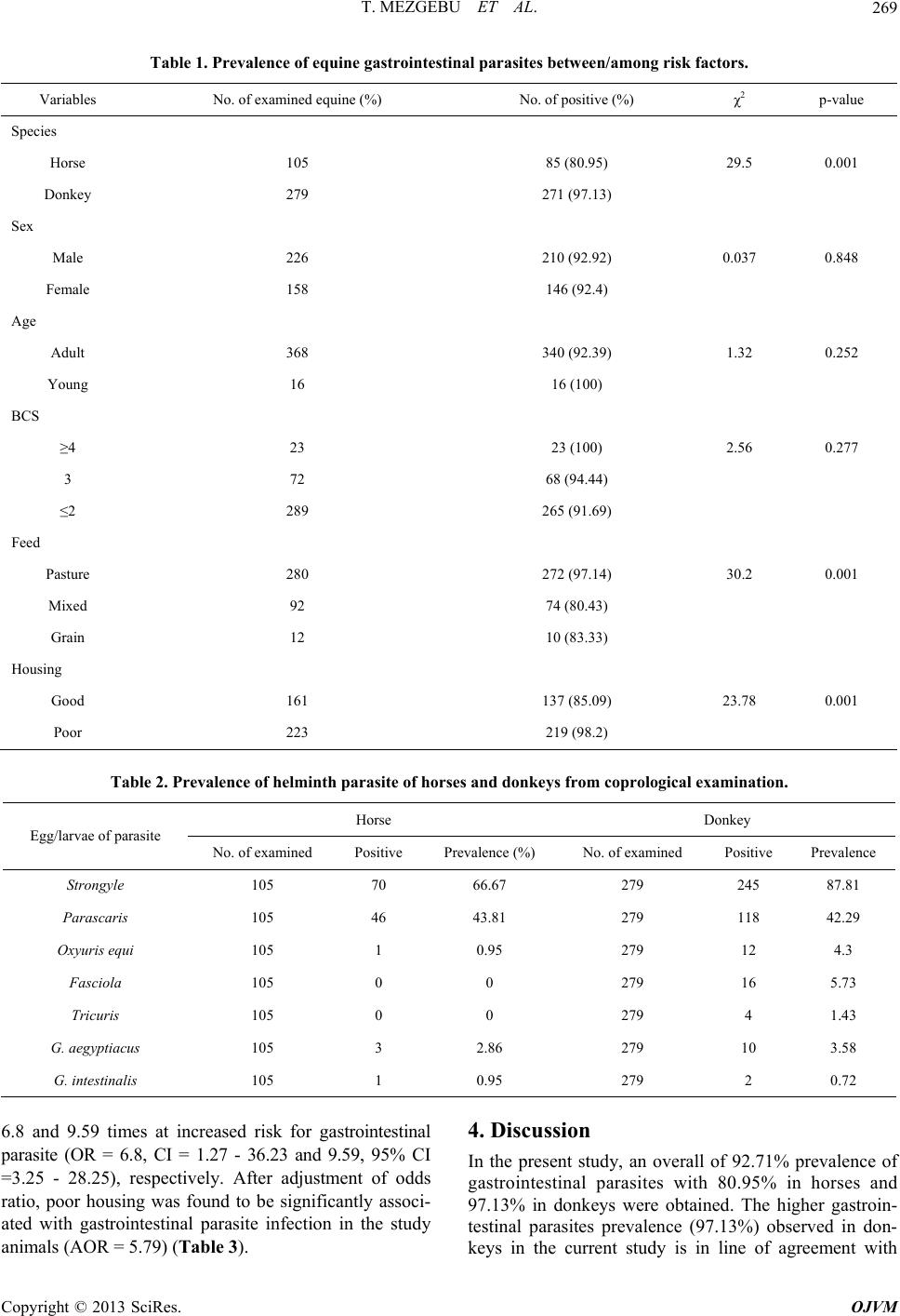 T. MEZGEBU ET AL. Copyright © 2013 SciRes. OJVM 269 Table 1. Prevalence of equine gastrointestinal parasites between/among risk factors. Variables No. of examined equine (%) No. of positive (%) χ2 p-value Species Horse 105 85 (80.95) 29.5 0.001 Donkey 279 271 (97.13) Sex Male 226 210 (92.92) 0.037 0.848 Female 158 146 (92.4) Age Adult 368 340 (92.39) 1.32 0.252 Young 16 16 (100) BCS ≥4 23 23 (100) 2.56 0.277 3 72 68 (94.44) ≤2 289 265 (91.69) Feed Pasture 280 272 (97.14) 30.2 0.001 Mixed 92 74 (80.43) Grain 12 10 (83.33) Housing Good 161 137 (85.09) 23.78 0.001 Poor 223 219 (98.2) Table 2. Prevalence of helminth parasite of horses and donkeys from coprological examination. Horse Donkey Egg/larvae of parasite No. of examined Positive Prevalence (%) No. of examined Positive Prevalence Strongyle 105 70 66.67 279 245 87.81 Parascaris 105 46 43.81 279 118 42.29 Oxyuris equi 105 1 0.95 279 12 4.3 Fasciola 105 0 0 279 16 5.73 Tricuris 105 0 0 279 4 1.43 G. aegyptiacus 105 3 2.86 279 10 3.58 G. intestinalis 105 1 0.95 279 2 0.72 6.8 and 9.59 times at increased risk for gastrointestinal parasite (OR = 6.8, CI = 1.27 - 36.23 and 9.59, 95% CI =3.25 - 28.25), respectively. After adjustment of odds ratio, poor housing was found to be significantly associ- ated with gastrointestinal parasite infection in the study animals (AOR = 5.79) (Table 3). 4. Discussion In the present study, an overall of 92.71% prevalence of gastrointestinal parasites with 80.95% in horses and 97.13% in donkeys were obtained. The higher gastroin- testinal parasites prevalence (97.13%) observed in don- keys in the current study is in line of agreement with 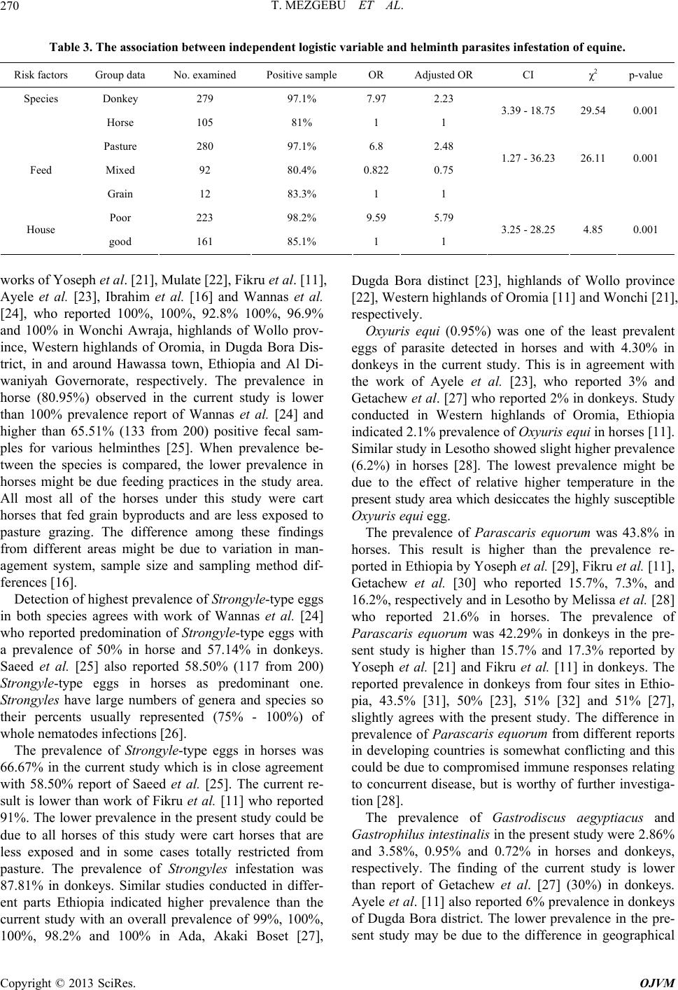 T. MEZGEBU ET AL. 270 Table 3. The association between independent logistic variable and helminth parasites infestation of equine. Risk factors Group data No. examined Positive sample OR Adjusted OR CI χ2 p-value Species Donkey 279 97.1% 7.97 2.23 Horse 105 81% 1 1 3.39 - 18.75 29.54 0.001 Pasture 280 97.1% 6.8 2.48 Mixed 92 80.4% 0.8220.75 1.27 - 36.23 26.11 0.001 Feed Grain 12 83.3% 1 1 Poor 223 98.2% 9.59 5.79 House good 161 85.1% 1 1 3.25 - 28.25 4.85 0.001 works of Yoseph et al. [21], Mulate [22], Fikru et al. [11], Ayele et al. [23], Ibrahim et al. [16] and Wannas et al. [24], who reported 100%, 100%, 92.8% 100%, 96.9% and 100% in Wonchi Awraja, highlands of Wollo prov- ince, Western highlands of Oromia, in Dugda Bora Dis- trict, in and around Hawassa town, Ethiopia and Al Di- waniyah Governorate, respectively. The prevalence in horse (80.95%) observed in the current study is lower than 100% prevalence report of Wannas et al. [24] and higher than 65.51% (133 from 200) positive fecal sam- ples for various helminthes [25]. When prevalence be- tween the species is compared, the lower prevalence in horses might be due feeding practices in the study area. All most all of the horses under this study were cart horses that fed grain byproducts and are less exposed to pasture grazing. The difference among these findings from different areas might be due to variation in man- agement system, sample size and sampling method dif- ferences [16]. Detection of highest prevalence of Strongyle-type eggs in both species agrees with work of Wannas et al. [24] who reported predomination of Strongyle-type eggs with a prevalence of 50% in horse and 57.14% in donkeys. Saeed et al. [25] also reported 58.50% (117 from 200) Strongyle-type eggs in horses as predominant one. Strongyles have large numbers of genera and species so their percents usually represented (75% - 100%) of whole nematodes infections [26]. The prevalence of Strongyle-type eggs in horses was 66.67% in the current study which is in close agreement with 58.50% report of Saeed et al. [25]. The current re- sult is lower than work of Fikru et al. [11] who reported 91%. The lower prevalence in the present study could be due to all horses of this study were cart horses that are less exposed and in some cases totally restricted from pasture. The prevalence of Strongyles infestation was 87.81% in donkeys. Similar studies conducted in differ- ent parts Ethiopia indicated higher prevalence than the current study with an overall prevalence of 99%, 100%, 100%, 98.2% and 100% in Ada, Akaki Boset [27], Dugda Bora distinct [23], highlands of Wollo province [22], Western highlands of Oromia [11] and Wonchi [21], respectively. Oxyuris equi (0.95%) was one of the least prevalent eggs of parasite detected in horses and with 4.30% in donkeys in the current study. This is in agreement with the work of Ayele et al. [23], who reported 3% and Getachew et al. [27] who reported 2% in donkeys. Study conducted in Western highlands of Oromia, Ethiopia indicated 2.1% prevalence of Oxyuris equi in horses [11]. Similar study in Lesotho showed slight higher prevalence (6.2%) in horses [28]. The lowest prevalence might be due to the effect of relative higher temperature in the present study area which desiccates the highly susceptible Oxyuris equi egg. The prevalence of Parascaris equorum was 43.8% in horses. This result is higher than the prevalence re- ported in Ethiopia by Yoseph et al. [29], Fikru et al. [11], Getachew et al. [30] who reported 15.7%, 7.3%, and 16.2%, respectively and in Lesotho by Melissa et al. [28] who reported 21.6% in horses. The prevalence of Parascaris equorum was 42.29% in donkeys in the pre- sent study is higher than 15.7% and 17.3% reported by Yoseph et al. [21] and Fikru et al. [11] in donkeys. The reported prevalence in donkeys from four sites in Ethio- pia, 43.5% [31], 50% [23], 51% [32] and 51% [27], slightly agrees with the present study. The difference in prevalence of Parascaris equorum from different reports in developing countries is somewhat conflicting and this could be due to compromised immune responses relating to concurrent disease, but is worthy of further investiga- tion [28]. The prevalence of Gastrodiscus aegyptiacus and Gastrophilus intestinalis in the present study were 2.86% and 3.58%, 0.95% and 0.72% in horses and donkeys, respectively. The finding of the current study is lower than report of Getachew et al. [27] (30%) in donkeys. Ayele et al. [11] also reported 6% prevalence in donkeys of Dugda Bora district. The lower prevalence in the pre- sent study may be due to the difference in geographical Copyright © 2013 SciRes. OJVM 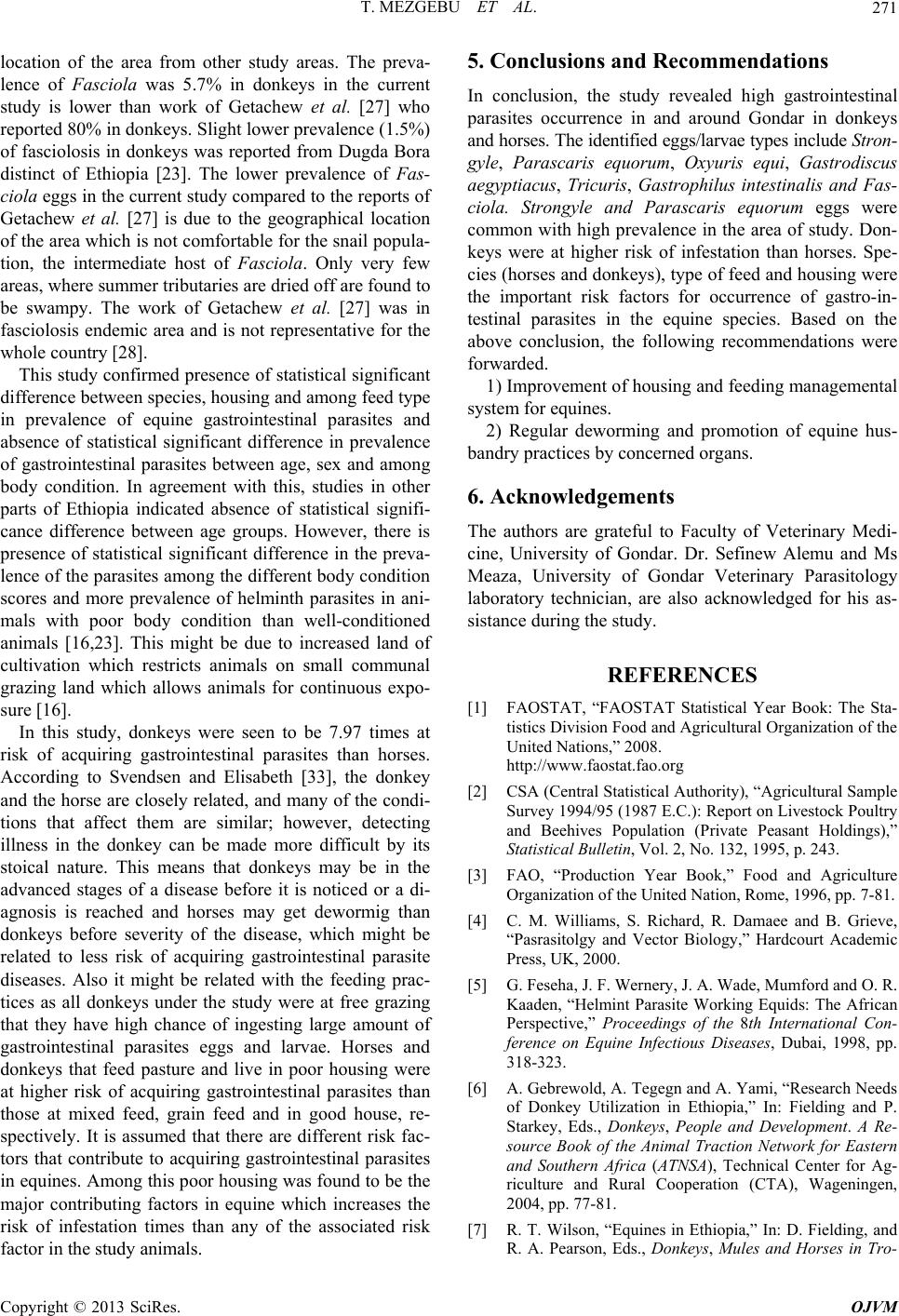 T. MEZGEBU ET AL. 271 location of the area from other study areas. The preva- lence of Fasciola was 5.7% in donkeys in the current study is lower than work of Getachew et al. [27] who reported 80% in donkeys. Slight lower prevalence (1.5%) of fasciolosis in donkeys was reported from Dugda Bora distinct of Ethiopia [23]. The lower prevalence of Fas- ciola eggs in the current study compared to the reports of Getachew et al. [27] is due to the geographical location of the area which is not comfortable for the snail popula- tion, the intermediate host of Fasciola. Only very few areas, where summer tributaries are dried off are found to be swampy. The work of Getachew et al. [27] was in fasciolosis endemic area and is not representative for the whole country [28]. This study confirmed presence of statistical significant difference between species, housing and among feed type in prevalence of equine gastrointestinal parasites and absence of statistical significant difference in prevalence of gastrointestinal parasites between age, sex and among body condition. In agreement with this, studies in other parts of Ethiopia indicated absence of statistical signifi- cance difference between age groups. However, there is presence of statistical significant difference in the preva- lence of the parasites among the different body condition scores and more prevalence of helminth parasites in ani- mals with poor body condition than well-conditioned animals [16,23]. This might be due to increased land of cultivation which restricts animals on small communal grazing land which allows animals for continuous expo- sure [16]. In this study, donkeys were seen to be 7.97 times at risk of acquiring gastrointestinal parasites than horses. According to Svendsen and Elisabeth [33], the donkey and the horse are closely related, and many of the condi- tions that affect them are similar; however, detecting illness in the donkey can be made more difficult by its stoical nature. This means that donkeys may be in the advanced stages of a disease before it is noticed or a di- agnosis is reached and horses may get dewormig than donkeys before severity of the disease, which might be related to less risk of acquiring gastrointestinal parasite diseases. Also it might be related with the feeding prac- tices as all donkeys under the study were at free grazing that they have high chance of ingesting large amount of gastrointestinal parasites eggs and larvae. Horses and donkeys that feed pasture and live in poor housing were at higher risk of acquiring gastrointestinal parasites than those at mixed feed, grain feed and in good house, re- spectively. It is assumed that there are different risk fac- tors that contribute to acquiring gastrointestinal parasites in equines. Among this poor housing was found to be the major contributing factors in equine which increases the risk of infestation times than any of the associated risk factor in the study animals. 5. Conclusions and Recommendations In conclusion, the study revealed high gastrointestinal parasites occurrence in and around Gondar in donkeys and horses. The identified eggs/larvae types include Stron- gyle, Parascaris equorum, Oxyuris equi, Gastrodiscus aegyptiacus, Tricuris, Gastrophilus intestinalis and Fas- ciola. Strongyle and Parascaris equorum eggs were common with high prevalence in the area of study. Don- keys were at higher risk of infestation than horses. Spe- cies (horses and donkeys), type of feed and housing were the important risk factors for occurrence of gastro-in- testinal parasites in the equine species. Based on the above conclusion, the following recommendations were forwarded. 1) Improvement of housing and feeding managemental system for equines. 2) Regular deworming and promotion of equine hus- bandry practices by concerned organs. 6. Acknowledgements The authors are grateful to Faculty of Veterinary Medi- cine, University of Gondar. Dr. Sefinew Alemu and Ms Meaza, University of Gondar Veterinary Parasitology laboratory technician, are also acknowledged for his as- sistance during the study. REFERENCES [1] FAOSTAT, “FAOSTAT Statistical Year Book: The Sta- tistics Division Food and Agricultural Organization of the United Nations,” 2008. http://www.faostat.fao.org [2] CSA (Central Statistical Authority), “Agricultural Sample Survey 1994/95 (1987 E.C.): Report on Livestock Poultry and Beehives Population (Private Peasant Holdings),” Statistical Bulletin, Vol. 2, No. 132, 1995, p. 243. [3] FAO, “Production Year Book,” Food and Agriculture Organization of the United Nation, Rome, 1996, pp. 7-81. [4] C. M. Williams, S. Richard, R. Damaee and B. Grieve, “Pasrasitolgy and Vector Biology,” Hardcourt Academic Press, UK, 2000. [5] G. Feseha, J. F. Wernery, J. A. Wade, Mumford and O. R. Kaaden, “Helmint Parasite Working Equids: The African Perspective,” Proceedings of the 8th International Con- ference on Equine Infectious Diseases, Dubai, 1998, pp. 318-323. [6] A. Gebrewold, A. Tegegn and A. Yami, “Research Needs of Donkey Utilization in Ethiopia,” In: Fielding and P. Starkey, Eds., Donkeys, People and Development. A Re- source Book of the Animal Traction Network for Eastern and Southern Africa (ATNSA), Technical Center for Ag- riculture and Rural Cooperation (CTA), Wageningen, 2004, pp. 77-81. [7] R. T. Wilson, “Equines in Ethiopia,” In: D. Fielding, and R. A. Pearson, Eds., Donkeys, Mules and Horses in Tro- Copyright © 2013 SciRes. OJVM 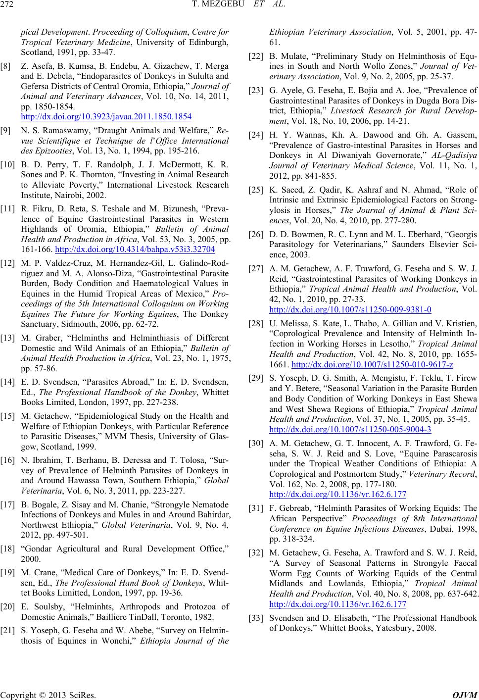 T. MEZGEBU ET AL. Copyright © 2013 SciRes. OJVM 272 pical Development. Proceeding of Colloquium, Centre for Tropical Veterinary Medicine, University of Edinburgh, Scotland, 1991, pp. 33-47. [8] Z. Asefa, B. Kumsa, B. Endebu, A. Gizachew, T. Merga and E. Debela, “Endoparasites of Donkeys in Sululta and Gefersa Districts of Central Oromia, Ethiopia,” Journal of Animal and Veterinary Advances, Vol. 10, No. 14, 2011, pp. 1850-1854. http://dx.doi.org/10.3923/javaa.2011.1850.1854 [9] N. S. Ramaswamy, “Draught Animals and Welfare,” Re- vue Scientifique et Technique de l’Office International des Epizooties, Vol. 13, No. 1, 1994, pp. 195-216. [10] B. D. Perry, T. F. Randolph, J. J. McDermott, K. R. Sones and P. K. Thornton, “Investing in Animal Research to Alleviate Poverty,” International Livestock Research Institute, Nairobi, 2002. [11] R. Fikru, D. Reta, S. Teshale and M. Bizunesh, “Preva- lence of Equine Gastrointestinal Parasites in Western Highlands of Oromia, Ethiopia,” Bulletin of Animal Health and Production in Africa, Vol. 53, No. 3, 2005, pp. 161-166. http://dx.doi.org/10.4314/bahpa.v53i3.32704 [12] M. P. Valdez-Cruz, M. Hernandez-Gil, L. Galindo-Rod- riguez and M. A. Alonso-Diza, “Gastrointestinal Parasite Burden, Body Condition and Haematological Values in Equines in the Humid Tropical Areas of Mexico,” Pro- ceedings of the 5th International Colloquium on Working Equines The Future for Working Equines, The Donkey Sanctuary, Sidmouth, 2006, pp. 62-72. [13] M. Graber, “Helminths and Helminthiasis of Different Domestic and Wild Animals of an Ethiopia,” Bulletin of Animal Health Production in Africa, Vol. 23, No. 1, 1975, pp. 57-86. [14] E. D. Svendsen, “Parasites Abroad,” In: E. D. Svendsen, Ed., The Professional Handbook of the Donkey, Whittet Books Limited, London, 1997, pp. 227-238. [15] M. Getachew, “Epidemiological Study on the Health and Welfare of Ethiopian Donkeys, with Particular Reference to Parasitic Diseases,” MVM Thesis, University of Glas- gow, Scotland, 1999. [16] N. Ibrahim, T. Berhanu, B. Deressa and T. Tolosa, “Sur- vey of Prevalence of Helminth Parasites of Donkeys in and Around Hawassa Town, Southern Ethiopia,” Global Veterinaria, Vol. 6, No. 3, 2011, pp. 223-227. [17] B. Bogale, Z. Sisay and M. Chanie, “Strongyle Nematode Infections of Donkeys and Mules in and Around Bahirdar, Northwest Ethiopia,” Global Veterinaria, Vol. 9, No. 4, 2012, pp. 497-501. [18] “Gondar Agricultural and Rural Development Office,” 2000. [19] M. Crane, “Medical Care of Donkeys,” In: E. D. Svend- sen, Ed., The Professional Hand Book of Donkeys, Whit- tet Books Limitted, London, 1997, pp. 19-36. [20] E. Soulsby, “Helminhts, Arthropods and Protozoa of Domestic Animals,” Bailliere TinDall, Toronto, 1982. [21] S. Yoseph, G. Feseha and W. Abebe, “Survey on Helmin- thosis of Equines in Wonchi,” Ethiopia Journal of the Ethiopian Veterinary Association, Vol. 5, 2001, pp. 47- 61. [22] B. Mulate, “Preliminary Study on Helminthosis of Equ- ines in South and North Wollo Zones,” Journal of Vet- erinary Association, Vol. 9, No. 2, 2005, pp. 25-37. [23] G. Ayele, G. Feseha, E. Bojia and A. Joe, “Prevalence of Gastrointestinal Parasites of Donkeys in Dugda Bora Dis- trict, Ethiopia,” Livestock Research for Rural Develop- ment, Vol. 18, No. 10, 2006, pp. 14-21. [24] H. Y. Wannas, Kh. A. Dawood and Gh. A. Gassem, “Prevalence of Gastro-intestinal Parasites in Horses and Donkeys in Al Diwaniyah Governorate,” AL-Qadisiya Journal of Veterinary Medical Science, Vol. 11, No. 1, 2012, pp. 841-855. [25] K. Saeed, Z. Qadir, K. Ashraf and N. Ahmad, “Role of Intrinsic and Extrinsic Epidemiological Factors on Strong- ylosis in Horses,” The Journal of Animal & Plant Sci- ences, Vol. 20, No. 4, 2010, pp. 277-280. [26] D. D. Bowmen, R. C. Lynn and M. L. Eberhard, “Georgis Parasitology for Veterinarians,” Saunders Elsevier Sci- ence, 2003. [27] A. M. Getachew, A. F. Trawford, G. Feseha and S. W. J. Reid, “Gastrointestinal Parasites of Working Donkeys in Ethiopia,” Tropical Animal Health and Production, Vol. 42, No. 1, 2010, pp. 27-33. http://dx.doi.org/10.1007/s11250-009-9381-0 [28] U. Melissa, S. Kate, L. Thabo, A. Gillian and V. Kristien, “Coprological Prevalence and Intensity of Helminth In- fection in Working Horses in Lesotho,” Tropical Animal Health and Production, Vol. 42, No. 8, 2010, pp. 1655- 1661. http://dx.doi.org/10.1007/s11250-010-9617-z [29] S. Yoseph, D. G. Smith, A. Mengistu, F. Teklu, T. Firew and Y. Betere, “Seasonal Variation in the Parasite Burden and Body Condition of Working Donkeys in East Shewa and West Shewa Regions of Ethiopia,” Tropical Animal Health and Production, Vol. 37, No. 1, 2005, pp. 35-45. http://dx.doi.org/10.1007/s11250-005-9004-3 [30] A. M. Getachew, G. T. Innocent, A. F. Trawford, G. Fe- seha, S. W. J. Reid and S. Love, “Equine Parascarosis under the Tropical Weather Conditions of Ethiopia: A Coprological and Postmortem Study,” Veterinary Record, Vol. 162, No. 2, 2008, pp. 177-180. http://dx.doi.org/10.1136/vr.162.6.177 [31] F. Gebreab, “Helminth Parasites of Working Equids: The African Perspective” Proceedings of 8th International Conference on Equine Infectious Diseases, Dubai, 1998, pp. 318-324. [32] M. Getachew, G. Feseha, A. Trawford and S. W. J. Reid, “A Survey of Seasonal Patterns in Strongyle Faecal Worm Egg Counts of Working Equids of the Central Midlands and Lowlands, Ethiopia,” Tropical Animal Health and Production, Vol. 40, No. 8, 2008, pp. 637-642. http://dx.doi.org/10.1136/vr.162.6.177 [33] Svendsen and D. Elisabeth, “The Professional Handbook of Donkeys,” Whittet Books, Yatesbury, 2008. |