

Paper Menu >>
Journal Menu >>
 Open Journal of Urology, 2013, 3, 223-226 http://dx.doi.org/10.4236/oju.2013.36042 Published Online October 2013 (http://www.scirp.org/journal/oju) Possible Origin of Aldosteronoma from Adrenohepatic Fusion Resulting in Intrahepatic Tumor* Takumi Takeuchi1, Takashi Inoue2, Keiko Kanemoto2, Kazuchika Hagiwara3, Koji Mikami1, Mami Hattori1, Masayoshi Zaitsu1, Akiko Tonooka4, Toshimasa Uekusa4, Susumu Uda2 1Department of Urology, Kanto Rosai Hospital, Kawasaki, Japan 2Nephrology, Kanto Rosai Hospital, Kawasaki, Japan 3Diagnostic Radiology, Kanto Rosai Hospital, Kawasaki, Japan 4Pathology, Kanto Rosai Hospital, Kawasaki, Japan Email: [email protected] Received August 9, 2013; revised August 28, 2013; accepted September 3, 2013 Copyright © 2013 Takumi Takeuchi et al. This is an open access article distributed under the Creative Commons Attribution License, which permits unrestricted use, distribution, and reproduction in any medium, provided the original work is properly cited. ABSTRACT A 69-year-old woman was diagnosed with primary aldosteronism. An enhanced computed tomography (CT) scan be- fore surgery indicated a right adrenal tumor outside the liver. Venous sampling tests revealed unilateral overproduction of aldosterone by the right adrenal gland. Separation of the right adrenal cortex from the liver parenchyma was imprac- tical during a laparoscopic right adrenalectomy because of the solid attachment between the two. Therefore, the exis- tence of adrenohepatic fusion was determined. An incision was made within the right adrenal gland, leaving completely the intrahepatic adrenal tissue on the inner side of the liver, because a partial hepatectomy was not preoperatively planned, and the patient was not informed of the consent before the surgery. Pathological examination did not reveal macro- or micro-adenomas in the resected right adrenal tissue. Aldosterone to renin ratio was as high as 1380 at 22 days following the surgery. Therefore, aldosteronoma originated from the adrenohepatic fusion that remained on the inner side of the liver was highly suspected. The patient’s blood pressure was well controlled, and she did not prefer hepatec- tomy to be further performed, and therefore, medical therapy was continued. When planning the type of surgery (laparoscopic or open) in these potentially confusing cases, it might be necessary to consider a possibility of the unex- pected intraoperative diagnosis and the immediate measures to be performed based on the diagnosis. Keywords: Adrenal; Tumor; Adrenohepatic Fusion 1. Introduction Primary aldosteronism is more frequent than previously speculated and it results in damaging the heart, blood vessels, and kidneys [1]. Majority of the hypertensive patients with primary aldosteronism, characterized by an autonomous secretion of aldosterone from the aldoster- onoma, are diagnosed with the introduction of advanced imaging, such as computed tomography (CT) scans, mag- netic resonance imaging (MRI), and ultrasound, as well as sophisticated advancements in endocrinology tests. Pri- mary aldosteronism among hypertensive patients is de- termined by the aldosterone to renin ratio, and then con- firmed by four testing procedures: oral sodium loading, saline infusion, fludrocortisone suppression, and capto- pril challenge. Furthermore, the laterality of aldosterono- mas is determined by interventional venous sampling techniques [2,3]. Those patients recently undergo laparoscopic adrena- lectomy for antihypertensive drugs to be discontinued or reduced and for the vital organs such as the brain, heart, kidneys, and vessels to be protected from further damages [4]. Here, we present a case of primary aldosteronism, where adrenohepatic fusion was identified during a laparo- scopic right adrenalectomy, and the aldosteronoma was speculated to be originated from adrenohepatic fusion. 2. Case Report A 69-year-old woman was diagnosed with primary al- dosteronism using endocrinology evaluation, following the cessation of antihypertensive drugs. The patient’s se- rum potassium level was 3.1 mEq/L, and the aldosterone to renin ratio was 406. After captopril challenge, the al- dosterone to renin ratio was 249 (positive if >200) con- *The authors declared no conflicts of interest. C opyright © 2013 SciRes. OJU 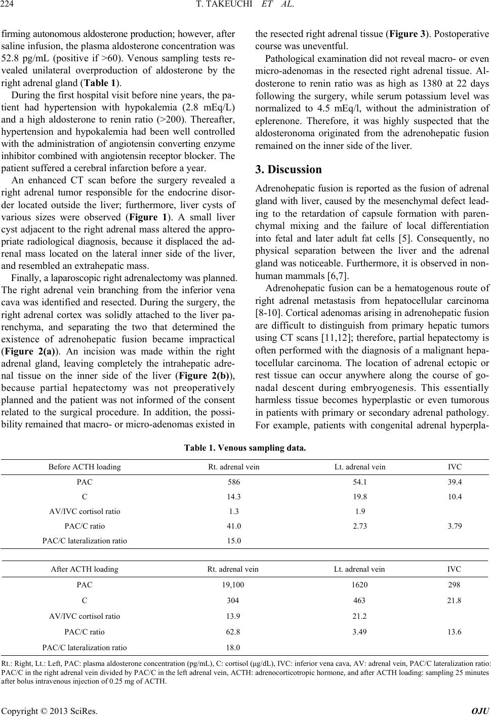 T. TAKEUCHI ET AL. 224 firming autonomous aldosterone production; however, after saline infusion, the plasma aldosterone concentration was 52.8 pg/mL (positive if >60). Venous sampling tests re- vealed unilateral overproduction of aldosterone by the right adrenal gland (Table 1). During the first hospital visit before nine years, the pa- tient had hypertension with hypokalemia (2.8 mEq/L) and a high aldosterone to renin ratio (>200). Thereafter, hypertension and hypokalemia had been well controlled with the administration of angiotensin converting enzyme inhibitor combined with angiotensin receptor blocker. The patient suffered a cerebral infarction before a year. An enhanced CT scan before the surgery revealed a right adrenal tumor responsible for the endocrine disor- der located outside the liver; furthermore, liver cysts of various sizes were observed (Figure 1). A small liver cyst adjacent to the right adrenal mass altered the appro- priate radiological diagnosis, because it displaced the ad- renal mass located on the lateral inner side of the liver, and resembled an extrahepatic mass. Finally, a laparoscopic right adrenalectomy was planned. The right adrenal vein branching from the inferior vena cava was identified and resected. During the surgery, the right adrenal cortex was solidly attached to the liver pa- renchyma, and separating the two that determined the existence of adrenohepatic fusion became impractical (Figure 2(a)). An incision was made within the right adrenal gland, leaving completely the intrahepatic adre- nal tissue on the inner side of the liver (Figure 2(b)), because partial hepatectomy was not preoperatively planned and the patient was not informed of the consent related to the surgical procedure. In addition, the possi- bility remained that macro- or micro-adenomas existed in the resected right adrenal tissue (Figure 3). Postoperative course was uneventful. Pathological examination did not reveal macro- or even micro-adenomas in the resected right adrenal tissue. Al- dosterone to renin ratio was as high as 1380 at 22 days following the surgery, while serum potassium level was normalized to 4.5 mEq/l, without the administration of eplerenone. Therefore, it was highly suspected that the aldosteronoma originated from the adrenohepatic fusion remained on the inner side of the liver. 3. Discussion Adrenohepatic fusion is reported as the fusion of adrenal gland with liver, caused by the mesenchymal defect lead- ing to the retardation of capsule formation with paren- chymal mixing and the failure of local differentiation into fetal and later adult fat cells [5]. Consequently, no physical separation between the liver and the adrenal gland was noticeable. Furthermore, it is observed in non- human mammals [6,7]. Adrenohepatic fusion can be a hematogenous route of right adrenal metastasis from hepatocellular carcinoma [8-10]. Cortical adenomas arising in adrenohepatic fusion are difficult to distinguish from primary hepatic tumors using CT scans [11,12]; therefore, partial hepatectomy is often performed with the diagnosis of a malignant hepa- tocellular carcinoma. The location of adrenal ectopic or rest tissue can occur anywhere along the course of go- nadal descent during embryogenesis. This essentially harmless tissue becomes hyperplastic or even tumorous in patients with primary or secondary adrenal pathology. For example, patients with congenital adrenal hyperpla- Table 1. Venous sampling data. Before ACTH loading Rt. adrenal vein Lt. adrenal vein IVC PAC 586 54.1 39.4 C 14.3 19.8 10.4 AV/IVC cortisol ratio 1.3 1.9 PAC/C ratio 41.0 2.73 3.79 PAC/C lateralization ratio 15.0 After ACTH loading Rt. adrenal vein Lt. adrenal vein IVC PAC 19,100 1620 298 C 304 463 21.8 AV/IVC cortisol ratio 13.9 21.2 PAC/C ratio 62.8 3.49 13.6 PAC/C lateralization ratio 18.0 Rt.: Right, Lt.: Left, PAC: plasma aldosterone concentration (pg/mL), C: cortisol (μg/dL), IVC: inferior vena cava, AV: adrenal vein, PAC/C lateralization ratio: PAC/C in the right adrenal vein divided by PAC/C in the left adrenal vein, ACTH: adrenocorticotropic hormone, and after ACTH loading: sampling 25 minutes after bolus intravenous injection of 0.25 mg of ACTH. Copyright © 2013 SciRes. OJU 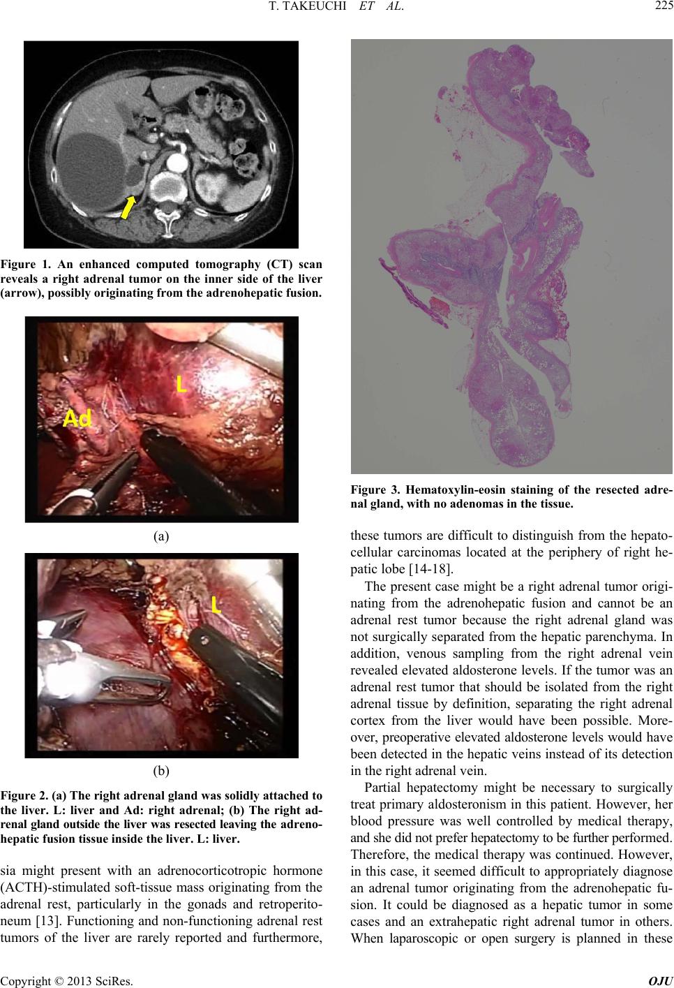 T. TAKEUCHI ET AL. 225 Figure 1. An enhanced computed tomography (CT) scan reveals a right adrenal tumor on the inner side of the liver (arrow), possibly originating from the adrenohepatic fusion. (a) (b) Figure 2. (a) The right adrenal gland was solidly attached to the liver. L: liver and Ad: right adrenal; (b) The right ad- renal gland outside the liver was resected leaving the a dre no- hepatic fusion tissue inside the liver. L: liver. sia might present with an adrenocorticotropic hormone (ACTH)-stimulated soft-tissue mass originating from the adrenal rest, particularly in the gonads and retroperito- neum [13]. Functioning and non-functioning adrenal rest tumors of the liver are rarely reported and furthermore, Figure 3. Hematoxylin-eosin staining of the resected adre- nal gland, with no adenomas in the tissue. these tumors are difficult to distinguish from the hepato- cellular carcinomas located at the periphery of right he- patic lobe [14-18]. The present case might be a right adrenal tumor origi- nating from the adrenohepatic fusion and cannot be an adrenal rest tumor because the right adrenal gland was not surgically separated from the hepatic parenchyma. In addition, venous sampling from the right adrenal vein revealed elevated aldosterone levels. If the tumor was an adrenal rest tumor that should be isolated from the right adrenal tissue by definition, separating the right adrenal cortex from the liver would have been possible. More- over, preoperative elevated aldosterone levels would have been detected in the hepatic veins instead of its detection in the right adrenal vein. Partial hepatectomy might be necessary to surgically treat primary aldosteronism in this patient. However, her blood pressure was well controlled by medical therapy, and she did not prefer hepatectomy to be further performed. Therefore, the medical therapy was continued. However, in this case, it seemed difficult to appropriately diagnose an adrenal tumor originating from the adrenohepatic fu- sion. It could be diagnosed as a hepatic tumor in some cases and an extrahepatic right adrenal tumor in others. When laparoscopic or open surgery is planned in these Copyright © 2013 SciRes. OJU  T. TAKEUCHI ET AL. 226 potentially confusing cases, it might be necessary to con- sider a possibility of the unexpected intraoperative diag- nosis and the immediate measures to be performed based on the diagnosis. According to the medical databases, there are no previous cases of patients where intrahepatic adrenal tumors originating from the adrenohepatic fusion were identified during the laparoscopic or open surgery. REFERENCES [1] G. P. Rossi, “A Comprehensive Review of the Clinical Aspects of Primary Aldosteronism,” Nature Reviews En- docrinology, Vol. 7, No. 8, 2011, pp. 485-495. http://dx.doi.org/10.1038/nrendo.2011.76 [2] M. Salvà, M. V. Cicala and F. Mantero, “Primary Aldos- teronism: The Role of Confirmatory Tests,” Hormone and Metabolic Research, Vol. 44, No. 3, 2012, pp. 177- 180. http://dx.doi.org/10.1055/s-0032-1304661 [3] P. Mulatero, S. Monticone, C. Bertello, G. Mengozzi, D. Tizzani, A. Iannaccone and F. Veglio, “Confirmatory Tests in the Diagnosis of Primary Aldosteronism,” Hor- mone and Metabolic Research, Vol. 42, No. 6, 2010,pp. 406-410. http://dx.doi.org/10.1055/s-0029-1246186 [4] O. Steichen, F. Zinzindohoué, P. F. Plouin and L. Amar, “Outcomes of Adrenalectomy in Patients with Unilateral Primary Aldosteronism: A Review,” Hormone and Meta- bolic Research, Vol. 44, No. 3, 2012, pp. 221-227. http://dx.doi.org/10.1055/s-0031-1299681 [5] L. H. Honoré and K. E. O’Hara, “Combined Adrenorenal Fusion and Adrenohepatic Adhesion: A Case Report with Review of the Literature and Discussion of Pathogene- sis,” Journal of Urology, Vol. 115, No. 3, 1976, pp. 323- 325. [6] R. Chamanza, H. A. Marxfeld, A. I. Blanco, S. W. Naylor and A. E. Bradley, “Incidences and Range of Spontane- ous Findings in Control Cynomolgus Monkeys (Macaca fascicularis) Used in Toxicity Studies,” Journal of Toxi- cologic Pathology, Vol. 38, No. 4, 2010, pp. 642-657. http://dx.doi.org/10.1177/0192623310368981 [7] O. Quesada-Canales, A. Suárez-Bonnet, G. A. Ramírez, M. Aguirre-Sanceledonio, M. Andrada, M. Rivero and A. Espinosa de Los Monteros, “Adrenohepatic Fusion in Do- mestic Ferrets (Mustela putorius furo),”Journal of Com- parative Pathology, Vol. 149, No. 2-3, 2013, pp. 314-317. http://dx.doi.org/10.1016/j.jcpa.2013.02.003 [8] S. Iwamoto, K. Okuda, N. Takeda, K. Sonoda and H. Sanefuji, “Case Report: Right-Sided Periadrenal Metasta- sis Supplied by the Hepatic Artery. Clue to the Genesis of Pedunculated Hepatocellular Carcinoma,” Journal of Ga- stroenterology and Hepatology, Vol. 12, No. 5, 1997, pp. 392-397. http://dx.doi.org/10.1111/j.1440-1746.1997.tb00449.x [9] K. Okuda, M. Arakawa, Y. Kubo, K. Sakata, M. Kage, S. Iwamoto, S. Takeda, K. Sonoda and H. Sanefuji, “Right- Sided Pedunculated Hepatocellular Carcinoma: A Form of Adrenal Metastasis,” Hepatology, Vol. 27, No. 1, 1998, pp. 81-85. http://dx.doi.org/10.1002/hep.510270114 [10] K. Okano, H. Usuki and H. Maeta, “Adrenal Metastasis from Hepatocellular Carcinoma through an ADRENO- HEPATIC Fusion,” Journal of Clinical Gastroenterology, Vol. 38, No. 10, 2004, p. 912. http://dx.doi.org/10.1097/00004836-200411000-00019 [11] H. S. Woo, K. H. Lee, S. Y. Park, H. S. Han, C. J. Yoon and Y. H. Kim, “Adrenal Cortical Adenoma in Adreno- hepatic Fusion Tissue: A Mimic of Malignant Hepatic Tumor at CT,” American Journal of Roentgenology, Vol. 188, No. 3, 2007, pp. W246-W248. http://dx.doi.org/10.2214/AJR.05.0498 [12] B. K. Park, C. K. Kim, B. C. Jung and Y. L. Suh, “Corti- cal Adenoma in Adrenohepatic Fusion Tissue: Clue to Making a Correct Diagnosis at Preoperative Computed Tomography Examination,” European Urology, Vol. 56, No. 6, 2009, pp. 1082-1085. http://dx.doi.org/10.1016/j.eururo.2009.05.007 [13] T. D. Barwick, A. Malhotra, J. A. Webb, M. O. Savage and R. H. Reznek, “Embryology of the Adrenal Glands and Its Relevance to Diagnostic Imaging,” Clinical Radi- ology, Vol. 60, No. 9, 2005, pp. 953-959. http://dx.doi.org/10.1016/j.crad.2005.04.006 [14] Y. M. Shin, “Hepatic Adrenal Rest Tumor Mimicking Hepatocellular Carcinoma,” Korean Journal of Hepatol- ogy, Vol. 16, No. 3, 2010, pp. 338-341. http://dx.doi.org/10.3350/kjhep.2010.16.3.338 [15] Y. Baba, T. Beppu, K. Imai, T. Masuda, K. Iyama, H. Sasano and H. Baba, “A Case of Adrenal Rest Tumor of the Liver: Radiological Imaging and Immunohistochemi- cal Study of Steroidogenic Enzymes,” Hepatology Re- search, Vol. 38, No. 11, 2008, pp. 1154-1158. http://dx.doi.org/10.1111/j.1872-034X.2008.00360.x [16] K. Arai, H. Muro, M. Suzuki, N. Oba, K. Ito and H. Sa- sano, “Adrenal Rest Tumor of the Liver: A Case Report with Immunohistochemical Investigation of Steroido- genesis,” Pathology International, Vol. 50, No. 3, 2000, pp. 244-248. http://dx.doi.org/10.1046/j.1440-1827.2000.01029.x [17] P. Contreras, E. Altieri, C. Liberman, A. Gac, A. Rojas, A. Ibarra, M. Ravanal, M. Serón-Ferré, “Adrenal Rest Tu- mor of the Liver Causing Cushing’s Syndrome: Therapy with Ketoconazole Preceding an Apparent Surgical Cure,” The Journal of Clinical Endocrinology & Metabolism, Vol. 60, No. 1, 1985, pp. 21-28. http://dx.doi.org/10.1210/jcem-60-1-21 [18] E. Z. Wallace, J. R. Leonidas, A. E. Stanek and A. Avra- mides, “Endocrine Studies in a Patient with Functioning Adrenal Rest Tumor of the Liver,” American Journal of Medicine, Vol. 70, No. 5, 1981, pp. 1122-1125. http://dx.doi.org/10.1016/0002-9343(81)90886-X Copyright © 2013 SciRes. OJU |