

Paper Menu >>
Journal Menu >>
 Surgical Science, 2013, 4, 415-420 http://dx.doi.org/10.4236/ss.2013.410081 Published Online October 2013 (http://www.scirp.org/journal/ss) The Role of Color Doppler Ultrasound Arterial Mapping for Decision Making in the Treatment of Patients with Lower Extremity Peripheral Arterial Disease Ali Babaei Jandaghi1, Zahra Mardanshahi1, Ahmad Alizadeh1, Iraj Baghi2, Hossein Hemmati3, Narges Tabarzan Baboli4, Shabnam Alizadeh Arasi4, Amin Keshavarzzirak1* 1Department of Radiology, Poursina Hospital, Guilan University of Medical Sciences, Rasht, Iran 2Department of Vascular Surgery, Poursina Hospital, Guilan University of Medical Sciences, Rasht, Iran 3Department of Vascular Surgery, Razi Hospital, Guilan University of Medical Sciences, Rasht, Iran 4Department of Cardiology, Heshmat Hospital, Guilan University of Medical Sciences, Rasht, Iran Email: *[email protected] Received August 9, 2013; revised August 31, 2013; accepted September 7, 2013 Copyright © 2013 Ali Babaei Jandaghi et al. This is an open access article distributed under the Creative Commons Attribution Li- cense, which permits unrestricted use, distribution, and reproduction in any medium, provided the original work is properly cited. ABSTRACT Purpose: To assess the efficacy of color Doppler imaging for decision making in the treatment of patients with lower extremity peripheral arterial disease (PAD) compared to digital subtraction angiography (DSA). Materials and Meth- ods: Color Doppler scan was done on patients suspected for lower limb PAD, a day prior to the DSA which was done by a vascular surgeon. Also, for the patients who were candidates for endovascular intervention based on the color Doppler arterial mapping results, endovascular interventions were performed at the same time if the DSA findings are correlated with the color Doppler map. The grading for evaluated segments was normal, insignificant stenosis (<50%), hemodynamically significant stenosis (≥50%) and occlusion. We yielded the diagnostic efficacy indices of Doppler for detecting arterial stenosis in each 18 different arterial segments below the renal arteries including, infrarenal aorta, common and external iliac, common femoral, superficial femoral (proximal, middle and distal segments), deep femoral, popliteal artery, tibioperoneal trunk, anterior and posterior tibial arteries (proximal, middle and distal segments) and peroneal artery (proximal and distal segments). Then, we yielded the kappa agreement between Doppler and DSA find- ings considering the grade of stenosis in 18 arterial segments separately. Results: Totally 115 lower extremities (2045 arterial segments) were evaluated in 90 patients [mean age: 60.8 ± 8.9 (range: 47 - 84 years old)] of which 68 (75.6%) were men. The sensitivity of color Doppler for all arterial segments was 90% or higher except for common iliac artery, distal segment of superficial femoral artery and proximal segments of anterior and posterior tibialis and peroneal arter- ies. However, the specificity was 89% or higher, in all arterial segments. Kappa agreement was 0.72 or higher in all segments (All P-Values < 0.001). Conclusion: This study suggests that considering excellent capability of color Dop- pler sonography in the evaluation of lower extremity arterial disease, color Doppler arterial mapping is sufficient for decision making in the treatment of these patients and can reduce the rate of diagnostic angiography. Keywords: Lower Extremity; Peripheral Arterial Disease; Color Doppler Sonography; Arterial Mapping; Digital Subtraction Angiography 1. Introduction Peripheral arterial disease (PAD) is a common problem in the elderly, especially among diabetic patients. When PAD limits the patients’ lifestyle, diagnostic imaging is performed to characterize the number, length, level and severity of the lesions. Although Doppler ultrasonogra- phy is safe, relatively inexpensive, reproducible, a non- ionizing method, non-invasive and widely available as an outpatient service, it is yet time consuming and operator dependent. On the other hand, digital subtraction an- giography (DSA) provides easily visualized images of arterial tree. It can be used for endovascular interventions. However, it’s of high cost, ionizing radiation, need for contrast agents and invasive nature, and makes this pro- cedure unsuitable for screening or for follow-up purposes [1-4]. Color Doppler imaging is not only a morphological but also a functional study, providing information about *Corresponding author. C opyright © 2013 SciRes. SS 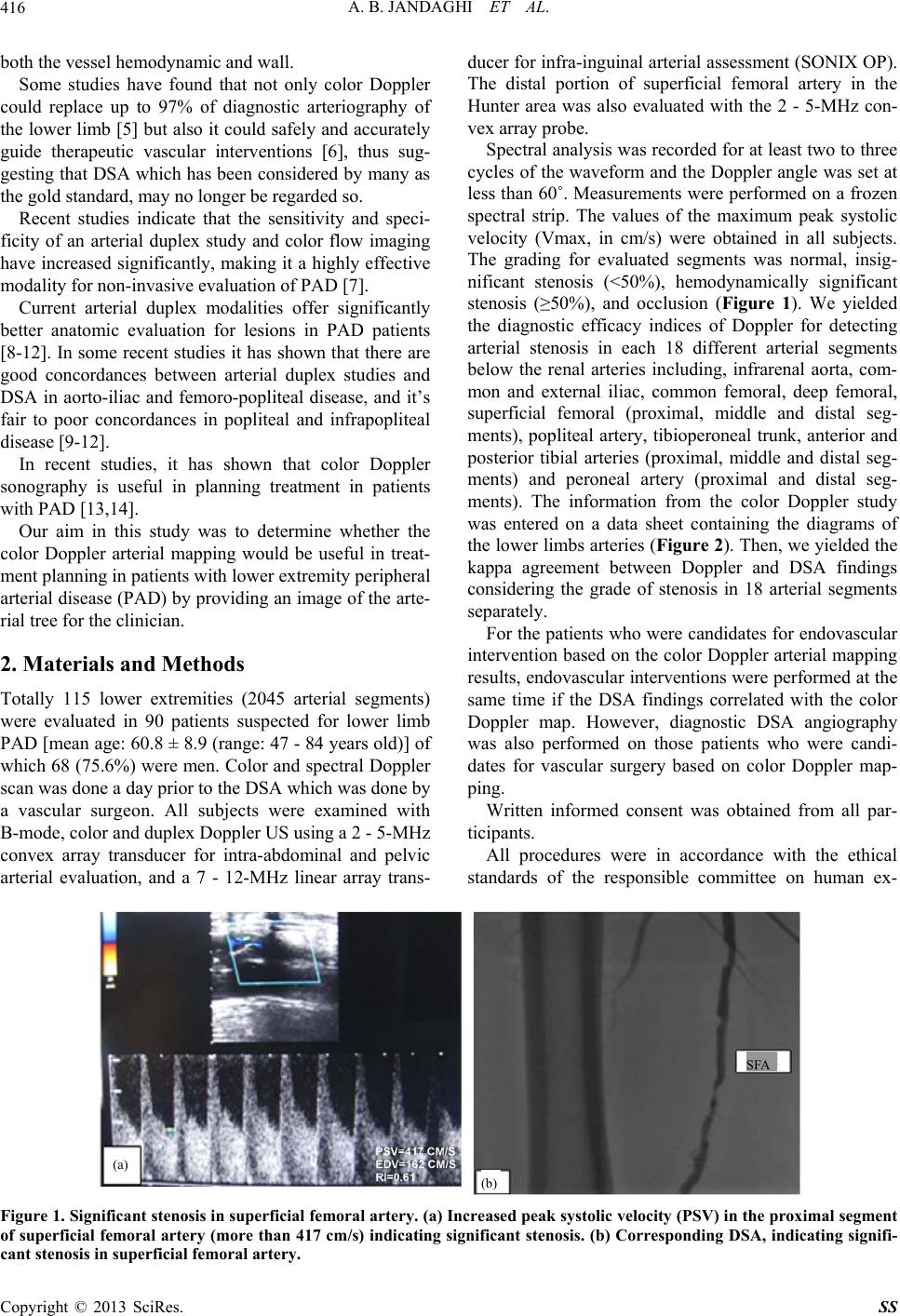 A. B. JANDAGHI ET AL. 416 both the vessel hemodynamic and wall. Some studies have found that not only color Doppler could replace up to 97% of diagnostic arteriography of the lower limb [5] but also it could safely and accurately guide therapeutic vascular interventions [6], thus sug- gesting that DSA which has been considered by many as the gold standard, may no longer be regarded so. Recent studies indicate that the sensitivity and speci- ficity of an arterial duplex study and color flow imaging have increased significantly, making it a highly effective modality for non-invasive evaluation of PAD [7]. Current arterial duplex modalities offer significantly better anatomic evaluation for lesions in PAD patients [8-12]. In some recent studies it has shown that there are good concordances between arterial duplex studies and DSA in aorto-iliac and femoro-popliteal disease, and it’s fair to poor concordances in popliteal and infrapopliteal disease [9-12]. In recent studies, it has shown that color Doppler sonography is useful in planning treatment in patients with PAD [13,14]. Our aim in this study was to determine whether the color Doppler arterial mapping would be useful in treat- ment planning in patients with lower extremity peripheral arterial disease (PAD) by providing an image of the arte- rial tree for the clinician. 2. Materials and Methods Totally 115 lower extremities (2045 arterial segments) were evaluated in 90 patients suspected for lower limb PAD [mean age: 60.8 ± 8.9 (range: 47 - 84 years old)] of which 68 (75.6%) were men. Color and spectral Doppler scan was done a day prior to the DSA which was done by a vascular surgeon. All subjects were examined with B-mode, color and duplex Doppler US using a 2 - 5-MHz convex array transducer for intra-abdominal and pelvic arterial evaluation, and a 7 - 12-MHz linear array trans- ducer for infra-inguinal arterial assessment (SONIX OP). The distal portion of superficial femoral artery in the Hunter area was also evaluated with the 2 - 5-MHz con- vex array probe. Spectral analysis was recorded for at least two to three cycles of the waveform and the Doppler angle was set at less than 60˚. Measurements were performed on a frozen spectral strip. The values of the maximum peak systolic velocity (Vmax, in cm/s) were obtained in all subjects. The grading for evaluated segments was normal, insig- nificant stenosis (<50%), hemodynamically significant stenosis (≥50%), and occlusion (Figure 1). We yielded the diagnostic efficacy indices of Doppler for detecting arterial stenosis in each 18 different arterial segments below the renal arteries including, infrarenal aorta, com- mon and external iliac, common femoral, deep femoral, superficial femoral (proximal, middle and distal seg- ments), popliteal artery, tibioperoneal trunk, anterior and posterior tibial arteries (proximal, middle and distal seg- ments) and peroneal artery (proximal and distal seg- ments). The information from the color Doppler study was entered on a data sheet containing the diagrams of the lower limbs arteries (Figure 2). Then, we yielded the kappa agreement between Doppler and DSA findings considering the grade of stenosis in 18 arterial segments separately. For the patients who were candidates for endovascular intervention based on the color Doppler arterial mapping results, endovascular interventions were performed at the same time if the DSA findings correlated with the color Doppler map. However, diagnostic DSA angiography was also performed on those patients who were candi- dates for vascular surgery based on color Doppler map- ping. Written informed consent was obtained from all par- ticipants. All procedures were in accordance with the ethical standards of the responsible committee on human ex- (a) (b) SF A Figure 1. Significant stenosis in superficial femoral artery. (a) Increased peak systolic velocity (PSV) in the proximal segment of superficial femoral artery (more than 417 cm/s) indicating significant stenosis. (b) Corresponding DSA, indicating signifi- cant stenosis in superficial femoral artery. Copyright © 2013 SciRes. SS 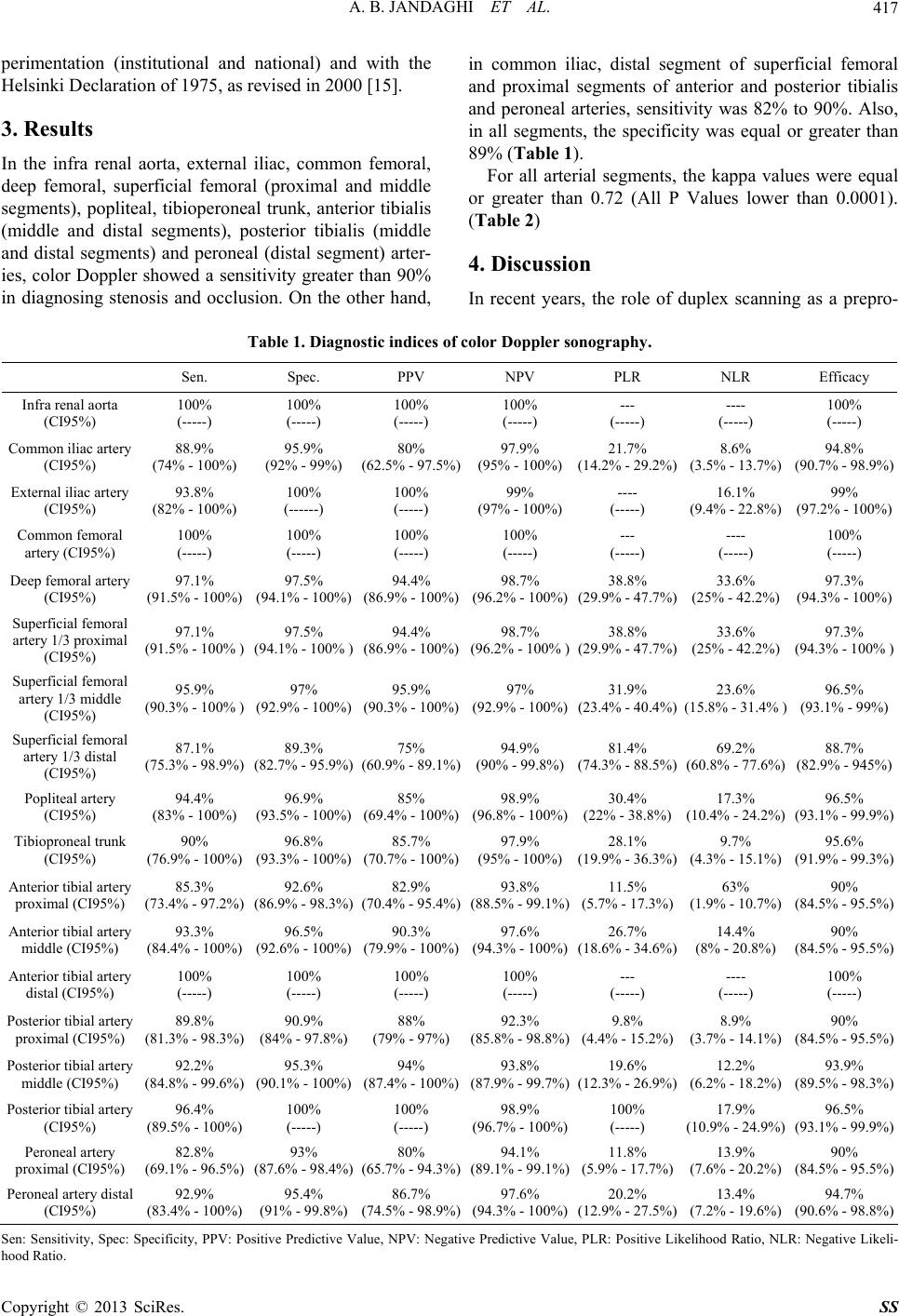 A. B. JANDAGHI ET AL. 417 perimentation (institutional and national) and with the Helsinki Declaration of 1975, as revised in 2000 [15]. 3. Results In the infra renal aorta, external iliac, common femoral, deep femoral, superficial femoral (proximal and middle segments), popliteal, tibioperoneal trunk, anterior tibialis (middle and distal segments), posterior tibialis (middle and distal segments) and peroneal (distal segment) arter- ies, color Doppler showed a sensitivity greater than 90% in diagnosing stenosis and occlusion. On the other hand, in common iliac, distal segment of superficial femoral and proximal segments of anterior and posterior tibialis and peroneal arteries, sensitivity was 82% to 90%. Also, in all segments, the specificity was equal or greater than 89% (Table 1). For all arterial segments, the kappa values were equal or greater than 0.72 (All P Values lower than 0.0001). (Table 2) 4. Discussion In recent years, the role of duplex scanning as a prepro- Table 1. Diagnostic indices of color Doppler sonography. Sen. Spec. PPV NPV PLR NLR Efficacy Infra renal aorta (CI95%) 100% (-----) 100% (-----) 100% (-----) 100% (-----) --- (-----) ---- (-----) 100% (-----) Common iliac artery (CI95%) 88.9% (74% - 100%) 95.9% (92% - 99%) 80% (62.5% - 97.5%) 97.9% (95% - 100%) 21.7% (14.2% - 29.2%) 8.6% (3.5% - 13.7%) 94.8% (90.7% - 98.9%) External iliac artery (CI95%) 93.8% (82% - 100%) 100% (------) 100% (-----) 99% (97% - 100%) ---- (-----) 16.1% (9.4% - 22.8%) 99% (97.2% - 100%) Common femoral artery (CI95%) 100% (-----) 100% (-----) 100% (-----) 100% (-----) --- (-----) ---- (-----) 100% (-----) Deep femoral artery (CI95%) 97.1% (91.5% - 100%) 97.5% (94.1% - 100%) 94.4% (86.9% - 100%) 98.7% (96.2% - 100%) 38.8% (29.9% - 47.7%) 33.6% (25% - 42.2%) 97.3% (94.3% - 100%) Superficial femoral artery 1/3 proximal (CI95%) 97.1% (91.5% - 100% ) 97.5% (94.1% - 100% ) 94.4% (86.9% - 100%) 98.7% (96.2% - 100% ) 38.8% (29.9% - 47.7%) 33.6% (25% - 42.2%) 97.3% (94.3% - 100% ) Superficial femoral artery 1/3 middle (CI95%) 95.9% (90.3% - 100% ) 97% (92.9% - 100%) 95.9% (90.3% - 100%) 97% (92.9% - 100%) 31.9% (23.4% - 40.4%) 23.6% (15.8% - 31.4% ) 96.5% (93.1% - 99%) Superficial femoral artery 1/3 distal (CI95%) 87.1% (75.3% - 98.9%) 89.3% (82.7% - 95.9%) 75% (60.9% - 89.1%) 94.9% (90% - 99.8%) 81.4% (74.3% - 88.5%) 69.2% (60.8% - 77.6%) 88.7% (82.9% - 945%) Popliteal artery (CI95%) 94.4% (83% - 100%) 96.9% (93.5% - 100%) 85% (69.4% - 100%) 98.9% (96.8% - 100%) 30.4% (22% - 38.8%) 17.3% (10.4% - 24.2%) 96.5% (93.1% - 99.9%) Tibioproneal trunk (CI95%) 90% (76.9% - 100%) 96.8% (93.3% - 100%) 85.7% (70.7% - 100%) 97.9% (95% - 100%) 28.1% (19.9% - 36.3%) 9.7% (4.3% - 15.1%) 95.6% (91.9% - 99.3%) Anterior tibial artery proximal (CI95%) 85.3% (73.4% - 97.2%) 92.6% (86.9% - 98.3%) 82.9% (70.4% - 95.4%) 93.8% (88.5% - 99.1%) 11.5% (5.7% - 17.3%) 63% (1.9% - 10.7%) 90% (84.5% - 95.5%) Anterior tibial artery middle (CI95%) 93.3% (84.4% - 100%) 96.5% (92.6% - 100%) 90.3% (79.9% - 100%) 97.6% (94.3% - 100%) 26.7% (18.6% - 34.6%) 14.4% (8% - 20.8%) 90% (84.5% - 95.5%) Anterior tibial artery distal (CI95%) 100% (-----) 100% (-----) 100% (-----) 100% (-----) --- (-----) ---- (-----) 100% (-----) Posterior tibial artery proximal (CI95%) 89.8% (81.3% - 98.3%) 90.9% (84% - 97.8%) 88% (79% - 97%) 92.3% (85.8% - 98.8%) 9.8% (4.4% - 15.2%) 8.9% (3.7% - 14.1%) 90% (84.5% - 95.5%) Posterior tibial artery middle (CI95%) 92.2% (84.8% - 99.6%) 95.3% (90.1% - 100%) 94% (87.4% - 100%) 93.8% (87.9% - 99.7%) 19.6% (12.3% - 26.9%) 12.2% (6.2% - 18.2%) 93.9% (89.5% - 98.3%) Posterior tibial artery (CI95%) 96.4% (89.5% - 100%) 100% (-----) 100% (-----) 98.9% (96.7% - 100%) 100% (-----) 17.9% (10.9% - 24.9%) 96.5% (93.1% - 99.9%) Peroneal artery proximal (CI95%) 82.8% (69.1% - 96.5%) 93% (87.6% - 98.4%) 80% (65.7% - 94.3%) 94.1% (89.1% - 99.1%) 11.8% (5.9% - 17.7%) 13.9% (7.6% - 20.2%) 90% (84.5% - 95.5%) Peroneal artery distal (CI95%) 92.9% (83.4% - 100%) 95.4% (91% - 99.8%) 86.7% (74.5% - 98.9%) 97.6% (94.3% - 100%) 20.2% (12.9% - 27.5%) 13.4% (7.2% - 19.6%) 94.7% (90.6% - 98.8%) Sen: Sensitivity, Spec: Specificity, PPV: Positive Predictive Value, NPV: Negative Predictive Value, PLR: Positive Likelihood Ratio, NLR: Negative Likeli- hood Ratio. Copyright © 2013 SciRes. SS 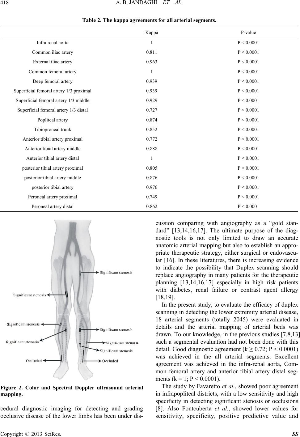 A. B. JANDAGHI ET AL. 418 Table 2. The kappa agreements for all arterial segments. Kappa P-value Infra renal aorta 1 P < 0.0001 Common iliac artery 0.811 P < 0.0001 External iliac artery 0.963 P < 0.0001 Common femoral artery 1 P < 0.0001 Deep femoral artery 0.939 P < 0.0001 Superficial femoral artery 1/3 proximal 0.939 P < 0.0001 Superficial femoral artery 1/3 middle 0.929 P < 0.0001 Superficial femoral artery 1/3 distal 0.727 P < 0.0001 Popliteal artery 0.874 P < 0.0001 Tibioproneal trunk 0.852 P < 0.0001 Anterior tibial artery proximal 0.772 P < 0.0001 Anterior tibial artery middle 0.888 P < 0.0001 Anterior tibial artery distal 1 P < 0.0001 posterior tibial artery proximal 0.805 P < 0.0001 posterior tibial artery middle 0.876 P < 0.0001 posterior tibial artery 0.976 P < 0.0001 Peroneal artery proximal 0.749 P < 0.0001 Peroneal artery distal 0.862 P < 0.0001 Figure 2. Color and Spectral Doppler ultrasound arterial mapping. cedural diagnostic imaging for detecting and grading occlusive disease of the lower limbs has been under dis- cussion comparing with angiography as a “gold stan- dard” [13,14,16,17]. The ultimate purpose of the diag- nostic tools is not only limited to draw an accurate anatomic arterial mapping but also to establish an appro- priate therapeutic strategy, either surgical or endovascu- lar [16]. In these literatures, there is increasing evidence to indicate the possibility that Duplex scanning should replace angiography in many patients for the therapeutic planning [13,14,16,17] especially in high risk patients with diabetes, renal failure or contrast agent allergy [18,19]. In the present study, to evaluate the efficacy of duplex scanning in detecting the lower extremity arterial disease, 18 arterial segments (totally 2045) were evaluated in details and the arterial mapping of arterial beds was drawn. To our knowledge, in the previous studies [7,8,13] such a segmental evaluation had not been done with this detail. Good diagnostic agreement (k ≥ 0.72; P < 0.0001) was achieved in the all arterial segments. Excellent agreement was achieved in the infra-renal aorta, Com- mon femoral artery and anterior tibial artery distal seg- ments (k = 1; P < 0.0001). The study by Favaretto et al., showed poor agreement in infrapopliteal districts, with a low sensitivity and high specificity in detecting significant stenosis or occlusions [8]. Also Fontcuberta et al., showed lower values for sensitivity, specificity, positive predictive value and Copyright © 2013 SciRes. SS  A. B. JANDAGHI ET AL. 419 negative predictive value in tibial arteries than for other sectors [13]. In our study, good agreement was achieved in all below the knee arterial segments (k ≥ 0.75; P < 0.0001); however, lower agreement was noted for proximal segments of anterior tibialis and peroneal arter- ies (k = 0.77 and 0.75, respectively). Also, distal portion of SFA in Hunter canal was difficult to be evaluated. So, we used convex probe to overcome the poor view of this arterial segment. Nevertheless, the lowest agreement (k = 0.72) in our study was related to this segment. Although duplex scanning has lower sensitivity and specificity in distal segment of superficial femoral and proximal seg- ments of anterior and posterior tibialis and peroneal ar- teries (Table 1), it is effective for drawing arterial map- ping and further clinical decision making. The arterial mapping in our study helped our vascular surgeon to be ready for possible interventional proce- dures such as need for stenting or angioplasty at the same time of diagnostic DSA angiography. This resulted in less patient’s costs and necessity for second interven- tional angiography. Moreover, the patients were satisfied by one step intervention. 5. Conclusion In conclusion, this study suggests that considering excel- lent capability of color Doppler sonography in the evaluation of lower extremity arterial disease, color Doppler arterial mapping is sufficient for decision mak- ing in treatment of these patients and can reduce the rate of diagnostic angiography. REFERENCES [1] K. A. Jager, D. J. Phillips, R. L. Martin, et al., “Noninva- sive Mapping of Lower Limb Arterial Lesions,” Ultra- sound in Medicine and Biology, Vol. 11, No. 3, 1985, pp. 515-521. http://dx.doi.org/10.1016/0301-5629(85)90164-4 [2] T. R. Kohler, D. R. Nance, M. M. Cramer, et al., “Duplex Scanning for Diagnosis of Aortoiliac and Femoropopliteal Disease: A Prospective Study,” Circulation, Vol. 76, No. 5, 1987, pp. 1074-1080. http://dx.doi.org/10.1161/01.CIR.76.5.1074 [3] B. L. Thiele and D. E. Strandness Jr., “Accuracy of An- giographic Quantification of Peripheral Atherosclerosis,” Progress in Cardiovascular Diseases, 1983, Vol. 26, No. 3, pp. 223-236. http://dx.doi.org/10.1016/0033-0620(83)90007-5 [4] H. B. Slot, L. Strijbosch and J. M. Greep, “Interobserver Variability in Single-Plane Aortography,” Surgery, Vol. 90, No. 3, 1981, pp. 497-503. [5] M. Pemberton, S. Nydahl, T. Hartshorne, et al., “Can Lower Limb Vascular Reconstruction Be Based on Color Duplex Imaging Alone?” European Journal of Vascular & Endovascular Surgery, Vol. 12, No. 4, 1996, pp. 452- 454. http://dx.doi.org/10.1016/S1078-5884(96)80013-X [6] B. H. Elsman, D. A. Legemate, F. H. van der Heijden, et al., “Impact of Ultrasonographic Duplex Scanning on Therapeutic Decision Making in Lower-Limb Arterial Disease,” British Journal of Surgery, Vol. 82, No. 5, 1995, pp. 630-633. [7] C. G. Koshy, B. R. Chacko and S. N. Keshava, “Diagnos- tic Accuracy of Color Doppler Imaging in the Evaluation of Peripheral Arterial Disease as Compared to Digital Subtraction Angiography,” Vascular Disease Manage- ment, Vol. 6, No. 1, 2009, pp. 2-9. [8] E. Favaretto, C. Pili, A. Amato, et al., “Analysis of Agreement between Duplex Ultrasound Scanning and Arteriography in Patients with Lower Limb Artery Dis- ease,” Journal of Cardiovascular Medicine, Vol. 8, No. 5, 2007, pp. 337-341. http://dx.doi.org/10.2459/01.JCM.0000268124.51543.b2 [9] A. Krnic, N. Vucic and Z. Sucic, “Duplex Scanning Com- pared with Intra-Arterial Angiography in Diagnosing Pe- ripheral Arterial Disease: Three Analytical Approaches,” Vasa, Vol. 35, No. 2, 2006, pp. 86-91. http://dx.doi.org/10.1024/0301-1526.35.2.86 [10] S. Aly, K. Sommerville, M. Adiseshiah, et al., “Com- parison of Duplex Imaging and Arteriography in the Evaluation of Lower Limb Arteries,” British Journal of Surgery, Vol. 85, No. 8, 1998, pp. 1099-1102. http://dx.doi.org/10.1046/j.1365-2168.1998.00786.x [11] T. Leiner, A. G. Kessels, P. J. Nelemans, et al., “Periph- eral Arterial Disease: Comparison of Color Duplex US and Contrast-Enhanced MR Angiography for Diagnosis,” Radiology, Vol. 235, 2005, pp. 699-708. http://dx.doi.org/10.1148/radiol.2352040089 [12] J. F. Polak, M. I. Karmel, J. A. Mannick, et al., “Deter- mination of the Extent of Lower-Extremity Peripheral Arterial Disease with Color-Assisted Duplex Sonography: Comparison with Angiography,” American Journal of Roentgenology, Vol. 155, No. 5, 1990, pp. 1085-1089. http://dx.doi.org/10.2214/ajr.155.5.2120939 [13] J. Fontcuberta, A. Flores, A. Orgaz, M. Doblas, J. Gil, I. Leal, R. Rodriguez, J. Maria and M. Dolores, “Reliability of Preoperative Duplex Scanning in Designing a Thera- peutic Strategy for Chronic Lower Limb Ischemia,” An- nals of Vascular Surgery, Vol. 23, No. 5, 2009, pp. 577- 582. http://dx.doi.org/10.1016/j.avsg.2008.07.011 [14] G. K. Chiramel, R. C. Binita, N. K. Shyamkumar, S. Edwin and A. Sunil, “Decision Making in the Treatment of Peripheral Arterial Disease—A Single-Institution Comparative Study Using Information from Color Dop- pler and Digital Subtraction Angiogram Studies,” Indian Journal of Radiology and Imaging, Vol. 21, No. 4, 2011, pp. 294-297. http://dx.doi.org/10.4103/0971-3026.90694 [15] World Medical Association, “Declaration of Helsinki- Ethical Principles for Medical Research Involving Human Subjects,” 2012. http://www.wma.net/en/30publications/10policies/b3/inde x.html [16] A.-M. Löfberg, S. Karacagil, A. Hellberg, A. Boström, C. Ljungman and G. Östholm, “The Role of Duplex Scan- ning in the Selection of Patients with Critical Lower-Limb Ischemia for Infrainguinal Percutaneous Transluminal Copyright © 2013 SciRes. SS  A. B. JANDAGHI ET AL. Copyright © 2013 SciRes. SS 420 Angioplasty,” CardioVascular and Interventional Radi- ology, Vol. 24, No. 4, 2001, pp. 229-232. http://dx.doi.org/10.1007/s00270-001-1786-7 [17] J. A. Grassbaugh, P. R. Nelson, E. M. Rzucidlo, M. L. Schermerhorn, M. F. Filinger, R. J. Powell, et al., “Blinded Comparison of Preoperative Duplex Ultrasound Scanning and Contrast Arteriography for Planning Re- vascularization at the Level of Tibia,” Journal of Vascu- lar Surgery, Vol. 37, No. 6, 2003, pp. 1186-1190. http://dx.doi.org/10.1016/S0741-5214(03)00328-8 [18] J. Q. Alexander, S. M. Leos and S. G. Katz, “Is Duplex Ultrasonography an Effective Single Modality for the Preoperative Evaluation of Peripheral Vascular Disease?” American Surgeon, Vol. 68, No. 12, 2002, pp. 1107-1110. [19] E. Ascher, A. Hingorani, N. Markevich, W. Yorkovich, R. H. Shutzer, T. Jacob, et al., “Role of Duplex Arteriogra- phy as the Sole Preoperative Imaging Modality Prior to Lower Extremity Revascularization Surgery in Diabetic and Renal Patients,” Annals of Vascular Surgery, Vol. 18, No. 4, 2004, pp. 433-439. http://dx.doi.org/10.1007/s10016-004-0058-x |