

Paper Menu >>
Journal Menu >>
 Open Journal of Urology, 2013, 3, 217-218 http://dx.doi.org/10.4236/oju.2013.35040 Published Online September 2013 (http://www.scirp.org/journal/oju) Inguinal Bladder Hernia: Case Report* Guo Liang Yong1, Mun Yee Siaw2, Amelia Jia Ling Yeoh3, George Eng Geap Lee2# 1University of Aberdeen, Aberdeen, UK 2Monash University Sunway Campus, Bandar Sunway, Malaysia 3University of Adelaide, Adelaide, Australia Email: #[email protected] Received June 17, 2013; revised July 16, 2013; accepted July 24, 2013 Copyright © 2013 Guo Liang Yong et al. This is an open access article distributed under the Creative Commons Attribution License, which permits unrestricted use, distribution, and reproduction in any medium, provided the original work is properly cited. ABSTRACT Sliding inguinal scrotal herniatio n of the bladder is rare but well-documented in the medical literatu re. The majority of the patients are asymptomatic and the hernia is usually diagnosed incidentally. Most authors had advocated surgically repair of the hernia with the reduction or resection of the bladder as the definitive management. We present an unusual case of inguinal scrotal herniation of the bladder presented as acute abdominal pain and treated with conservatively with urethral catheterization. Keywords: Inguinal Bladder Hernia; Conservative Management 1. Introduction Inguinal hernia is one of the most common surgical pre- sentations, and approximately 20 million surgical repairs are performed annually worldwide [1]. Involvement of the urinary bladder as a sliding inguinal scrotal hernia is rare. The majority of patients with such condition are asymptomatic and the urinary bladder herniation is usu- ally diagnosed intra-operatively [2]. The surgical reduc- tion or resection of the bladder is usually carried out during the hernia repair as the defin itive treatment for the sliding hernia. We present a rare case of a sliding ingui- nal bladder hernia presented as acute abdominal pain mimicking renal colic. After the Computed Tomography Urogram (CTU) demonstrating the sliding urinary blad- der inguinal herniation, the patient was successfully treated with conservative management of catheterization. 2. Case Report A previously well 79-year-old man presented to the Ac- cident and Emergency Department with sudden onset of right iliac fossa pain consistent with renal colic. The pa- tient also complained of voiding and storage lower uri- nary tract symptoms three days prior to the admission. Examination revealed a palpable urinary bladder and a non-tender mass arising from the right inguinal region extending into the scrotu m, consisten t with non-reducible ilio-inguinal hernia. The patient was able to urinate and the urine microscopy demonstrated microscopic heama- turia. In view of the symptoms of renal colic, a CTU was conducted which demonstrated a right inguinal hernia with the sliding contents of the right-sided wall of the urinary bladder and omental fat (Figure 1). The bladder demonstrated post void residual and no bowel loops were visible in the hernia. The patient was treated with urinary bladder catheteri- zation which drained 300 mL of clear urine. Upon cathe- terization, the ilio-inguinal hernia reduced and the patient experienced instant pain relieved. After 24 hours of oral Tamsulosin (400 mcg OD), the patient had a successful trial of void and became symptom-free. One year fol- lowing the initial presentation, he attended the follow-up with no furth er recurrence of the hernia 3. Discussion Herniation of the urinary bladder accounts for 1% to 3% of all inguinal hernias [3]. It is frequently unilateral, on the right side with a 70% male predominance [4]. Other risk factors include older age, obesity and history of her- niorrhaphy [5]. Two main factors in development of bladder hernia are the presence of lower urinary tract obstruction which leads to bladder distension together with weakening of both the abdominal and the bladder wall which enables it to slide thro ugh the dilated inguin al ring, especially with constant increase in the pelvic *Competing Interests: The authors declare that they have no competing interest. #Corresponding author. C opyright © 2013 SciRes. OJU 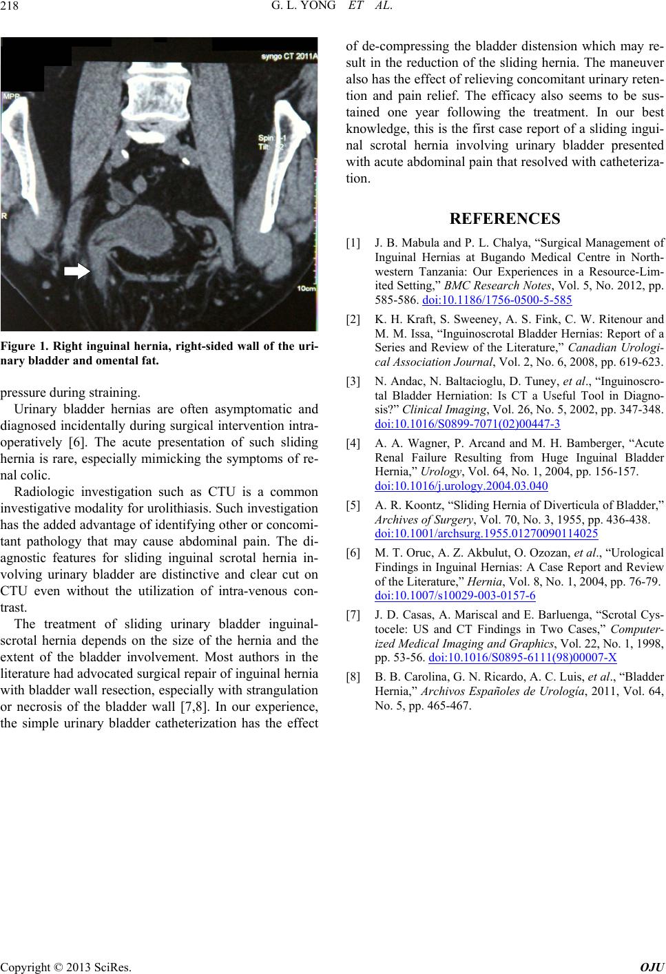 G. L. YONG ET AL. 218 Figure 1. Right inguinal hernia, right-sided wall of the uri- nary bladder and omental fat. pressure during straining. Urinary bladder hernias are often asymptomatic and diagnosed incidentally during surgical intervention intra- operatively [6]. The acute presentation of such sliding hernia is rare, especially mimicking the symptoms of re- nal colic. Radiologic investigation such as CTU is a common investigative modalit y for urolithiasis. Such inve stigation has the added advantage of identifying other or concomi- tant pathology that may cause abdominal pain. The di- agnostic features for sliding inguinal scrotal hernia in- volving urinary bladder are distinctive and clear cut on CTU even without the utilization of intra-venous con- trast. The treatment of sliding urinary bladder inguinal- scrotal hernia depends on the size of the hernia and the extent of the bladder involvement. Most authors in the literature had advocated surg ical repair of inguinal h ernia with bladder wall resection, especially with strangulation or necrosis of the bladder wall [7,8]. In our experience, the simple urinary bladder catheterization has the effect of de-compressing the bladder distension which may re- sult in the reduction of the sliding hernia. The maneuver also has the effect of relieving concomitant urinary reten- tion and pain relief. The efficacy also seems to be sus- tained one year following the treatment. In our best knowledge, this is the first case report of a sliding ingui- nal scrotal hernia involving urinary bladder presented with acute abdominal pain that resolved with cathe teriza- tion. REFERENCES [1] J. B. Mabula and P. L. Chalya, “Surgical Management of Inguinal Hernias at Bugando Medical Centre in North- western Tanzania: Our Experiences in a Resource-Lim- ited Setting,” BMC Research Notes, Vol. 5, No. 2012, pp. 585-586. doi:10.1186/1756-0500-5-585 [2] K. H. Kraft, S. Sweeney, A. S. Fink, C. W. Ritenour and M. M. Issa, “Inguinoscrotal Bladder Hernias: Report of a Series and Review of the Literature,” Canadian Urologi- cal Association Journal , Vol. 2, No. 6, 2008, pp. 619-623. [3] N. Andac, N. Baltacioglu, D. Tuney, et al., “Inguinoscro- tal Bladder Herniation: Is CT a Useful Tool in Diagno- sis?” Clinical Imaging, Vol. 26, No. 5, 2002, pp. 347-348. doi:10.1016/S0899-7071(02)00447-3 [4] A. A. Wagner, P. Arcand and M. H. Bamberger, “Acute Renal Failure Resulting from Huge Inguinal Bladder Hernia,” Urology, Vol. 64, No. 1, 2004, pp. 156-157. doi:10.1016/j.urology.2004.03.040 [5] A. R. Koontz, “Sliding Hernia of Diverticula of Bladder,” Archives of Surgery, Vol. 70, No. 3, 1955, pp. 436-438. doi:10.1001/archsurg.1955.01270090114025 [6] M. T. Oruc, A. Z. Akbulut, O. Ozozan, et al., “Urological Findings in Inguinal Hernias: A Case Report and Review of the Literature,” Hernia, Vol. 8, No. 1, 2004, pp. 76-79. doi:10.1007/s10029-003-0157-6 [7] J. D. Casas, A. Mariscal and E. Barluenga, “Scrotal Cys- tocele: US and CT Findings in Two Cases,” Computer- ized Medical Imaging and Graphics, Vol. 22, No. 1, 1998, pp. 53-56. doi:10.1016/S0895-6111(98)00007-X [8] B. B. Carolina, G. N. Ricardo, A. C. Luis, et al., “Bladder Hernia,” Archivos Españoles de Urología, 2011, Vol. 64, No. 5, pp. 465-467. Copyright © 2013 SciRes. OJU |