

Paper Menu >>
Journal Menu >>
 International Journal of Otolaryngology and Head & Neck Surgery, 2013, 2, 207-210 http://dx.doi.org/10.4236/ijohns.2013.25043 Published Online September 2013 (http://www.scirp.org/journal/ijohns) The Presenting Symptom of Metastatic Prostate Carcinoma: Case of a Large Supraclavicular Mass and Review of Literature Cory A. Vaughn1, Kaitlin R. Jaqua1, Ryan K. Meacham2, Fransisco Vieira2 1MD Candidate, University of Tennessee Health Science Cen t e r, Memphis, USA 2Department of Otolaryngology, Head & Neck Surgery, University of Tennessee, Memphis, USA Email: [email protected] Received June 23, 2013; revised July 24, 2013; accepted August 9, 2013 Copyright © 2013 Cory A. Vaughn et al. This is an open access article distributed under the Creative Commons Attribution License, which permits unrestricted use, distribution, and reproduction in any medium, provided the original work is properly cited. ABSTRACT Prostate cancer is the most common noncutaneous malignancy of American males and typically presents with genitou- rinary symptoms, however, head and neck extension is a rare finding. We present a case of a 53-year-old male initially presenting with a large supraclavicular mass as the only complaint. After an initial non-diagnostic FNA biopsy, PSA levels, histology and immunohistochemical findings were consistent with metastatic prostatic adenocarcinoma. The patient was started on anti-hormonal treatment after diagnosis. Due to the increasing reports of such cases, we suggest PSA levels and appropriate immunohistochemical staining should be obtained on all unknown neck masses. Keywords: Neck Mass; Prostate; Adenocarcinoma; Metastasis 1. Introduction Non-tender, mobile masses in the various neck regions induce a concern for metastatic squamous cell carci- noma for any otolaryngologist. The presence of a metas- tatic neoplasm to the neck changes the stage of disease, thus altering the treatment and prognosis. Adenocarci- noma uncommonly metastasizes to the neck, thus is often excluded from initial differential diagno ses. There are in- creasing numbers of reports documenting adenocarci- noma dissemination to the head and neck region present- ing as non-tender, mobile masses relatively small in size. We describe a case of metastatic prostate adenocarci- noma with initial presentation as large supraclavicular lymphadenopathy. 2. Case Report A 53-year-old African-American male presented with complaint of a large left neck mass that had been pro- gressively enlargingfor approximately ten months. The patient was completely asymptomatic from the mass. Physical exam revealed a 4 × 7 cm, fluctuant, non-tender, non-adherent mass in the left supraclaviuclar fossa. The remainder of the physical exam was unremarkable but did not include a genital or rectal exam. An ultrasound revealed a multilobulated left neck mass. The patient was prescribed Naprosyn and discharged from the Emergency Department with scheduled follow up to the PCP. The patient was subsequently lost to fo llow up for four months. The patient complained of further growth of the left neck mass and weight loss. The left supraclavicular mass now measured 8 × 7 cm. Laryngo scopy revealed no abnormal findings. With concerns of lymphoma, due to the size of the mass, a fine need aspiration (FNA) was performed. Cytology and flow cytometry results were non-diagn ost i c , ho wever rule out lymphoma. The patient was scheduled for an incisional biopsy re- vealing low-power eviden ce of prostatic adenocarcinoma (Figure 1(a)) and histologic evidence of cribriform pat- tern of cells (Figure 1(b)), prominent solitary nucleoli, and a uniform population of cells with presence of ve- sicular chormatin (Figure 1(c)). Immunohistochemical evaluation of the specimen was positive for CDX2, Ber- EP4, Vimentin, prostate specific antigen (PSA), and cy- tokeratin. Histologic and immunohistochemical findings were diagnostic for metastatic prostatic adenocarcinoma. At this time a genitourinary exam revealed a normal phallus and scrotum. On rectal exam, the prostate was nodular and tender. Initial PSA level was 990. The pa- tient now had complaints of pain localized to the left C opyright © 2013 SciRes. IJOHNS 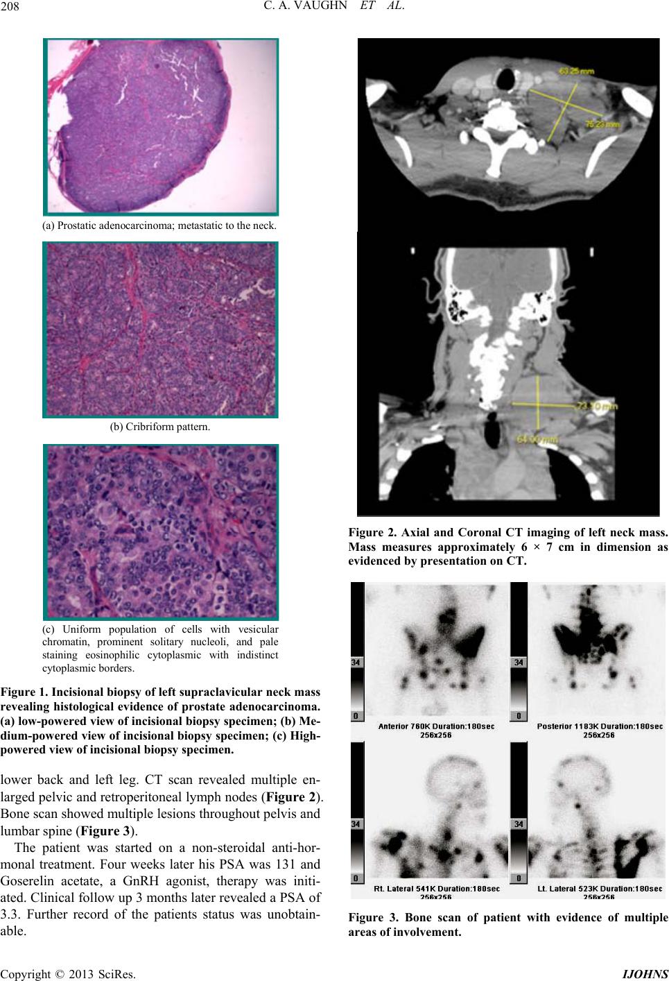 C. A. VAUGHN ET AL. 208 (a) Prostatic adenocarcinoma; metastatic to the neck. (b) Cribriform pattern. (c) Uniform population of cells with vesicula r chromatin, prominent solitary nucleoli, and pale staining eosinophilic cytoplasmic with indistinct cyto pl asm ic borde r s. Figure 1. Incisional biopsy of left supraclavicular neck mass revealing histological evidence of prostate adenocarcinoma. (a) low-powered view of incisional biopsy specimen; (b) Me- dium-powered view of incisional biopsy specimen; (c) High- powered view of incisional biopsy specimen. lower back and left leg. CT scan revealed multiple en- larged pelvic and retroperitoneal lymph nodes (Figure 2). Bone scan showed multiple lesions througho ut pelvis and lumbar spine (Figure 3). The patient was started on a non-steroidal anti-hor- monal treatment. Four weeks later his PSA was 131 and Goserelin acetate, a GnRH agonist, therapy was initi- ated. Clinical follow up 3 months later revealed a PSA of 3.3. Further record of the patients status was unobtain- able. Figure 2. Axial and Coronal CT imaging of left neck mass. Mass measures approximately 6 × 7 cm in dimension as evidenced by presentation on CT. Figure 3. Bone scan of patient with evidence of multiple areas of involvement. Copyright © 2013 SciRes. IJOHNS 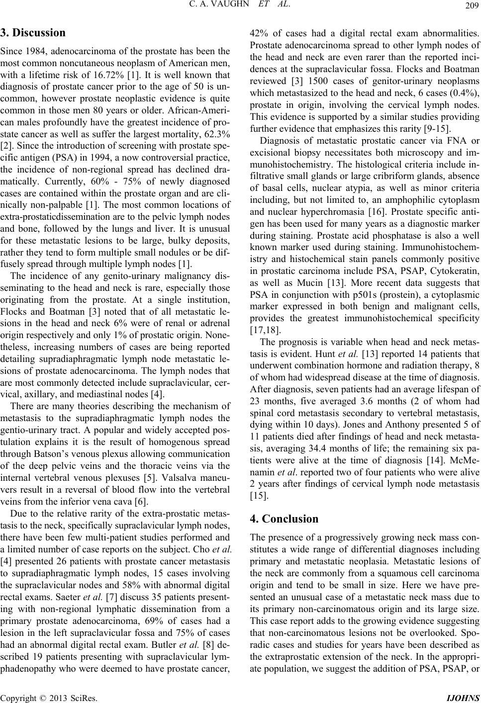 C. A. VAUGHN ET AL. 209 3. Discussion Since 1984, adenocarcinoma of the prostate has been the most common noncutaneous neoplasm of American men, with a lifetime risk of 16.72% [1]. It is well known that diagnosis of prostate cancer prior to the age of 50 is un- common, however prostate neoplastic evidence is quite common in those men 80 years or older. African-Ameri- can males profoundly have the greatest incidence of pro- state cancer as well as suffer the largest mortality, 62.3% [2]. Since the in trodu ction of screening with p ros tate sp e- cific antigen (PSA) in 1994, a now controversial practice, the incidence of non-regional spread has declined dra- matically. Currently, 60% - 75% of newly diagnosed cases are contained within the prostate organ and are cli- nically non-palpable [1]. The most common locations of extra-prostaticdissemination are to the pelvic lymph nodes and bone, followed by the lungs and liver. It is unusual for these metastatic lesions to be large, bulky deposits, rather they tend to form multiple small nodules or be dif- fusely spread through multiple lymph nodes [1]. The incidence of any genito-urinary malignancy dis- seminating to the head and neck is rare, especially those originating from the prostate. At a single institution, Flocks and Boatman [3] noted that of all metastatic le- sions in the head and neck 6% were of renal or adrenal origin respectively and only 1% of prostatic origin. None- theless, increasing numbers of cases are being reported detailing supradiaphragmatic lymph node metastatic le- sions of prostate adenocarcinoma. The lymph nodes that are most commonly detected include supraclavicular, cer - vical, axillary, and mediastinal nodes [4]. There are many theories describing the mechanism of metastasis to the supradiaphragmatic lymph nodes the gentio-urinary tract. A popular and widely accepted pos- tulation explains it is the result of homogenous spread through Batson’s venous plexus allowing communication of the deep pelvic veins and the thoracic veins via the internal vertebral venous plexuses [5]. Valsalva maneu- vers result in a reversal of blood flow into the vertebral veins from the inferior vena cava [6]. Due to the relative rarity of the extra-prostatic metas- tasis to the neck, specifically supraclavicu lar lymph nod es, there have been few multi-patient studies performed and a limited number of case reports on the subject. Cho et al. [4] presented 26 patients with prostate cancer metastasis to supradiaphragmatic lymph nodes, 15 cases involving the supraclavicular nodes and 58% with abnormal digital rectal exams. Saeter et al. [7] discuss 35 patients present- ing with non-regional lymphatic dissemination from a primary prostate adenocarcinoma, 69% of cases had a lesion in the left supraclavicular fossa and 75% of cases had an abnormal digital rectal exam. Butler et al. [8] de- scribed 19 patients presenting with supraclavicular lym- phadenopathy who were deemed to have prostate cancer, 42% of cases had a digital rectal exam abnormalities. Prostate adenocarcinoma spread to other lymph nodes of the head and neck are even rarer than the reported inci- dences at the supraclavicular fossa. Flocks and Boatman reviewed [3] 1500 cases of genitor-urinary neoplasms which metastasized to the head and neck, 6 cases (0.4%), prostate in origin, involving the cervical lymph nodes. This evidence is supported by a similar studies providing further evidence that emphasizes this rarity [9-15]. Diagnosis of metastatic prostatic cancer via FNA or excisional biopsy necessitates both microscopy and im- munohistochemistry. The histological criteria include in- filtrative small glands or large cribriform glands, absence of basal cells, nuclear atypia, as well as minor criteria including, but not limited to, an amphophilic cytoplasm and nuclear hyperchromasia [16]. Prostate specific anti- gen has been used for many years as a diagnostic marker during staining. Prostate acid phosphatase is also a well known marker used during staining. Immunohistochem- istry and histochemical stain panels commonly positive in prostatic carcinoma include PSA, PSAP, Cytokeratin, as well as Mucin [13]. More recent data suggests that PSA in conjunction with p501s (prostein), a cytoplasmic marker expressed in both benign and malignant cells, provides the greatest immunohistochemical specificity [17,18]. The prognosis is variable when head and neck metas- tasis is evident. Hunt et al. [13] reported 14 patients that underwent combination hormone and radiation therapy, 8 of whom had widespread disease at the time of diagnosis. After diagnosis, seven patients h ad an average lif espan of 23 months, five averaged 3.6 months (2 of whom had spinal cord metastasis secondary to vertebral metastasis, dying within 10 days). Jon es and Anthony pr esented 5 of 11 patients died after findings of head and neck metasta- sis, averaging 34.4 months of life; the remaining six pa- tients were alive at the time of diagnosis [14]. McMe- namin et al. reported two of four patients who were alive 2 years after findings of cervical lymph node metastasis [15]. 4. Conclusion The presence of a progressively growing neck mass con- stitutes a wide range of differential diagnoses including primary and metastatic neoplasia. Metastatic lesions of the neck are commonly from a squamous cell carcinoma origin and tend to be small in size. Here we have pre- sented an unusual case of a metastatic neck mass due to its primary non-carcinomatous origin and its large size. This case report adds to the growing evidence suggesting that non-carcinomatous lesions not be overlooked. Spo- radic cases and studies for years have been described as the extraprostatic extension of the neck. In the appropri- ate population, we sugg est the addition of PSA, PSAP, or Copyright © 2013 SciRes. IJOHNS  C. A. VAUGHN ET AL. Copyright © 2013 SciRes. IJOHNS 210 p501s immunohistochemical stains to FNA or excisional biopies of all neck masses concerning for cancer to pre- vent delay in diagno sis and to i mprove progn osis th rough earlier therapy. REFERENCES [1] A. J. Wein, L. R. Kavoussi, A. C. Novick, A. W. Partin and C. A. Peters, “Campbell-Walsh Urology,” 10th Edi- tion, Elsevier Inc., Philadelphia, 2012, pp. 2704-2725. [2] American Cancer Society, “Cancer Facts & Figures 2008,” American Cancer Society, Atlanta, 2008. [3] R. H. Flocks and D. L. Boatman, “Incidence of Head and Neck Metastases from Genitor-Urinary Neoplasms,” La- ryngoscope, Vol. 83, 1973, pp. 1527-1539. doi:10.1288/00005537-197309000-00011 [4] K. R. Cho and J. I. Epstein, “Metastatic Prostate Carci- noma to Supradiaphragmtic Lymph Nodes. A Clinicopa- thologic and Immunohistochemical Study,” American Journal of Surgical Pathology, Vol. 11, No. 6, 1987, pp. 457-463. doi:10.1097/00000478-198706000-00006 [5] B. Copeland, J. M. Clark, A. Sura, S. E. Kilpatrick, W. Shockley and S. Meredith, “Prostate Carcinoma Metas- tatic to the Cervical Lymph Nodes: Report of Two Cases and Review of the Literature,” American Journal of Oto- laryngology, Vol. 22, No. 6, 2001, pp. 420-423. doi:10.1053/ajot.2001.28066 [6] W. T. Couldwell, P. T. Chandrasoma and M. H. Weiss, “Pituitary Gland Metastasis from Adenocarcinoma of the Prostate. Case Report,” Journal of Neurosurgery, Vol. 71, No. 1, 1989, pp. 138-140. doi:10.3171/jns.1989.71.1.0138 [7] G. Saeter, S. D. Fossa, S. Ous, G. P. Blom and O. Kaal- hus, “Carcinoma of the Prostate with Soft Tissue or Non- Regional Lymphatic Metastases at the Time of Diagnosis: A Review of 47 Cases,” British Journal of Urology, Vol. 56, 1984, pp. 385-390. doi:10.1111/j.1464-410X.1984.tb05827.x [8] J. J. Butler, C. Howe and D. Johnson, “Enlargement of Supraclavicular Lymph Nodes as the Initial Sign of Pros- tate Carcinoma,” Cancer, Vol. 27, 1971, pp. 1055-1063. doi:10.1002/1097-0142(197105)27:5<1055::AID-CNCR2 820270509>3.0.CO;2-T [9] H. Saitoh, M. Hida, T. Shimbo, et al., “Metastatic Pat- terns of Prostatic Cancer,” Cancer, Vol. 54, No. 12, 1984, pp. 3078-3084. doi:10.1002/1097-0142(19841215)54:12<3078::AID-CN CR2820541245>3.0.CO;2-U [10] M. Elkin and H. P. Mueller, “Metastases from Cancer of the Prostate,” Cancer, Vol. 7, No. 6, 1954, pp. 1246- 1248. doi:10.1002/1097-0142(195411)7:6<1246::AID-CNCR28 20070616>3.0.CO;2-C [11] H. L. Abrams, R. Sprio and N. Goldstein, “Metastases in carcinoma,” Cancer, Vol. 3, No. 1, 1950, pp. 74-85. doi:10.1002/1097-0142(1950)3:1<74::AID-CNCR282003 0111>3.0.CO;2-7 [12] E. R. Mintz and G. G. Smith, “Autopsy Findings in 100 Cases of Prostate Cancer,” New England Journal of Medicine, Vol. 211, 1934, pp. 479-487. doi:10.1056/NEJM193409132111101 [13] J. L. Hunt, J. E. Tomaszewski and K. T. Montone, “Pros- tatic Adenocarcinoma Metastatic to the Head and Neck and the Workup of an Unknown Epithelioid Neoplasm,” Head & Neck, Vol. 26, No. 2, 2004, pp. 171-178. doi:10.1002/hed.10353 [14] H. Jones and P. P. Anthony, “Metastatic Prostatic Carci- noma Presenting as Left-Sided Cervical Lymphadenopath: A Series of 11 Cases,” Histopathology, Vol. 21, No. 2, 1992, pp. 149-154. doi:10.1111/j.1365-2559.1992.tb00363.x [15] P. G. McMenamin, J. D. Anderson and J. J. Baker, “Pros- tatic Carcinoma Presenting in Neck Metastasis,” Journal of Otolaryngology, Vol. 3, 1989, pp. 119-124. [16] P. A. Humphrey, “Diagnosis of Adenocarcinoma in Pros- tate Needle Biopsy Tissue,” Journal of Clinical Pathol- ogy, Vol. 60, No. l, 2007, pp. 35-42. doi:10.1136/jcp.2005.036442 [17] T. Sheridan, M. Herawi, J. I. Epstein and P. B. Illei, “The Role of P501S and PSA in the Diagnosis of Metastatic Adenocarcinoma of the Prostate,” American Journal of Surgical Pathology, Vol. 31, No. 9, 2007, pp. 1351-1355. doi:10.1097/PAS.0b013e3180536678 [18] M. Yin, R. Dhir and A. V. Parwani, “Diagnostic Utility of p501s (Prostein) in Comparison to Prostate Specific An- tigen (PSA) for the Detection of Metastatic Prostatic Ade- nocarcinoma,” Diagnostic Pathology, Vol. 27, No. 2, 2007, p. 41. |