

Paper Menu >>
Journal Menu >>
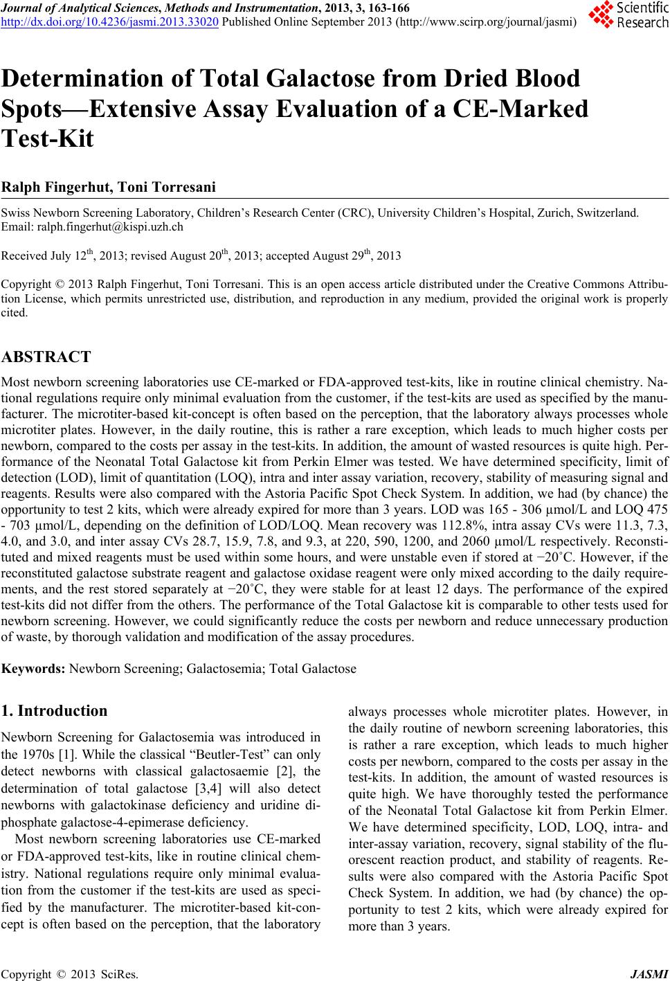 Journal of Analytical Sciences, Methods and Instrumentation, 2013, 3, 163-166 http://dx.doi.org/10.4236/jasmi.2013.33020 Published Online September 2013 (http://www.scirp.org/journal/jasmi) 163 Determination of Total Galactose from Dried Blood Spots—Extensive Assay Evaluation of a CE-Marked Test-Kit Ralph Fingerhut, Toni Torresani Swiss Newborn Screening Laboratory, Children’s Research Center (CRC), University Children’s Hospital, Zurich, Switzerland. Email: [email protected] Received July 12th, 2013; revised August 20th, 2013; accepted August 29th, 2013 Copyright © 2013 Ralph Fingerhut, Toni Torresani. This is an open access article distributed under the Creative Commons Attribu- tion License, which permits unrestricted use, distribution, and reproduction in any medium, provided the original work is properly cited. ABSTRACT Most newborn screening laboratories use CE-marked or FDA-approved test-kits, like in routine clinical chemistry. Na- tional regulations require only minimal evaluation from the customer, if the test-kits are used as specified by the manu- facturer. The microtiter-based kit-concept is often based on the perception, that the laboratory always processes whole microtiter plates. However, in the daily routine, this is rather a rare exception, which leads to much higher costs per newborn, compared to the costs per assay in the test-kits. In addition, the amount of wasted resources is quite high. Per- formance of the Neonatal Total Galactose kit from Perkin Elmer was tested. We have determined specificity, limit of detection (LOD), limit of quantitation (LOQ), intra and inter assay variation, recovery, stability of measuring signal and reagents. Results were also compared with the Astoria Pacific Spot Check System. In addition, we had (by chance) the opportunity to test 2 kits, which were already expired for more than 3 years. LOD was 165 - 306 µmol/L and LOQ 475 - 703 µmol/L, depending on the definition of LOD/LOQ. Mean recovery was 112.8%, intra assay CVs were 11.3, 7.3, 4.0, and 3.0, and inter assay CVs 28.7, 15.9, 7.8, and 9.3, at 220, 590, 1200, and 2060 µmol/L respectively. Reconsti- tuted and mixed reagents must be used within some hours, and were unstable even if stored at −20˚C. However, if the reconstituted galactose substrate reagent and galactose oxidase reagent were only mixed according to the daily require- ments, and the rest stored separately at −20˚C, they were stable for at least 12 days. The performance of the expired test-kits did not differ from the others. The performance of the Total Galactose kit is comparable to other tests used for newborn screening. However, we could significantly reduce the costs per newborn and reduce unnecessary production of waste, by thorough validation and modification of the assay procedures. Keywords: Newborn Screening; Galactosemia; Total Galactose 1. Introduction Newborn Screening for Galactosemia was introduced in the 1970s [1]. While the classical “Beutler-Test” can only detect newborns with classical galactosaemie [2], the determination of total galactose [3,4] will also detect newborns with galactokinase deficiency and uridine di- phosphate galactose-4-epimerase deficiency. Most newborn screening laboratories use CE-marked or FDA-approved test-kits, like in routine clinical chem- istry. National regulations require only minimal evalua- tion from the customer if the test-kits are used as speci- fied by the manufacturer. The microtiter-based kit-con- cept is often based on the perception, that the laboratory always processes whole microtiter plates. However, in the daily routine of newborn screening laboratories, this is rather a rare exception, which leads to much higher costs per newborn, compared to the costs per assay in the test-kits. In addition, the amount of wasted resources is quite high. We have thoroughly tested the performance of the Neonatal Total Galactose kit from Perkin Elmer. We have determined specificity, LOD, LOQ, intra- and inter-assay variation, recovery, signal stability of the flu- orescent reaction product, and stability of reagents. Re- sults were also compared with the Astoria Pacific Spot Check System. In addition, we had (by chance) the op- portunity to test 2 kits, which were already expired for more than 3 years. Copyright © 2013 SciRes. JASMI 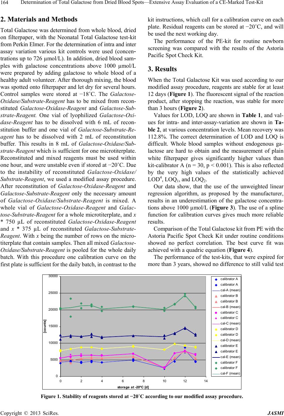 Determination of Total Galactose from Dried Blood Spots—Extensive Assay Evaluation of a CE-Marked Test-Kit 164 2. Materials and Methods Total Galactose was determined from whole blood, dried on filterpaper, with the Neonatal Total Galactose test-kit from Perkin Elmer. For the determination of intra and inter assay variation various kit controls were used (concen- trations up to 726 µmol/L). In addition, dried blood sam- ples with galactose concentrations above 1000 µmol/L were prepared by adding galactose to whole blood of a healthy adult volunteer. After thorough mixing, the blood was spotted onto filterpaper and let dry for several hours. Control samples were stored at −18˚C. The Galactose- Oxidase/Substrate-Reagent has to be mixed from recon- stituted Galactose-Oxidase-Reagent and Galactose-Sub- strate-Reagent. One vial of lyophilized Galactose-Oxi- dase-Reagent has to be dissolved with 6 mL of recon- stitution buffer and one vial of Galactose-Substrate-Re- agent has to be dissolved with 2 mL of reconstitution buffer. This results in 8 mL of Galactose-Oxidase/Sub- strate-Reagent which is sufficient for one microtiterplate. Reconstituted and mixed reagents must be used within one hour, and were unstable even if stored at −20˚C. Due to the instability of reconstituted Galactose-Oxidase/ Substrate-Reagent, we used a modified assay procedure. After reconstitution of Galactose-Oxidase-Reagent and Galactose- Substrate-Reagent only the necessary amount of Galactose-Oxidase/Substrate-Reagent is mixed. A whole vial of Galactose-Oxidase-Reagent and Galac- tose-Substrate-Reagent for a whole microtiterplate, and x * 750 µL of reconstituted Galactose-Oxidase-Reagent and x * 375 µL of reconstituted Galactose-Substrate- Reagent. With x being the number of rows on the micro- titerplate that contain samples. Then all mixed Galactose- Oxidase/Substrate-Reagent is pooled for the whole daily batch. With this procedure one calibration curve on the first plate is sufficient for the daily batch, in contrast to the kit instructions, which call for a calibration curve on each plate. Residual reagents can be stored at −20˚C, and will be used the next working day. The performance of the PE-kit for routine newborn screening was compared with the results of the Astoria Pacific Spot Check Kit. 3. Results When the Total Galactose Kit was used according to our modified assay procedure, reagents are stable for at least 12 days (Figure 1). The fluorescent signal of the reaction product, after stopping the reaction, was stable for more than 3 hours (Figure 2). Values for LOD, LOQ are shown in Table 1, and val- ues for intra- and inter-assay-variation are shown in Ta- ble 2, at various concentration levels. Mean recovery was 112.8%. The correct determination of LOD and LOQ is difficult. Whole blood samples without endogenous ga- lactose are hard to obtain and the measurement of plain white filterpaper gives significantly higher values than kit-calibrator A (n = 30, p < 0.001). This is also reflected by the very high values of the statistically achieved LOD4, LOQ1, and LOQ2. Our data show, that the use of the unweighted linear regression algorithm, as proposed by the manufacturer, results in an underestimation of the galactose concentra- tions above 1000 µmol/L (Figure 3). The use of a spline function for calibration curves gives much more reliable results. Comparison of the Total Galactose kit from PE with the Astoria Pacific Spot Check Kit under routine conditions showed no perfect correlation. The best curve fit was achieved with a quadric equation (Figure 4). The performance of the test-kits, that were expired for more than 3 years, showed no difference to still valid test 0 5000 10000 15000 20000 25000 30000 02468101214 storage at -20ºC [d] [counts] c ali brator A c ali brator A c al-A (mean) c ali brator B c ali brator B c al-B (mean) c ali brator C c ali brator C c al-C (mean) c ali brator D c ali brator D c al-D (mean) c ali brator E c ali brator E c al-E (mean) c ali brator F c ali brator F c al -F (m ea n) Figure 1. Stability of reagents stored at −20˚C according to our modified assay procedure. Copyright © 2013 SciRes. JASMI 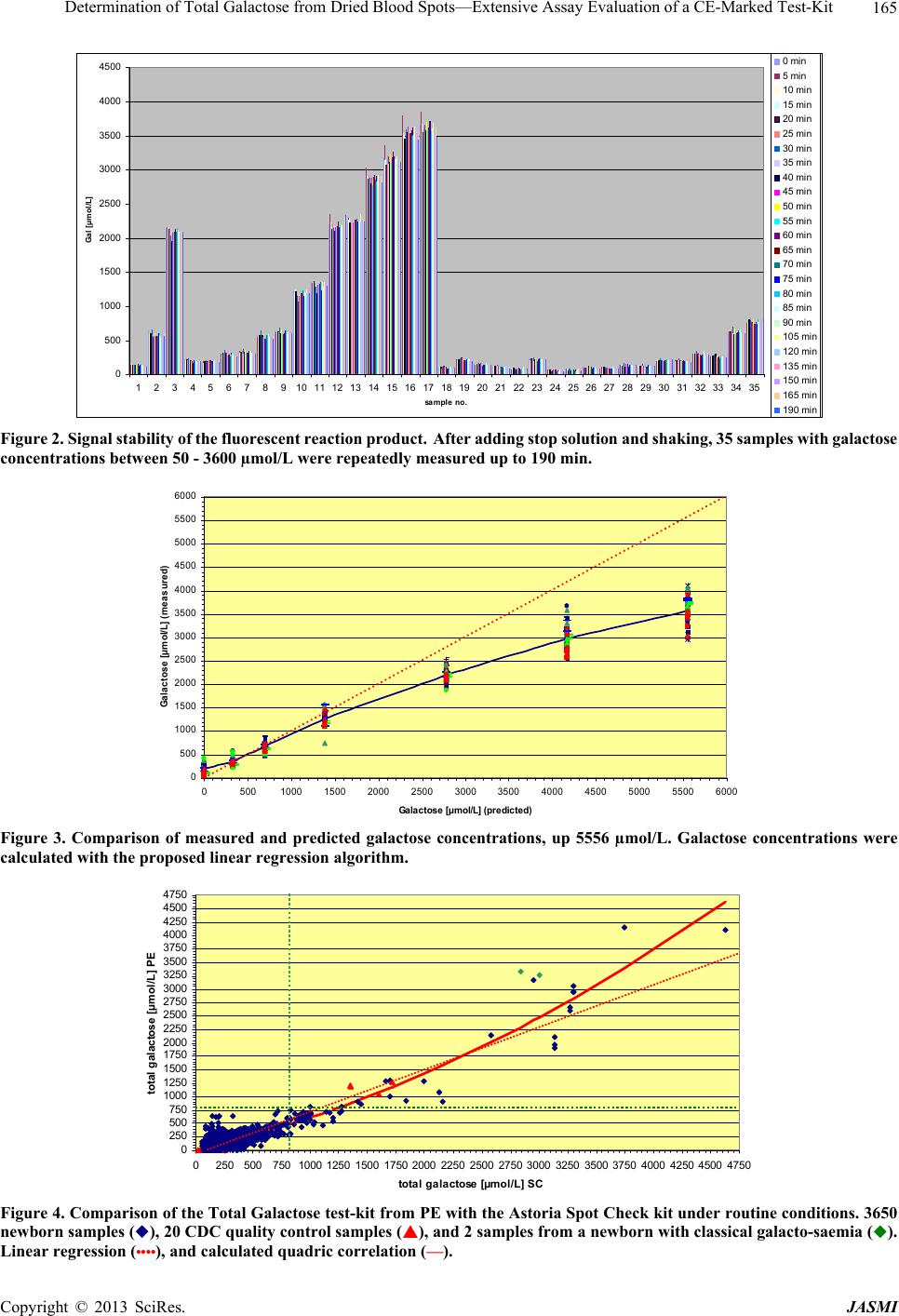 Determination of Total Galactose from Dried Blood Spots—Extensive Assay Evaluation of a CE-Marked Test-Kit 165 0 500 1000 1500 2000 2500 3000 3500 4000 4500 1234567891011121314151617181920212223242526272829303132333435 sample no. Gal [µmol/L] 0 min 5 min 10 m in 15 m in 20 m in 25 m in 30 m in 35 m in 40 m in 45 m in 50 m in 55 m in 60 m in 65 m in 70 m in 75 m in 80 m in 85 m in 90 m in 105 m in 120 m in 135 m in 150 m in 165 m in 190 m in Figure 2. Signal stability of the fluorescent reaction product. After adding stop solution and shaking, 35 samples with galactose concentrations between 50 - 3600 µmol/L were repeatedly measured up to 190 min. 0 500 1000 1500 2000 2500 3000 3500 4000 4500 5000 5500 6000 05001000 1500 2000 2500 30003500 4000 4500 5000 5500 6000 Galactose [µ mol/L] ( pr edict e d) G alactose [µmol/L] (m easured) Figure 3. Comparison of measured and predicted galactose concentrations, up 5556 µmol/L. Galactose concentrations were calculated with the proposed linear regression algorithm. 0 250 500 750 1000 1250 1500 1750 2000 2250 2500 2750 3000 3250 3500 3750 4000 4250 4500 4750 025050075010001250 150017502000 22502500 275030003250 350037504000 425045004750 total galactose [µmol/L] SC total galactose [µmol / L] PE Figure 4. Comparison of the Total Galactose test-kit from PE with the Astoria Spot Check kit under routine conditions. 3650 newborn samples (), 20 CDC quality control samples (), and 2 samples from a newborn with classical galacto-saemia (). Linear regression (••••), and calculated quadric correlation (—). Copyright © 2013 SciRes. JASMI 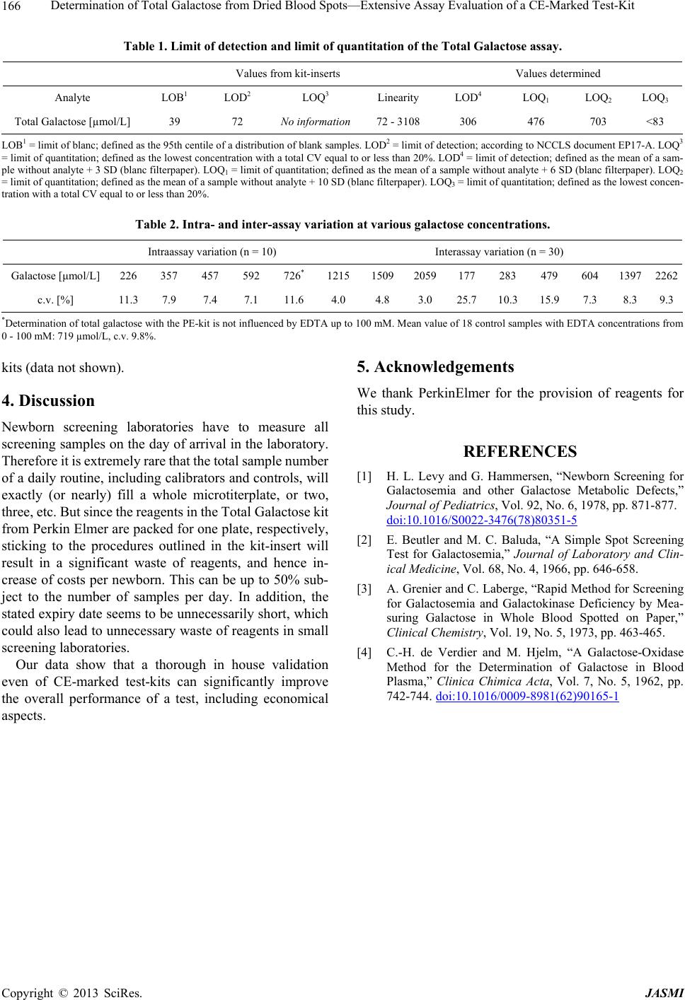 Determination of Total Galactose from Dried Blood Spots—Extensive Assay Evaluation of a CE-Marked Test-Kit Copyright © 2013 SciRes. JASMI 166 Table 1. Limit of detection and limit of quantitation of the Total Galactose assay. Values from kit-inserts Values determined Analyte LOB1 LOD2 LOQ3 Linearity LOD4 LOQ1 LOQ2 LOQ3 Total Galactose [µmol/L] 39 72 No information 72 - 3108 306 476 703 <83 LOB1 = limit of blanc; defined as the 95th centile of a distribution of blank samples. LOD2 = limit of detection; according to NCCLS document EP17-A. LOQ3 = limit of quantitation; defined as the lowest concentration with a total CV equal to or less than 20%. LOD4 = limit of detection; defined as the mean of a sam- ple without analyte + 3 SD (blanc filterpaper). LOQ1 = limit of quantitation; defined as the mean of a sample without analyte + 6 SD (blanc filterpaper). LOQ2 = limit of quantitation; defined as the mean of a sample without analyte + 10 SD (blanc filterpaper). LOQ3 = limit of quantitation; defined as the lowest concen- tration with a total CV equal to or less than 20%. Table 2. Intra- and inter-assay variation at various galactose concentrations. Intraassay variation (n = 10) Interassay variation (n = 30) Galactose [μmol/L] 226 357 457 592 726* 1215 15092059177 283 479 604 13972262 c.v. [%] 11.3 7.9 7.4 7.1 11.6 4.0 4.8 3.0 25.710.3 15.9 7.3 8.3 9.3 *Determination of total galactose with the PE-kit is not influenced by EDTA up to 100 mM. Mean value of 18 control samples with EDTA concentrations from 0 - 100 mM: 719 µmol/L, c.v. 9.8%. kits (data not shown). 4. Discussion Newborn screening laboratories have to measure all screening samples on the day of arrival in the laboratory. Therefore it is extremely rare that the total sample number of a daily routine, including calibrators and controls, will exactly (or nearly) fill a whole microtiterplate, or two, three, etc. But since the reagents in the Total Galactose kit from Perkin Elmer are packed for one plate, respectively, sticking to the procedures outlined in the kit-insert will result in a significant waste of reagents, and hence in- crease of costs per newborn. This can be up to 50% sub- ject to the number of samples per day. In addition, the stated expiry date seems to be unnecessarily short, which could also lead to unnecessary waste of reagents in small screening laboratories. Our data show that a thorough in house validation even of CE-marked test-kits can significantly improve the overall performance of a test, including economical aspects. 5. Acknowledgements We thank PerkinElmer for the provision of reagents for this study. REFERENCES [1] H. L. Levy and G. Hammersen, “Newborn Screening for Galactosemia and other Galactose Metabolic Defects,” Journal of Pediatrics, Vol. 92, No. 6, 1978, pp. 871-877. doi:10.1016/S0022-3476(78)80351-5 [2] E. Beutler and M. C. Baluda, “A Simple Spot Screening Test for Galactosemia,” Journal of Laboratory and Clin- ical Medicine, Vol. 68, No. 4, 1966, pp. 646-658. [3] A. Grenier and C. Laberge, “Rapid Method for Screening for Galactosemia and Galactokinase Deficiency by Mea- suring Galactose in Whole Blood Spotted on Paper,” Clinical Chemistry, Vol. 19, No. 5, 1973, pp. 463-465. [4] C.-H. de Verdier and M. Hjelm, “A Galactose-Oxidase Method for the Determination of Galactose in Blood Plasma,” Clinica Chimica Acta, Vol. 7, No. 5, 1962, pp. 742-744. doi:10.1016/0009-8981(62)90165-1 |