

Paper Menu >>
Journal Menu >>
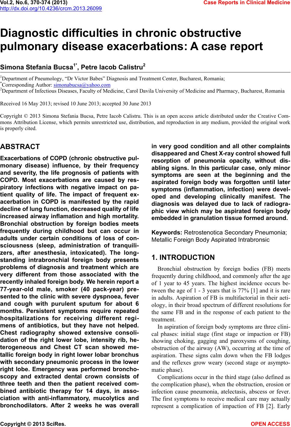 Vol.2, No.6, 370-374 (2013) Case Reports in Clinical Medicine http://dx.doi.org/10.4236/crcm.2013.26099 Diagnostic difficulties in chronic obstructive pulmonary disease exacerbations: A case report Simona Stefania Bucsa1*, Petre Iacob Calistru2 1Department of Pneumology, “Dr Victor Babes” Diagnosis and Treatment Center, Bucharest, Romania; *Corresponding Author: [email protected] 2Department of Infectious Diseases, Faculty of Medicine, Carol Davila University of Medicine and Pharmacy, Bucharest, Romania Received 16 May 2013; revised 10 June 2013; accepted 30 June 2013 Copyright © 2013 Simona Stefania Bucsa, Petre Iacob Calistru. This is an open access article distributed under the Creative Com- mons Attribution License, which permits unrestricted use, distribution, and reproduction in any medium, provided the original work is properly cited. ABSTRACT Exacerbations of COPD (chronic obstructive pul- monary disease) influence, by their frequency and severity, the life prognosis of patients with COPD. Most exacerbations are caused by res- piratory infections with negative impact on pa- tient quality of life. The impact of frequent ex- acerbation in COPD is manifested by the rapid decline of lung function, decreased quality of life increased airway inflamation and high mortality. Bronchial obstruction by foreign bodies meets frequently during childhood but can occur in adults under certain conditions of loss of con- sciousness (sleep, administration of tranquili- zers, after anesthesia, intoxicated). The long- standing intrabronchial foreign body presents problems of diagnosis and treatment which are very different from those associated with the recently inhaled foreign body. W e herein repor t a 77-year-old male, smoker (40 pack-year) pre- sented to the clinic with severe dyspnoea, fever and cough with purulent sputum for about 6 months. Persistent symptoms require repeated hospitalizations for receiving different regi- mens of antibiotics, but they have not helped. Chest radiography showed extensive consoli- dation of the right lower lobe, intensity rib, he- terogeneous and Chest CT scan showed me- tallic foreign body in right lower lobar bronchus with secondary pneumonic process in the lower right lobe. Emergency was performed broncho- scopy and extracted dental crown consists of three teeth and then the patient received com- bined antibiotic therapy for 14 days, in asso- ciation with anti-inflammatory, mucolytics and bronchodilators. After 2 weeks he was overall in very good condition and all other complaints disappeared and C hest X-ray control sho wed ful l resorption of pneumonia opacity, without dis- abling signs. In this particular case, only minor symptoms are seen at the beginning and the aspirated foreign body was forgotten until later symptoms (inflammation, infection) were devel- oped and developing clinically manifest. The diagnosis was delayed due to lack of radiogra- phic view which may be aspirated foreign body embedded in granulation tissue formed around. Keywords: Retrostenotica Secondary Pneumonia; Metallic Foreign Body Aspirated Intrabronsic 1. INTRODUCTION Bronchial obstruction by foreign bodies (FB) meets frequently during childhood, and commonly after the age of 1 year to 45 years. The highest incidence occurs be- tween the age of 1 - 3 years that is 77% [1] and it is rare in adults. Aspiration of FB is multifactorial in their aeti- ology, in their broad spectrum of different resolutions for the same FB and in the response of each patient to the treatment. In aspiration of foreign body symptoms are three clini- cal phases: initial stage (first stage or impaction or FB) showing choking, gagging and paroxysms of coughing, obstruction of the airway (AW), occurring at the time of aspiration. These signs calm down when the FB lodges and the reflexes grow weary (second stage or asympto- matic phase). Complications occur in the third stag e (also defined as the complication phase), when the obstruction, erosion or infection cause pneumonia, atelectasis, abscess or fever. The first symptoms to receive medical care may actually represent a complication of impaction of FB [2]. Early Copyright © 2013 SciRes. OPEN ACCESS 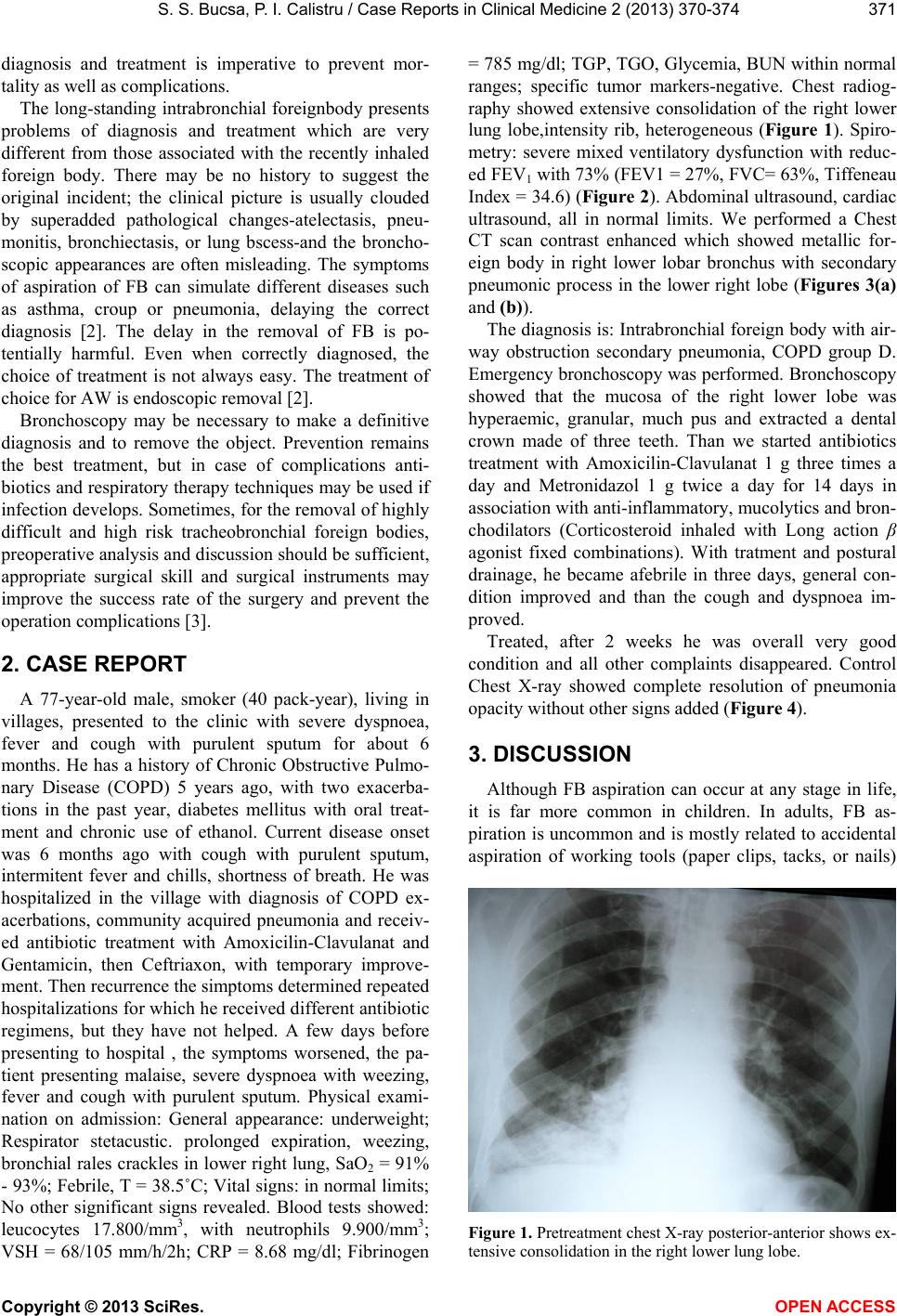 S. S. Bucsa, P. I. Calistru / Case Reports in Clinical Medicine 2 (2013) 370-374 371 diagnosis and treatment is imperative to prevent mor- tality as well as complications. The long-standing intrabronchial foreignbody presents problems of diagnosis and treatment which are very different from those associated with the recently inhaled foreign body. There may be no history to suggest the original incident; the clinical picture is usually clouded by superadded pathological changes-atelectasis, pneu- monitis, bronchiectasis, or lung bscess-and the broncho- scopic appearances are often misleading. The symptoms of aspiration of FB can simulate different diseases such as asthma, croup or pneumonia, delaying the correct diagnosis [2]. The delay in the removal of FB is po- tentially harmful. Even when correctly diagnosed, the choice of treatment is not always easy. The treatment of choice for AW is endoscopic removal [2]. Bronchoscopy may be necessary to make a definitive diagnosis and to remove the object. Prevention remains the best treatment, but in case of complications anti- biotics and resp iratory therapy techniques may b e used if infection develops. Sometimes, for the removal of highly difficult and high risk tracheobronchial foreign bodies, preoperative analysis and discussion should be sufficient, appropriate surgical skill and surgical instruments may improve the success rate of the surgery and prevent the operation complications [3]. 2. CASE REPORT A 77-year-old male, smoker (40 pack-year), living in villages, presented to the clinic with severe dyspnoea, fever and cough with purulent sputum for about 6 months. He has a history of Chronic Obstructive Pulmo- nary Disease (COPD) 5 years ago, with two exacerba- tions in the past year, diabetes mellitus with oral treat- ment and chronic use of ethanol. Current disease onset was 6 months ago with cough with purulent sputum, intermitent fever and chills, shortness of breath. He was hospitalized in the village with diagnosis of COPD ex- acerbations, community acquired pneumonia and receiv- ed antibiotic treatment with Amoxicilin-Clavulanat and Gentamicin, then Ceftriaxon, with temporary improve- ment. Then recurrence the simptoms determined repeated hospitalizations for which he received different antibiotic regimens, but they have not helped. A few days before presenting to hospital , the symptoms worsened, the pa- tient presenting malaise, severe dyspnoea with weezing, fever and cough with purulent sputum. Physical exami- nation on admission: General appearance: underweight; Respirator stetacustic. prolonged expiration, weezing, bronchial rales crackles in lower right lung, SaO2 = 91% - 93%; Febrile, T = 38 .5˚C; Vital signs: in normal limits; No other significant signs revealed. Blood tests showed: leucocytes 17.800/mm3, with neutrophils 9.900/mm3; VSH = 68/105 mm/h/2h; CRP = 8.68 mg/dl; Fibrinogen = 785 mg/dl; TGP, TGO, Glycemia, BUN within normal ranges; specific tumor markers-negative. Chest radiog- raphy showed extensive consolidation of the right lower lung lobe,intensity rib, heterogeneous (Figure 1). Spiro- metry: severe mixed ventilatory dysfunction with reduc- ed FEV1 with 73% (FEV1 = 27%, FVC= 63%, Tiffeneau Index = 34.6) (Figure 2). Abdominal ultrasound, cardiac ultrasound, all in normal limits. We performed a Chest CT scan contrast enhanced which showed metallic for- eign body in right lower lobar bronchus with secondary pneumonic process in the lower right lobe (Figures 3(a) and (b)). The diagnosis is: Intrabron chial foreign body with air- way obstruction secondary pneumonia, COPD group D. Emergency bronchoscopy was performed. Bronchoscopy showed that the mucosa of the right lower lobe was hyperaemic, granular, much pus and extracted a dental crown made of three teeth. Than we started antibiotics treatment with Amoxicilin-Clavulanat 1 g three times a day and Metronidazol 1 g twice a day for 14 days in association with anti-inflammatory, mucolyti cs and bron- chodilators (Corticosteroid inhaled with Long action β agonist fixed combinations). With tratment and postural drainage, he became afebrile in three days, general con- dition improved and than the cough and dyspnoea im- proved. Treated, after 2 weeks he was overall very good condition and all other complaints disappeared. Control Chest X-ray showed complete resolution of pneumonia opacity without other signs added (Figure 4). 3. DISCUSSION Although FB aspiration can occur at any stage in life, it is far more common in children. In adults, FB as- piration is uncommon and is mostly related to accidental aspiration of working tools (paper clips, tacks, or nails) Figure 1. Pretreatment chest X-ray posterior-anterior shows ex- ensive consolidation in the right lower lung lobe. t Copyright © 2013 SciRes. OPEN ACCESS 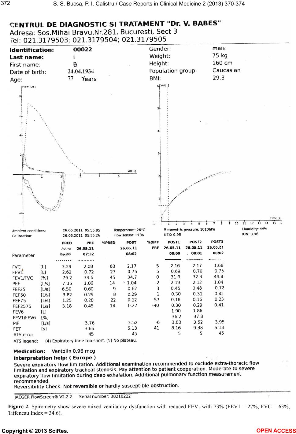 S. S. Bucsa, P. I. Calistru / Case Reports in Clinical Medicine 2 (2013) 370-374 Copyright © 2013 SciRes. 372 Figure 2. Spirometry show severe mixed ventilatory dysfunction with reduced FEV1 with 73% (FEV1 = 27%, FVC = 63%, Tiffeneau Index = 34.6). OPEN ACCESS 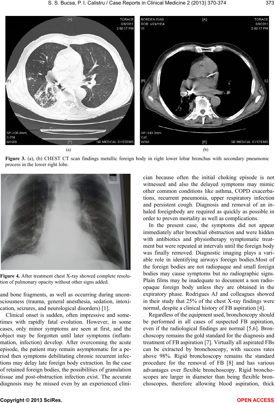 S. S. Bucsa, P. I. Calistru / Case Reports in Clinical Medicine 2 (2013) 370-374 373 (a) (b) Figure 3. (a), (b) CHEST CT scan findings metallic foreign body in right lower lobar bronchus with secondary pneumonic process in the lower right lobe. Figure 4. After treatment chest X-ray showed complete resolu- tion of pulmonary opacity without other signs added. and bone fragments, as well as occurring during uncon- sciousness (trauma, general anesthesia, sedation, intoxi- cation, seizures, and neurological disorders) [1]. Clinical onset is sudden, often impressive and some- times with rapidly fatal evolution. However, in some cases, only minor symptoms are seen at first, and the object may be forgotten until later symptoms (inflam- mation, infection) develop. After overcoming the acute episode, the patient may remain asymptomatic for a pe- riod then symptoms debilitating chronic recurrent infec- tions may delay late foreign body extraction. In the case of retained foreign bod ies, the p ossibilities o f granu lation tissue and post-obstruction infection exist. The accurate diagnosis may be missed even by an experienced clini- cian because often the initial choking episode is not witnessed and also the delayed symptoms may mimic other common conditions like asthma, COPD exacerba- tions, recurrent pneumonia, upper respiratory infection and persistent cough. Diagnosis and removal of an in- haled foreignbody are required as quickly as possible in order to preven mortality as well as complications. In the present case, the symptoms did not appear immediately after bronchial obstruction and were hidden with antibiotics and physiotherapy symptomatic treat- ment but were repeated at intervals until th e foreign body was finally removed. Diagnostic imaging plays a vari- able role in identifying airways foreign bodies.Most of the foreign bodies are not radiopaque and small foreign bodies may cause symptoms but no radiographic signs. Plain films may be inadequate to document a non radio- opaque foreign body unless they are obtained in the expiratory phase. Rodrigues AJ and colleagues showed in their study that 25% of the chest X-ray findings were normal, despite a clinical history of FB aspiration [4]. Regardless of the equipment used, bronchosc opy should be performed in all cases of suspected FB aspiration, even if the radiological findings are normal [5,6]. Bron- choscopy remains the gold standard for the diagnosis and treatment of FB aspiration [7]. Virtually all aspirated FBs can be extracted by bronchoscopy, with success rates above 98%. Rigid bronchoscopy remains the standard procedure for the removal of FB [8] and has various advantages over flexible bronchoscopy. Rigid broncho- scopes are larger in diameter than being flexible bron- choscopes, therefore allowing blood aspiration, thick Copyright © 2013 SciRes. OPEN ACCESS  S. S. Bucsa, P. I. Calistru / Case Reports in Clinical Medicine 2 (2013) 370-374 374 secretion aspiration, and patient ventilation. Although rigid bronchoscopy is considered the gold standard for the removal of foreign bodies from the air- ways, exist studys that demonstrated that flexible bron- choscopy can be safely and effectively used in the dia- gnosis and treatment of stable adult patients [4]. In the rare instances in which bronchoscopic removal fails, surgical bronchotomy or segmental resection is indicated. The presence of chronic bronchial obstruction, together with bronchiectasis, lung abscess, and paren- chymal destruction, might be an indication for segmental or lobar resection [9]. In this particular case, the diagnosis was delayed due to lack of radiographic visualization of the foreign body intrabronsic with persistent symptoms of infection des- pite antibiotic treatment. 4. ACKNOWLEDGEMENTS This paper is supported by the Sectoral Operational Programme Hu- man Resources Development (SOP HRD) 2007-2013, financed from the European Social Fund and by the Romanian Government under the contract number POSDRU/107/1.5/S/82839. REFERENCES [1] Qureshi, A. and Behzadi, A. (2008) Foreign-body aspira- tion in an adult. Canadian Journal of Surgery, 51, E69- E70. [2] Rodríguez, H., Passali, G.C., Gregori, D., Chinski, A., Tiscornia, C., Botto, H., Nieto, M., Zanetta, A., Passali, D. and Cuestas, G. (2012) Management of foreign bodies in the airway and oesophagus. International Journal of Pe- diatric Otorhinolaryngology, 76, S84-S91. doi:10.1016/j.ijporl.2012.02.010 [3] Xu, E.M., Xu, Z.Q., Wang, Z.N., Wang, Y., Chen, P. Zhang, Y.M., Xia, F and Zhang, Y. (2012) Experience in the removal of difficult and high risk trachea bronchial foreign body by bronchoscopy. Chinese Journal of Oto- rhinolaryngology Head and Neck Surgery, 47, 982-986. [4] Rodrigues, A.J., Oliveira, E.Q., Scordamaglio, P.R., Gre- gório, M.G., Jacomelli, M. And Figueiredo, V.R. (2012) Flexible bronchoscopy as the first-choice method of re- moving foreign bodies from the airways of adults. Jornal Brasileiro de Pneumologia, 38, 315-320. doi:10.1590/S1806-37132012000300006 [5] Orji, F.T. and Akpeh, J.O. (2010) Tracheobronchial for- eign body aspiration in children: How reliable are clinical and radiological signs in the diagnosis? Clinical Otolar- yngology, 35, 479-485. doi:1 0.1111 /j. 1749-4486.2010.02214.x [6] Korlacki, W., Korecka, K. and Dzielicki, J. (2011) For- eign body aspiration in children: Diagnostic and thera- peutic role of bronchoscopy. Pediatric Surgery Interna- tional, 27, 833-837. doi:10.1007/s00383-011-2874-8 [7] Shlizerman, L., Mazzawi, S., Rakover, Y. and Ashkenazi, D. (2010) Foreign body aspiration in children: The effects of delayed diagnosis. American Journal of Otolaryngol- ogy, 31, 320-324. doi:10.1016/j.amjoto.2009.03.007 [8] Grover, S., Bansal, A. and Singhi, S.C. (2011) Airway foreign body aspiration. Indian Journal of Pediatrics, 78, 1401-1403. doi:10.1007/s12098-011-0488-8 [9] Isherwood, J. and Firmin, R. (2011) Late presentation of foreign body aspiration requiring extracorporeal mem- brane oxygenation support for surgical management. In- teractive CardioVasc Thoracic Surgery, 12, 631-632. Copyright © 2013 SciRes. OPEN ACCESS |