

Paper Menu >>
Journal Menu >>
 Journal of Biomaterials and Nanobiotechnology, 2013, 4, 351-356 http://dx.doi.org/10.4236/jbnb.2013.44044 Published Online October 2013 (http://www.scirp.org/journal/jbnb) 351 Molecular Modeling of Cell Adhesion Peptides on Hydroxyapatite and TiO2 Surfaces: Implication in Biomedical Implant Devices Subhashis Biswas1, Udo Becker2 1Department of Chemistry, Narula Institute of Technology, Westbengal University of Technology, Kolkata, India; 2Department of Earth and Environmental Sciences, University of Michigan, Ann Arbor, USA. Email: biswass.[email protected] Received May 28th, 2013; revised June 29th, 2013; accepted July 15th, 2013 Copyright © 2013 Subhashis Biswas, Udo Becker. This is an open access article distributed under the Creative Commons Attribution License, which permits unrestricted use, distribution, and reproduction in any medium, provided the original work is properly cited. ABSTRACT Molecular modeling as a tool in studying peptide-substrate interactions provides insight on peptide adsorption confor- mation, adsorption energy, and stability of the peptide-inorganic interface. This work investigates the hydration and interaction of cell-adhesion peptides, specifically RGD and YIGSR, with the hydroxyapatite surface and TiO2 surface in cluster and periodic boundary condition approaches. The comparison of adsorption energies of RGD and YIGSR on both Hydroxyapatite (HA) and TiO2 surfaces reveals the similarities in adsorption energy and orientation pattern of pep- tides on both surfaces. The models demonstrate that initial peptide orientation affects adsorption energy for both. YIGSR is consistently more strongly adsorbed to HA-(001) surfaces and steps than RGD for both the surfaces. In addi- tion, RGD maintained its “hairpin”-like structure during adsorption on a flat HA-(001) surface, and a slightly “relaxed hairpin” structure on TiO2 (110) surface. Adsorption energies of RGD on TiO2 (110) surface are significantly more fa- vorable compared to HA-(001) surface, suggesting potential role of TiO2 as biomedical implants when tissue regenera- tion occurs via cell signaling. Keywords: Hydroxyapatite; Nanobiomaterial; Bone-Tissue Engineering; RGD; TiO2 1. Introduction The structure-function properties in bone and teeth are based on organic-inorganic interactions at the surface, and these properties are used to design developing func- tional tissue engineered bone substitutes. Specifically, the organic-inorganic microenvironment of a biomaterial surface can influence cellular behavior positively by en- hancing cell adhesion, spreading, and growth or nega- tively by promoting cell death, or apoptosis [1]. Bioac- tive surfaces can incorporate extracellular proteins, cell- mediated synthetic proteins, or bio-engineered motifs, all of which interact with the substrate material. This bio- logical molecule-inorganic mineral interaction can dic- tate a series of cellular events, including cell-adhesion. The main goal of bone tissue engineering is to restore functionality of the damaged tissue. Interactions of or- ganic molecules such as proteins with inorganic biomate- rials such as apatite have already been studied exten- sively. The major challenge is to design a biocompatible medical device to be inserted inside the damaged tissue whose growth pattern will be similar to that of protein- hydroxyapatite interactions. It is well known that Ti shows a mechanically stable interface towards bone. The good biological properties are due to the beneficial properties of the native oxide (TiO2) that forms on Ti when exposed to oxygen. The native titanium oxide on Ti is usually amorphous and very thin, 2 - 7 nm. In addition to being stable in the physiological environment, titanium oxides increase cal- cium ion interactions, which are important for protein and subsequent osteoblast adhesion (osseointegration). It is therefore essential to know the interaction of protein molecules with TiO2 which is the starting point of cell adhesion mechanism that finally leads to bone tissue en- gineering. In the previous works, interaction of RGD and YIGSR with hydroxyapatite (001) surface and surface steps has been studied extensively. It was determined that initial orientation of the peptide is important regarding final adsorption energies on the mineral surface. For example, orientation of the peptides can dictate the sur- Copyright © 2013 SciRes. JBNB  Molecular Modeling of Cell Adhesion Peptides on Hydroxyapatite and TiO2 Surfaces: Implication in Biomedical Implant Devices 352 face charge of material surfaces, influencing whether cells will begin to form focal adhesions. The cell-adhe- sion peptides used in this study were YIGSR (Tyr-Ile- Gly-Ser-Arg), derived from laminin [2-4], and RGD (Arg-Gly-Asp), a ubiquitous adhesion peptide [5,6]. The RGD portion of the hydrated fusion peptide can change orientation on the hydroxyapatite surface as the fusion peptide itself binds strongly to the hydroxyapatite surface during the cell adhesion process. However, the orienta- tion of the peptide and interaction with surface ions when adsorbed on the TiO2 surface are unknown. Thus it is important to calculate the adsorption energies of RGD and YIGSR with TiO2 surface and compare the results with the interactions of the same peptides with hy- droxyapatite surface. As we know, Computational molecular modeling pro- vides an alternative to the experimental limitations, and can provide alternative views of organic-inorganic inter- actions. Molecular modeling has proven useful for inves- tigating protein-substrate and peptide-substrate interac- tions in calcite, calcium oxalate, and apatite systems [7-12]. Various parameters regarding the interactions of oligopeptides with biominerals can be deduced from this type of modeling, such as adsorption conformation, ad- sorption energies, and stability of the organic-inorganic interface. Molecular modeling is employed to study in- teractions between cell adhesion peptides and the hy- droxyapatite and TiO2 system. The introduction of sur- face steps can also be included to simulate imperfections in natural or man-made hydroxyapatite/TiO2 materials. The goal of this work was to utilize molecular model- ing methods to investigate the influence of peptide ori- entation on adsorption of peptides to a hydroxyapatite mineral surface and TiO2 surface, with the intent to com- pare the adsorption energies for the interaction of cell- adhesion peptides with the hydroxyapatite (001) surface and surface steps along with TiO2 (110) surface. This was investigated using both a cluster approach and a pe- riodic boundary condition approach. Specifically, the aims of this work are to determine the orientation of RGD and YIGSR when adsorbed to the (110) TiO2 sur- face and then compare the results with (001) hydroxya- patite surface with a [010] step. Empirical methods were used to study the adsorption of these peptides on hy- droxyapatite surfaces and TiO2 surface using clusters and periodic surfaces, in addition to studying the hydration energies of the peptides on hydroxyapatite and TiO2. 2. Computational Modeling Procedures 2.1. Molecular Modeling Set-Up Using the Cerius2 software, hydroxyapatite (001) sur- faces and surface steps parallel to [010] were created. The HYDROXYAPATITE force field was developed and used to investigate the interactions within the organic molecules and interactions between each organic mole- cule and the substrate. This HYDROXYAPATITE force field is derived from the UNIVERSAL1.02 force field [13] for the interactions within the peptides, but was al- tered to accommodate interactions relevant to hydroxya- patite surfaces and organic molecules. Suitable parame- ters for three bond intra-atomic potentials, Morse intra bond potentials and Buckingham interatomic potentials were obtained by fitting potential parameters to the ex- perimental structure and physical properties of HA using GULP [14]. The HYDROXYAPATITE force-field parameters con- trol the interaction between P and O atoms in the phos- phate group and the O-P-O angle in the tetrahedral phos- phate group. These parameters also account for the bond stretching action between the P and O atoms and intera- tomic van-der-Waals interaction. The UNIVERSAL1.02 force field does not contain off-diagonal van der Waals force terms that are necessary to represent the interac- tions between non-bonded atoms in both the inorganic and organic layer. Ca2+ ions, P and O atoms in 3 4 PO , O and H atoms in OH− have non-bonded van der Waals interactions between them. In order to represent these interactions correctly, non-bonded van der Waals terms are generated by fitting Lenard-Jones potentials to ex- perimental constants for these respective atoms. The above measures are similar to the previous work done in [15], where adsorption energies of RGD and YIGSR on hydroxyapatite surface and surface steps are calculated. Here in this study, interactions of the same peptides on TiO2 is calculated with TITANIA force field. The nature of TITANIA and HYDROXYAPATITE force-fields are almost identical except for few terms that did not require presence of Ca and P. The Buckingham potential involv- ing Ca-O, Morse potential involving O-P and the Three- body potential involving O-P-O is not included in TI- TANIA force-field, as the interaction of RGD with TiO2 does not require any interactions involving the above- mentioned atom pairs. The charge distribution within the peptide molecules, RGD and YIGSR, was calculated using the QEq charge equilibration scheme for the neutral peptide molecule [16]. Molecular dynamics simulations at 300 K were performed to avoid trapping the adsorbate in a local en- ergy minimum before and during optimization. These dynamics simulations were performed using a constant NVE ensemble. The dynamic time step was 0.001 ps, and for each molecular dynamics simulation 50,000 steps were run. These parameters are also similar to the previ- ous work of RGD-YIGSR interaction with HA, to main- tain similarity between HA and TiO2 interactions with Copyright © 2013 SciRes. JBNB 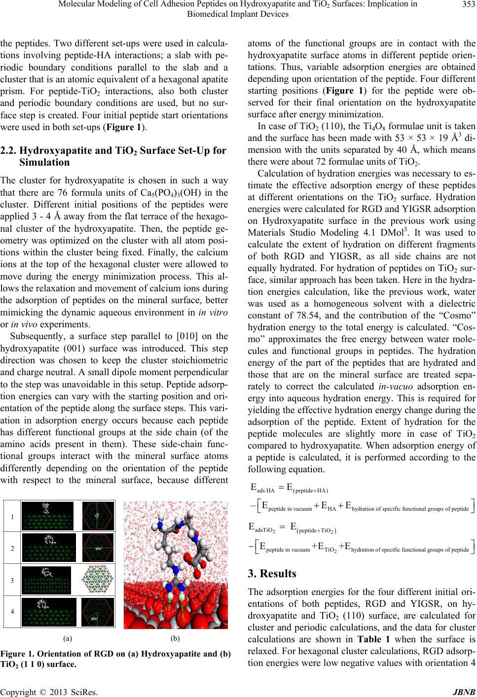 Molecular Modeling of Cell Adhesion Peptides on Hydroxyapatite and TiO2 Surfaces: Implication in Biomedical Implant Devices 353 the peptides. Two different set-ups were used in calcula- tions involving peptide-HA interactions; a slab with pe- riodic boundary conditions parallel to the slab and a cluster that is an atomic equivalent of a hexagonal apatite prism. For peptide-TiO2 interactions, also both cluster and periodic boundary conditions are used, but no sur- face step is created. Four initial peptide start orientations were used in both set-ups (Figure 1). 2.2. Hydroxyapatite and TiO2 Surface Set-Up for Simulation The cluster for hydroxyapatite is chosen in such a way that there are 76 formula units of Ca5(PO4)3(OH) in the cluster. Different initial positions of the peptides were applied 3 - 4 Å away from the flat terrace of the hexago- nal cluster of the hydroxyapatite. Then, the peptide ge- ometry was optimized on the cluster with all atom posi- tions within the cluster being fixed. Finally, the calcium ions at the top of the hexagonal cluster were allowed to move during the energy minimization process. This al- lows the relaxation and movement of calcium ions during the adsorption of peptides on the mineral surface, better mimicking the dynamic aqueous environment in in vitro or in vivo experiments. Subsequently, a surface step parallel to [010] on the hydroxyapatite (001) surface was introduced. This step direction was chosen to keep the cluster stoichiometric and charge neutral. A small dipole moment perpendicular to the step was unavoidable in this setup. Peptide adsorp- tion energies can vary with the starting position and ori- entation of the peptide along the surface steps. This vari- ation in adsorption energy occurs because each peptide has different functional groups at the side chain (of the amino acids present in them). These side-chain func- tional groups interact with the mineral surface atoms differently depending on the orientation of the peptide with respect to the mineral surface, because different 1 2 3 4 1 2 3 4 (a) (b) Figure 1. Orientation of RGD on (a) Hydroxyapatite and (b) TiO2 (1 1 0) surface. atoms of the functional groups are in contact with the hydroxyapatite surface atoms in different peptide orien- tations. Thus, variable adsorption energies are obtained depending upon orientation of the peptide. Four different starting positions (Figure 1) for the peptide were ob- served for their final orientation on the hydroxyapatite surface after energy minimization. In case of TiO2 (110), the Ti4O8 formulae unit is taken and the surface has been made with 53 × 53 × 19 Å3 di- mension with the units separated by 40 Å, which means there were about 72 formulae units of TiO2. Calculation of hydration energies was necessary to es- timate the effective adsorption energy of these peptides at different orientations on the TiO2 surface. Hydration energies were calculated for RGD and YIGSR adsorption on Hydroxyapatite surface in the previous work using Materials Studio Modeling 4.1 DMol3. It was used to calculate the extent of hydration on different fragments of both RGD and YIGSR, as all side chains are not equally hydrated. For hydration of peptides on TiO2 sur- face, similar approach has been taken. Here in the hydra- tion energies calculation, like the previous work, water was used as a homogeneous solvent with a dielectric constant of 78.54, and the contribution of the “Cosmo” hydration energy to the total energy is calculated. “Cos- mo” approximates the free energy between water mole- cules and functional groups in peptides. The hydration energy of the part of the peptides that are hydrated and those that are on the mineral surface are treated sepa- rately to correct the calculated in-vacuo adsorption en- ergy into aqueous hydration energy. This is required for yielding the effective hydration energy change during the adsorption of the peptide. Extent of hydration for the peptide molecules are slightly more in case of TiO2 compared to hydroxyapatite. When adsorption energy of a peptide is calculated, it is performed according to the following equation. ads HA(peptideHA) peptide in vacuumHAhydration of specific functional groups of peptide EE –EE E 22 2 adsTiO peptide TiO peptide in vacuumTiOhydration of specific functional groups of peptide E E E+E+E 3. Results The adsorption energies for the four different initial ori- entations of both peptides, RGD and YIGSR, on hy- droxyapatite and TiO2 (110) surface, are calculated for cluster and periodic calculations, and the data for cluster calculations are shown in Table 1 when the surface is relaxed. For hexagonal cluster calculations, RGD adsorp- tion energies were low negative values with orientation 4 Copyright © 2013 SciRes. JBNB 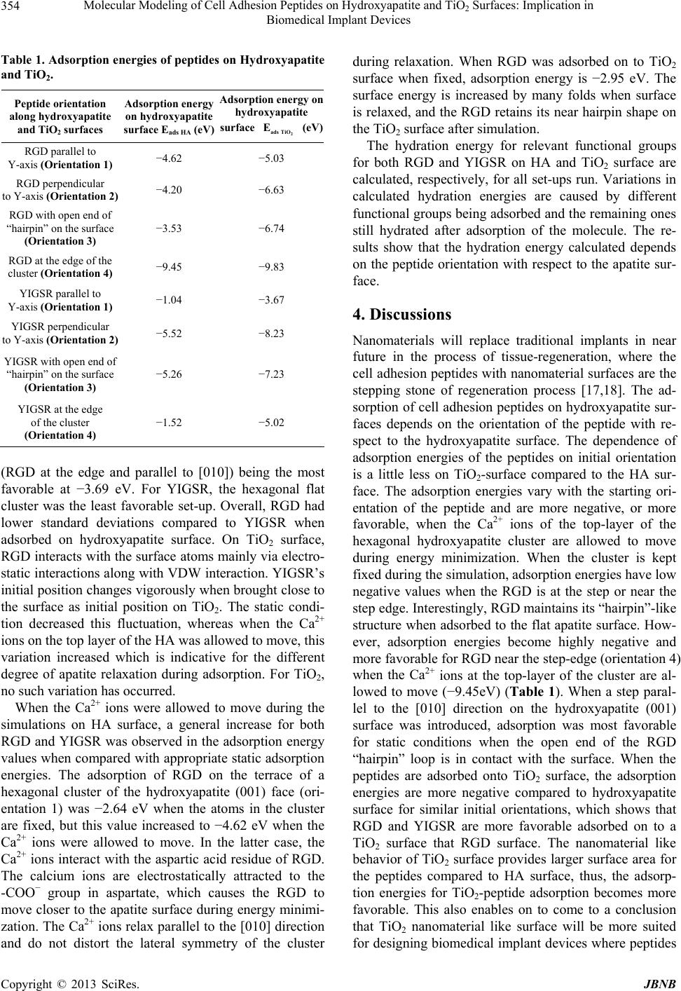 Molecular Modeling of Cell Adhesion Peptides on Hydroxyapatite and TiO2 Surfaces: Implication in Biomedical Implant Devices 354 Table 1. Adsorption energies of peptides on Hydroxyapatite and TiO2. Peptide orientation along hydroxyapatite and TiO2 surfaces Adsorption energy on hydroxyapatite surface Eads HA (eV) Adsorption energy on hydroxyapatite surface eV) 2 ads TiO E ( RGD parallel to Y-axis (Orientation 1) −4.62 −5.03 RGD perpendicular to Y-axis (Orientation 2) −4.20 −6.63 RGD with open end of “hairpin” on the surface (Orientation 3) −3.53 −6.74 RGD at the edge of the cluster (Orientation 4) −9.45 −9.83 YIGSR parallel to Y-axis (Orientation 1) −1.04 −3.67 YIGSR perpendicular to Y-axis (Orientation 2) −5.52 −8.23 YIGSR with open end of “hairpin” on the surface (Orientation 3) −5.26 −7.23 YIGSR at the edge of the cluster (Orientation 4) −1.52 −5.02 (RGD at the edge and parallel to [010]) being the most favorable at −3.69 eV. For YIGSR, the hexagonal flat cluster was the least favorable set-up. Overall, RGD had lower standard deviations compared to YIGSR when adsorbed on hydroxyapatite surface. On TiO2 surface, RGD interacts with the surface atoms mainly via electro- static interactions along with VDW interaction. YIGSR’s initial position changes vigorously when brought close to the surface as initial position on TiO2. The static condi- tion decreased this fluctuation, whereas when the Ca2+ ions on the top layer of the HA was allowed to move, this variation increased which is indicative for the different degree of apatite relaxation during adsorption. For TiO2, no such variation has occurred. When the Ca2+ ions were allowed to move during the simulations on HA surface, a general increase for both RGD and YIGSR was observed in the adsorption energy values when compared with appropriate static adsorption energies. The adsorption of RGD on the terrace of a hexagonal cluster of the hydroxyapatite (001) face (ori- entation 1) was −2.64 eV when the atoms in the cluster are fixed, but this value increased to −4.62 eV when the Ca2+ ions were allowed to move. In the latter case, the Ca2+ ions interact with the aspartic acid residue of RGD. The calcium ions are electrostatically attracted to the -COO− group in aspartate, which causes the RGD to move closer to the apatite surface during energy minimi- zation. The Ca2+ ions relax parallel to the [010] direction and do not distort the lateral symmetry of the cluster during relaxation. When RGD was adsorbed on to TiO2 surface when fixed, adsorption energy is −2.95 eV. The surface energy is increased by many folds when surface is relaxed, and the RGD retains its near hairpin shape on the TiO2 surface after simulation. The hydration energy for relevant functional groups for both RGD and YIGSR on HA and TiO2 surface are calculated, respectively, for all set-ups run. Variations in calculated hydration energies are caused by different functional groups being adsorbed and the remaining ones still hydrated after adsorption of the molecule. The re- sults show that the hydration energy calculated depends on the peptide orientation with respect to the apatite sur- face. 4. Discussions Nanomaterials will replace traditional implants in near future in the process of tissue-regeneration, where the cell adhesion peptides with nanomaterial surfaces are the stepping stone of regeneration process [17,18]. The ad- sorption of cell adhesion peptides on hydroxyapatite sur- faces depends on the orientation of the peptide with re- spect to the hydroxyapatite surface. The dependence of adsorption energies of the peptides on initial orientation is a little less on TiO2-surface compared to the HA sur- face. The adsorption energies vary with the starting ori- entation of the peptide and are more negative, or more favorable, when the Ca2+ ions of the top-layer of the hexagonal hydroxyapatite cluster are allowed to move during energy minimization. When the cluster is kept fixed during the simulation, adsorption energies have low negative values when the RGD is at the step or near the step edge. Interestingly, RGD maintains its “hairpin”-like structure when adsorbed to the flat apatite surface. How- ever, adsorption energies become highly negative and more favorable for RGD near the step-edge (orientation 4) when the Ca2+ ions at the top-layer of the cluster are al- lowed to move (−9.45eV) (Table 1). When a step paral- lel to the [010] direction on the hydroxyapatite (001) surface was introduced, adsorption was most favorable for static conditions when the open end of the RGD “hairpin” loop is in contact with the surface. When the peptides are adsorbed onto TiO2 surface, the adsorption energies are more negative compared to hydroxyapatite surface for similar initial orientations, which shows that RGD and YIGSR are more favorable adsorbed on to a TiO2 surface that RGD surface. The nanomaterial like behavior of TiO2 surface provides larger surface area for the peptides compared to HA surface, thus, the adsorp- tion energies for TiO2-peptide adsorption becomes more favorable. This also enables on to come to a conclusion that TiO2 nanomaterial like surface will be more suited for designing biomedical implant devices where peptides Copyright © 2013 SciRes. JBNB  Molecular Modeling of Cell Adhesion Peptides on Hydroxyapatite and TiO2 Surfaces: Implication in Biomedical Implant Devices 355 can be adsorbed directly toinitiate the cell-adhesion pro- cess during bone tissue regeneration process. The water molecules occupy the adsorption sites on the hydrophilic TiO2 surfaces, i.e. the water oxygen at- oms bond to the surface titanium atoms to form the stable first hydration layer and interact with the surface oxygen atoms to form the second hydration layer. Besides being in competition with RGD for the adsorption sites, the ad- sorbed water layers also play an intermediary role, form- ing HB interactions with the hydrophilic groups of RGD. The guanido (NH2), amino (NH3) and carboxyl groups (COO-) of tripeptide RGD are the main groups bonding to TiO2 surface by electrostatic and VDW interactions. The extent of hydration of different functional groups on each peptide was also considered. Some of the func- tional groups in the peptide are still hydrated after ad- sorption and the others are bonded to the mineral surface. The final adsorbate structure on the mineral surface eva- luates which functional groups were bonded to the min- eral surface, and hydration energies of these functional groups were subtracted from the peptide vacuum adsorp- tion energy to obtain the effective adsorption energies. The NH2- with peptide bond elements are more strongly hydrated than other functional groups (−1.465 eV hydra- tion energy), followed by the amide functional group (C(NH)=NH-NH2) of arginine (R) (−1.106 eV). The COO- groups have hydration energies of ~−0.2 eV to −0.5 eV. When the peptide is kept parallel or perpen- dicular to the [010] direction (orientations 1 and 2), the amide and carboxylic groups interact with the mineral surface, allowing the peptide to maintain its “hairpin” position. The above hydration strategies were used in exactly same way when adsorption of peptide is calculated only on HA surface. Adsorption of peptides on TiO2 surface followed similar course and the hydration energies for responsible functional groups on TiO2 surface has been deducted accordingly to calculate the effective adsorp- tion energy of RGD, YIGSR on TiO2. The retention of hairpin like structure of RGD on TiO2 surface is of great importance in showing similarity of behaviour of RGD on both HA and TiO2-surface in aqueous environment. 5. Conclusion The adsorption energies of RGD and YIGSR on Hy- droxyapatite surface and TiO2 surface reveal similar trends. Comparing the adsorption energies for both the surfaces, we can conclude that RGD behaves similarly on both the surfaces, with positioning of the peptide near the edge of the cluster (Orientation 4) yielding most favor- able adsorption energy. This gives us a clear idea about the peptide orientation during the tissue-engineering process. When artificial inclusion of Titania-based nano- particles is required inside the tissue, it is important to know how the peptides are going to interact with TiO2 surface. The adsorption energies and favorable confor- mations of peptides on TiO2 assist researches to design biomedical implants making them appropriately keep in mind that the most favorable surface for peptide adsorp- tion on TiO2 is (1 1 0). Future studies will concentrate on more TiO2 surfaces and more peptides to develop a more general approach for designing of biomedical implants to facilitate bone-tissue engineering. 6. Acknowledgements This work has been partially funded by National Science Foundation, USA, Tissue Engineering at University of Michigan, NSF Grant of Prof. Udo Becker at University of Michigan. REFERENCES [1] M. Woo, J. Seo, R. Y. Zhang and P. X. Ma, “Suppression of Apoptosis by Enhanced Protein Adsorption on Poly- mer,” Biomaterials, Vol. 28, No. 16, 2007, pp. 2622- 2630. [2] S. P. Massia and J. A. Hubbell, “Covalent Surface Immo- bilization of Arg-Gly-Asp-and Tyr-Ile-Gly-Ser-Arg-Con- taining Peptides to Obtain Well-Defined Cell-Adhesive Substrates,” Analytical Biochemistry, Vol. 187, No. 2, 1990, pp. 292-301. [3] Y. Iwamoto, F. A. Robey, J. Graf, M. Sasaki, H. K. Kleinman, Y. Yamada and G. R. Martin, “YIGSR, a Syn- thetic Lamininpentapeptide, Inhibits Experimental Me- tastasis Formation,” Science, Vol. 238, No. 4830, 1987, pp. 1132-1134. [4] W. H. Kim, H. W. Schnaper, M. Nomizu, Y. Yamada and H. K. Kleinman, “Apoptosis in Human Fibrosarcoma Cells Is Induced by a Multimeric Synthetic Tyr-Ile-Gly-Ser- Arg (YIGSR)-Containing Polypeptide from Laminin,” Can- cer Research, Vol. 54, No. 18, 1994, pp. 5005-5010. [5] E. Ruoslahti, “RGD and Other Recognition Sequences for Integrins,” Annual Review of Cell and Developmental Bi- ology, Vol. 12, 1996, pp. 697-715. http://dx.doi.org/10.1146/annurev.cellbio.12.1.697 [6] M. Kantlehner, D. Finsinger, J. Meyer, P. Schaffner, A. Jonczyk, B. Diefenbach, B. Nies and H. Kessler, “Selec- tive RGD-Mediated Adhesion of Osteoblasts at Surfaces of Implants,” Angewandte Chemie International Edition, Vol. 38, No. 4, 1999, pp. 560-562. http://dx.doi.org/10.1002/(SICI)1521-3773(19990215)38: 4<560::AID-ANIE560>3.0.CO;2-F [7] M. Gilbert, W. J. Shaw, J. R. Long, K. Nelson, G. P. Drobny, C. M. Giachelli and P. S. Stayton, “Chimeric Peptides of Statherin and Osteopontin That Bind Hydro- xyapatite and Mediate Cell Adhesion,” Journal of Bio- logical Chemistry, Vol. 275, No. 21, 2000, pp. 16213- 16218. [8] U. Becker, S. Biswas, T. Kendall, P. Risthaus, C. V. Put- Copyright © 2013 SciRes. JBNB  Molecular Modeling of Cell Adhesion Peptides on Hydroxyapatite and TiO2 Surfaces: Implication in Biomedical Implant Devices Copyright © 2013 SciRes. JBNB 356 nis and C. M. Pina, “Interactions between Mineral Sur- faces and Dissolved Species: From Monovalent Ions to Complex Organic Molecules,” American Journal of Sci- ence, Vol. 305, No. 6-8, 2005, pp. 791-825. [9] C. Boiziau, S. Leroy, C. Reynaud, G. Lecayon, C. Le- gressus and P. Viel, “Elementary Mechanisms in the In- teraction of Organic-Molecules with Mineral Surfaces,” Journal of Adhesion, Vol. 23, No. 1, 1987, pp. 21-44. [10] A. Wierzbicki and H. S. Cheung, “Molecular Modeling of Inhibition of Hydroxyapatite by Phosphocitrate,” Journal of Molecular Structure: Theochem, Vol. 529, No. 1-3, 2000, pp. 73-82. [11] N. H. de Leeuw, “A Computer Modeling Study of the Up- take and Segregation of Fluoride Ions at the Hydrated Hy- droxyapatite (0001) Surface: Introducing a Ca10(PO4)6OH2 Potential Model,” Physical Chemistry Chemical Physics, Vol. 6, No. 8, 2004, pp. 1860-1866. [12] D. Mkhonto and N. H. de Leeuw, “A Computer Modeling Study of the Effect of Water on the Surface Structure and Morphology of Fluorapatite: Introducing a Ca10(PO4)6F2 Potential Model,” Journal of Materials Chemistry, Vol. 12, No. 9, 2002, pp. 2633-2642. [13] A. K. Rappe, C. J. Casewit, K. S. Colwell, W. A. God- dard and W. M. Skiff, “UFF, a Full Periodic-Table Force Field for Molecular-Mechanics and Molecular-Dynamics Simulations,” Journal of the American Chemical Society, Vol. 114, No. 25, 1992, pp. 10024-10035. http://dx.doi.org/10.1021/ja00051a040 [14] J. D. Gale and A. L. Rohl, “The General Utility Lattice Program (GULP),” Molecular Simulation, Vol. 29, No. 5, 2003, pp. 291-341. [15] A. K. Rappe and W. A. Goddard, “Charge Equilibration for Molecular-Dynamics Simulations,” Journal of Physi- cal Chemistry, Vol. 95, No. 8, 1991, pp. 3358-3363. [16] S. Biswas and U. Becker, “Molecular Modeling of Cell Adhesion Peptides on Hydroxyapatite Surfaces and Sur- face Steps: Application in Bone Tissue Engineering and Biomimetics,” 3rd International Conference on Chemical, Biological and Environment Sciences (ICCEBS’2013), Kuala Lumpur, 8-9 January 2013, pp. 59-63. [17] L. Zhang and T. J. Webster, “Nanotechnology and Nano- materials: Promises for Improved Tissue Regeneration,” Nano Today, Vol. 4, No. 1, 2009, pp. 66-80. http://dx.doi.org/10.1016/j.nantod.2008.10.014 [18] M. J. Webber, J. A. Kessler and S. I. Stupp, “Emerging Peptide Nanomedicine to Regenerate Tissues and Or- gans,” Journal of Internal Medicine, Vol. 267, No. 1, 2010, pp. 71-88. http://dx.doi.org/10.1111/j.1365-2796.2009.02184.x |