

Paper Menu >>
Journal Menu >>
 International Journal of Otolaryngology and Head & Neck Surgery, 2013, 2, 201-206 http://dx.doi.org/10.4236/ijohns.2013.25042 Published Online September 2013 (http://www.scirp.org/journal/ijohns) Audiological Evaluation in Goitrous Hypotyhroidism M. K. Aggarwal1, Gautam Bir Singh2*, Ranjan K. Nag1, S. K. Singh1, Rajesh Kumar1, Mayank Yadav2 1Institute of Medical Sciences, BHU, Varanasi, India 2Lady Hardinge Medical College & Associated Hospitals, New Delhi, India Email: [email protected] Received May 26, 2013; revised June 25, 2013; accepted July 10, 2013 Copyright © 2013 M. K. Aggarwal et al. This is an open access article distributed under the Creative Commons Attribution License, which permits unrestricted use, distribution, and reproduction in any medium, provided the original work is properly cited. ABSTRACT Obje ctive: To determine the incidence of deafness in patients suffering from goiterous hypothyroidism exclusively and to evaluate the role of L-thyroxine therapy in improving the hearing in this group of patients. Study Design: A prospec- tive cohort study. Materials and Methods: Hearing status was evaluated in a sample size of 100 consecutive patients reporting to the ENT/Endocrinology department of Institute of Medical Sciences, Banaras Hindu University, Varanasi, UP with the diagnosis of goitrous hypothyroidism. The study group included patients in the age group of 5 to 65 years belonging to either sex. Patients with detected hearing loss were categorized into group A, and all other patients were designated group B. L-Thyroxine treatment for goitrous hypothyroidism was initiated in all the cases. At the end of 6 months, a repeat audiogram was done in all the patients in order to evaluate the efficacy of the said treatment protocol on the hearing in these patients. The data were tabulated and statistically analysed using Paired Students “t” test. Results: An overall 39% hearing loss was observed in patients with goitrous hypothyroidism. 15% cases had sensorineural hear- ing loss, 13% had mixed hearing loss and 8% had a conductive hearing loss. A statistically significant hearing improve- ment was recorded in this study by L-thyroxine treatment in group-A, and no deterioration of hearing was recorded in group-B. Conclusions: The incidence of sensorineural hearing is less in patients with goitrous hypothyroidism (15%) as compared with the overall incidence of sensorineural hearing loss reported for hypothyroidism (30% - 40%). Further, there is a definitive improvement in hearing with the use of L-thyroxine treatment of goitrous hypothyroidism. Keywords: Hypothyroidism; Goitre; Hearing Loss; L-Thyroxine Treatment 1. Introduction Hearing loss was first reported in acquired hypothyroid- ism in 1907 [1]. Over a period of time, a distinct associa- tion between hypothyroidism and auditory system dys- function has been reported in medical text [2], though there are studies which have failed to elucidate a defini- tive relationship between hypothyroidism and deafness [3-5]. The medical literature now mentions vertigo, hear- ing loss, tinnitus and pruritic external auditory canal as important vestibular and audiological symptoms of hy- vpothyroidism [2]. Hypothyroidism is associated with all types of deaf- ness: sensorineural, mixed and conductive, however, the real incidence and pathophysiology of this hearing loss in these patients is still uncertain. This is attributed to the marked paucity of literature on the cited subject. The in- cidence of hearing loss varies from 25% to 50% with a higher incidence in congenital hypothyroidism [6,7]. Moreover, the results of audiological evaluation of pa- tients with hypothyroidism under treatment with L-Thy- roxine [LT] are conflicting. There are studies which have highlighted the importance of this modality of treatment in improving hearing in hypothyroid patients [8-12], but literature is also replete with studies which have found no correlation between the two [3-5,13,14]. Considering this, in order to broaden the studies in this line of research a prospective study was initiated exclusively in goitrous hypothyroid patients with the aim to: 1) Determine the incidence of hearing loss in these pa- tients; 2) Evaluate the effect of LT therapy on this hearing loss in these patients. To our knowledge, the current study is unique, in that audiological evaluation of goitrous hypothyroidism has not been previously reported in medical literature. *Corresponding author. C opyright © 2013 SciRes. IJOHNS  M. K. AGGARWAL ET AL. 202 2. Materials & Methods A prospective outcome analysis study was carried out in the departments of Otorhinolaryngology & Endocrinol- ogy, Institute of Medical Sciences, Banaras Hindu Uni- versity, Varanasi, UP, India from September 2002 to September 2005. For the purpose of this study 100 con- secutive patients of either sex suffering from goitre with hypothyroidism were recruited in the study design. The study was approved by the University Board of Studies & Ethics committee, and an informed consent was man- datory for recruitment in the study design. Paediatric pa- tients were also recruited in the study design. All the patients of goitre reporting either to the otorhi- nolaryngology department or endocrinology department were subjected to detailed history taking and clinical ex- amination. A FNAC (fine needle aspiration cytology), USG (ultrasound) of the enlarged thyroid and screening for thyroid function was done in all the cases to confirm the nature of the goitre and the hypothyroidism. The pa- tients were specifically screened for thyroid dysfunction by T4 and TSH levels. Thyroid function test were done by RIA [Radio Immune Assay], the kit for which was supplied by Bhabha Atomic Research Centre, Mumbai, India. Only patients with proven hypothyroid status were enrolled in the study: ● Serum thyroxine level (T4): normal range 4 to 13 mi- crograms/dl; ● Serum thyrotropin (TSH): normal range 0.3 to 6 mi- cro IU/ml. Their particulars, detailed history of thyroid swelling and disease along with clinical examination were entered in a Performa. Tests to diagnose aetiology of hypothy- roidism and goitre were also done. A special note of deafness, vertigo, and tinnitus was also made. The pa- tients of deafness were further evaluated by tuning fork test and pure tone audiogram [PTA]. Alps advanced di- agnostic audiometer AD100 with the following standards was used for PTA testing: ● Tone audiometer: EN60645-1/ANSI S3.6, Type 2; ● Speech audiometer: EN60645-2/ANSI S3.6, Type B or B-E. It would be prudent to note that hypothyroid patients with deafness due to any other causes like: ● Chronic suppurative otitis media; ● Meningitis; ● Head Injury; ● Presbyacusis (patients above 60 years were not re- cruited in this study); ● Ototoxic drugs etc were excluded from the study de- sign. Finally, these patients with audiogram proven deafness were categorized in a designated special group-A, all other patients were enrolled in group-B. Patients in both the groups continued to receive thyroxine treatment un- der the supervision of endocrinology department and a regular monthly follow-up was maintained. At the end of 6 months a repeat audiogram was done and the hearing status was once again evaluated. A 10 db improvement in two consecutive frequencies was regarded as an audi- ological improvement [15,16]. The data was tabulated and statistically analysed by paired students “t” test. In this study patient with Hashimoto’s thyroiditis, io- dine deficiency, thyroid malignancy, irradiated thyroid malignancy, Pendred’s Syndrome and hyperthyroid pa- tients treated with drugs/radioiodine etc were all included in the study design. Distinguishing congenital hearing loss due to “Pendred’s Syndrome” and other causes of congenital hearing loss was difficult as “Perchlorate Test” was not available in our institution. Thus all the cases of congenital hearing loss with hypothyroidism and goitre were clinically regarded as “Pendred Syndrome”. Al- though we recruited cases with diagnosis of hypothy- roidism with goitre which reported for the first time to ENT/endocrinology department OPD of our institution, however during the course of study it was revealed that some of these patients had taken replacement in the past for short duration. This was probably due to the rural background, poor socio-economic status and illiteracy of our patient profile. 3. Results Out of a total of 100 patients with goitre and hypothy- roidism, 70 were females and 30 were males. The age group of patients ranged from 5 years to 64 years. There were 12 paediatric patients. The age distribution of these patients is given in Table 1. A total of 39 patients had an audiological proven deaf- ness. However over a period of time 3 patients were lost in follow-up, thus for statistical interpretation and dis- cussion in this study the results in 36 patients were taken into consideration. Out of these 36 patients 8 patients had conductive hearing loss, 15 patients had sensorineural hearing loss and 13 patients had a mixed hearing loss. The distribution of these cases in accordance with WHO classification is shown in Table 2. The relationship of this hearing loss with the levels of T4 is also given in Table 2. The mean T4 levels which were associated with normal hearing are 2.7 micrograms. From the table it is clearly evident that the severity of the hearing loss is directly proportional to the decreasing value of T4 levels. The hearing thresholds were again measured 6 months later in euthyroid state post thyroxine therapy. The Table 3 shows the audiological gains after L-thyroxine therapy in each ear separately and the overall gain in all the 36 patients. The analysis of the data revealed: 1) The 10 db hearing improvement or more is seen in 9 Copyright © 2013 SciRes. IJOHNS 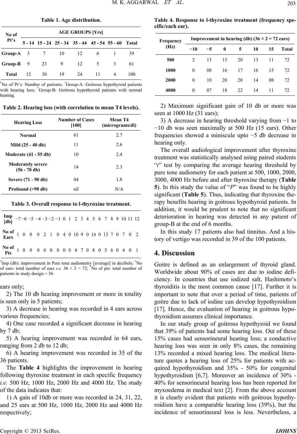 M. K. AGGARWAL ET AL. 203 Table 1. Age distribution. AGE GROUPS [Yrs] No of Pt’s 5 - 14 15 - 24 25 - 34 35 - 44 45 - 54 55 - 60Total Group-A 3 7 10 12 6 1 39 Group-B 9 23 9 12 5 3 61 Total 12 30 19 24 11 4 100 *No of Pt’s: Number of patients; *Group-A: Goitrous hypothyroid patients with hearing loss; *Group-B: Goitrous hypothyroid patients with normal hearing. Table 2. Hearing loss (with correlation to mean T4 levels). Hearing Loss Number of Cases [100] Mean T4 (micrograms/dl) Normal 61 2.7 Mild (25 - 40 db) 11 2.6 Moderate (41 - 55 db) 10 2.4 Moderately severe (56 - 70 db) 14 2.3 Severe (71 - 90 db) 04 1.8 Profound (>90 db) nil N/A Table 3. Overall response to l-thyroxine treatment. Imp [db] −7 −6 −5 −4 −3 −2 −1 0 1 23 4 5 6 7 8 9101112 No of Ears 1 0 0 0 2 1 0 4 0 109 0 16 0 13 7 0702 No of Pts 1 0 0 0 0 0 0 0 0 4 7 0 8 0 5 6 0401 *Imp (db): improvement in Pure tone audiometry [average] in decibels; *No of ears: total number of ears i.e. 36 × 2 = 72; *No of pts: total number of patients in study design = 36. ears only; 2) The 10 db hearing improvement or more in totality is seen only in 5 patients; 3) A decrease in hearing was recorded in 4 ears across various frequencies; 4) One case recorded a significant decrease in hearing by 7 db; 5) A hearing improvement was recorded in 64 ears, ranging from 2 db to 12 db; 6) A hearing improvement was recorded in 35 of the 36 patients. The Table 4 highlights the improvement in hearing following thyroxine treatment in each specific frequency i.e. 500 Hz, 1000 Hz, 2000 Hz and 4000 Hz. The study of the data indicates that: 1) A gain of 10db or more was recorded in 24, 31, 22, and 25 ears at 500 Hz, 1000 Hz, 2000 Hz and 4000 Hz respectively; Table 4. Response to l-thyroxine treatment (frequency spe- cific/each ear). Improvement in hearing (db) (36 × 2 = 72 ears) Frequency (Hz) −10 −5 0 5 10 15 Total 500 2 13 13 20 13 11 72 1000 0 08 16 17 16 15 72 2000 0 10 20 20 14 08 72 4000 0 07 18 22 14 11 72 2) Maximum significant gain of 10 db or more was seen at 1000 Hz (31 ears); 3) A decrease in hearing threshold varying from −1 to −10 db was seen maximally at 500 Hz (15 ears). Other frequencies showed a miniscule upto −5 db decrease in hearing only. The overall audiological improvement after thyroxine treatment was statistically analysed using paired students “t” test by comparing the average hearing threshold by pure tone audiometry for each patient at 500, 1000, 2000, 3000, 4000 Hz before and after thyroxine therapy (Table 5). In this study the value of “P” was found to be highly significant (Table 5). Thus, indicating that thyroxine the- rapy benefits hearing in goitrous hypothyroid patients. In addition, it would be prudent to note that no significant deterioration in hearing was detected in any patient of group-B at the end of 6 months. In this study 17 patients also had tinnitus. And a his- tory of vertigo was recorded in 39 of the 100 patients. 4. Discussion Goitre is defined as an enlargement of thyroid gland. Worldwide about 90% of cases are due to iodine defi- ciency. In countries that use iodized salt, Hashimoto’s thyroiditis is the most common cause [17]. Further it is important to note that over a period of time, patients of goitre due to lack of iodine can develop hypothyroidism [17]. Hence, the evaluation of hearing in goitrous hypo- thyroidism assumes clinical importance. In our study group of goitrous hypothyroid we found that 39% of patients had some hearing loss. Out of these 15% cases had sensorineural hearing loss; a conductive hearing loss was seen in only 8% cases, the remaining 13% recorded a mixed hearing loss. The medical litera- ture quotes a hearing loss of 25% for patients with ac- quired hypothyroidism and 35% - 50% for congenital hypothyroidism [6,7]. Moreover an incidence of 30% - 40% for sensorineural hearing loss has been reported for myxoedema in medical text [2]. From the above account it is clearly evident that patients with goitrous hypothy- roidism have a comparable hearing loss (39%), but the incidence of sensorineural loss is less. Nevertheless, a Copyright © 2013 SciRes. IJOHNS 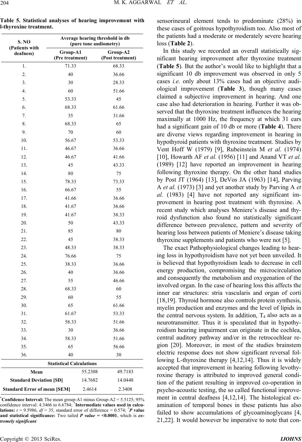 M. K. AGGARWAL ET AL. 204 Table 5. Statistical analyses of hearing improvement with l-thyroxine treatment. Average hearing threshold in db (pure tone audiometry) S. NO (Patients with deafness) Group-A1 (Pre treatment) Group-A2 (Post treatment) 1. 71.33 68.33 2. 40 36.66 3. 30 28.33 4. 60 51.66 5. 53.33 45 6. 68.33 61.66 7. 35 31.66 8. 68.33 65 9. 70 60 10. 56.67 53.33 11. 46.67 36.66 12. 46.67 41.66 13. 45 43.33 14. 80 75 15. 78.33 73.33 16. 66.67 55 17. 41.66 36.66 18. 41.67 36.66 19. 41.67 38.33 20. 50 43.33 21. 85 80 22. 45 38.33 23. 48.33 38.33 24. 76.66 75 25. 38.33 36.66 26. 40 36.66 27. 55 46.66 28. 68.33 60 29. 60 55 30. 65 61.66 31. 61.67 53.33 32. 58.33 51.66 33. 30 36.66 34. 58.33 51.66 35. 65 56.66 36. 40 30 Statistical Calculations Mean 55.2308 49.7183 Standard Deviation [SD] 14.7682 14.0448 Standard Error of mean [SEM] 2.4614 2.3408 *Confidence Interval: The mean group-A1 minus Group-A2 = 5.5125; 95% confidence interval: 4.3466 to 6.6784; *Intermediate values used in calcu- lations: t = 9.5986, df = 35, standard error of difference = 0.574; *P value and statistical significance: Two tailed P value = <0.0001, which is ex- tremely significant. sensorineural element tends to predominate (28%) in these cases of goitrous hypothyroidism too. Also most of the patients had a moderate or moderately severe hearing loss (Table 2). In this study we recorded an overall statistically sig- nificant hearing improvement after thyroxine treatment (Table 5). But the author’s would like to highlight that a significant 10 db improvement was observed in only 5 cases i.e. only about 13% cases had an objective audi- ological improvement (Table 3), though many cases claimed a subjective improvement in hearing. And one case also had deterioration in hearing. Further it was ob- served that the thyroxine treatment influences the hearing maximally at 1000 Hz, the frequency at which 31 ears had a significant gain of 10 db or more (Table 4). There are diverse views regarding improvement in hearing in hypothyroid patients with thyroxine treatment. Studies by Vent Hoff W (1979) [9], Rubeinstein M et al. (1974) [10], Howarth AF et al. (1956) [11] and Anand VT et al. (1989) [12] have reported an improvement in hearing following thyroxine therapy. On the other hand studies by Post JT (1964) [13], DeVos JA (1963) [14], Parving A et al. (1973) [3] and yet another study by Parving A et al. (1983) [4] have not reported any significant im- provement in hearing post treatment with thyroxine. A recent study which analyses Meniere’s disease and thy- roid dysfunction also found no statistically significant difference between prevalence, pattern and severity of hearing loss between patients of Meniere’s disease taking thyroxine supplements and patients who were not [5]. The exact Pathophysiological changes leading to hear- ing loss in hypothyroidism have not yet been unveiled. It is believed that hypothyroidism leads to decrease in cell energy production, compromising the microcirculation and consequently the metabolism and oxygenation of the involved organ. In the case of hearing loss this affects the inner ear structures: stria vascularis and organ of corti [18,19]. Thyroid hormone also controls protein synthesis, myelin production and enzymes and the level of lipids in the central nervous system. In addition, T4 also acts as a neurotransmitter. Thus it is speculated that in hypothy- roidism hearing impairment can originate in the cochlea, central auditory pathway and/or in the retrocochlear re- gion [20]. Moreover, in most of the studies brainstem electric response does not show significant reversal fol- lowing L-thyroxine therapy [4,12,14]. Thus it is widely accepted that improvement in hearing following levothy- roxine therapy is attributed to improved general condi- tion of the patient resulting in improved co-operation in psycho-acoustic testing, the so called functional improve- ment in central deafness [4,12,14]. The histological ex- amination of temporal bones in these patients has also failed to show accumulations of glycoaminoglycans [4, 21,22]. It would however be imperative to note that con- Copyright © 2013 SciRes. IJOHNS  M. K. AGGARWAL ET AL. 205 ductive hearing loss in hypothyroidism is secondary to Eustachian tube mucosal oedema [23]. Although it was not the endeavour of this study to analyse the vestibular changes in goitrous hypothyroid- ism, the authors would like to highlight that almost 39% cases had a history of vertigo, out of which 26 patients had an subjective improvement after levothyroxine ther- apy at the end of 6 months. In this context it would be important to note that the medical text mentions that al- most 66% of patients of hypothyroidism suffer from ver- tigo [2]. Tinnitus too was present in 17 cases. Nine of these cases recorded a subjective improvement in form of decrease in the duration and intensity of the tinnitus. Thus a joint involvement of cochlear and vestibular sys- tem is also seen in goitrous hypothyroidism as has been reported for other metabolic disorders [24]. The authors would also like to highlight that no case of Meniere’s disease i.e. the classical triad of vertigo, deafness with tinnitus was recorded in this case series, though the lit- erature reports an intrinsic relationship between hypo- thyroidism and Meniere’s disease (a recent study quotes a prevalence of 32%) [5]. Interpretations of these results must take into consid- eration the limitations of our analysis. As data from a single tertiary health care centre was used, it reflects the experience of our geographical area and may not be gen- eralized. Information from observational studies can be subject to potential biases (e.g. selection bias) and con- founding. Critics may contend that 6 months follow-up period is short. Furthermore, the results were not ascer- tained blindly. Last but not the least; we were unable to discern the reason for this decreased sensorineural hear- ing loss in goitrous hypothyroidism. This could be due to the demographic profile of our patients. But then the au- thors would like to emphasise that the very cause of hearing loss in hypothyroidism is debatable and contro- versial. Nevertheless, the strength of this study lies in its prospective character and independent statistical valida- tion, which allowed for accurate assessment of data with- out depending upon recalled information in accordance with evidence based medicine. The authors would like to highlight that this study represents the largest series of patients on hypothyroidism. The true value of this study in context of existing literature lies in the audiological evaluation of patients belonging to the subgroup of goi- trous hypothyroid, hitherto unreported in medical litera- ture. 5. Conclusion In conclusion, the patients of goitrous hypothyroidism have a lower rate of sensorineural hearing loss as com- pared with other patients of hypothyroidism. Further, this study delineates a definitive role of L-thyroxine therapy in improvement of hearing in patients suffering from goitrous hypothyroidism. We believe that conclusions of this study can serve as a guide for future research on the cited subject. REFERENCES [1] W. R. Kemp, “Deafness in Myxoedema,” British Medical Journal, Vol. 1, 1907, p. 375. [2] J. M. Morgan and T. McCaffrey, “Head and Neck Mani- festations of Endocrine Disease,” In: M. Gleeson, Ed., Scott-Brown’s Otorhinolaryngology, Head and Neck Sur- gery, Hodder Arnold, London, 2008, p. 400. [3] A. Parving, H. Parving and J. Lyngsoe, “Hearing Sensi- tivity in Patients with Myxoedema before and after Treat- ment with L-Thyroxine,” Acta Oto-Laryngologica (Stock- holm), Vol. 95, 1983, pp. 315-321. doi:10.3109/00016488309130948 [4] A. Parving, B. Ostri, P. Bretlau, et al., “Audiological and Temporal Bone Findings in Myxoedema,” Annals of Oto- logy, Rhinology, and Laryngology, Vol. 95, 1986, pp. 278-283. [5] M. Brenner, D. L. Hoistad and T. C. Hain, “Prevalence of Thyroid Dysfunction in Patients with Meniere’s Disease,” Archives of Otolaryngology—Head and Neck Surgery, Vol. 130, No. 2, 2004, pp. 226-228. doi:10.1001/archotol.130.2.226 [6] R. Ben-Tovim, Y. Zohar, N. Laurian, et al., “Auditory Brainstem Response in Experimentally Induced Hypo- thyroidism in Albino Rats,” Laryngoscope, Vol. 95, 1985, pp. 982-986. doi:10.1288/00005537-198508000-00020 [7] M. Vanasse, C. Fisher, F. Berthezene, et al., “Normal Auditory Evoked Potentials in Adult Hypothyroidism,” Laryngoscope, Vol. 99, No. 3, 1989, pp. 302-306. doi:10.1288/00005537-198903000-00012 [8] Y. Sachdev and R. Hall, “Effusions into Body Cavities in Hypothyroidism,” Lancet, Vol. 1, 1975, pp. 564-565. doi:10.1016/S0140-6736(75)91571-8 [9] W. Vant Hoff and D. W. Stuart, “Deafness in Myxoe- dema,” Quarterly Journal of Medicine, Vol. 48, 1979, pp. 361-367. [10] M. Rubenstein, C. Rubenstein and R. Theodor, “Hearing Dysfunction Associated with Congenital Sporadic Hypo- thyroidism,” Annals of Otology, Rhinology, and Laryn- gology, Vol. 83, 1974, pp. 814-819. [11] A. F. Howarth and H. E. Lloyd, “Perceptive Deafness in Hypothyroidism,” British Medical Journal, Vol. 1, 1956, pp. 431-433. doi:10.1136/bmj.1.4964.431 [12] V. T. Anand, S. B. S. Mann, R. J. Dash, et al., “Auditory Investigation in Hypothyroidism,” Acta Oto-Laryngolo- gica (Stockh olm), Vol. 108, No. 1-2, 1989, pp. 83-87. doi:10.3109/00016488909107396 [13] J. T. Post, “Hypothyroid Deafness,” Laryngoscope, Vol. 74, 1964, pp. 221-232. [14] J. A. DeVos, “Deafness in Hypothyroidism,” Journal of Laryngology & Otology, Vol. 77, 1963, pp. 390-414. doi:10.1017/S0022215100060795 [15] N. Umapathy and P. J. Dekker, “Myringoplasty Is It Copyright © 2013 SciRes. IJOHNS  M. K. AGGARWAL ET AL. Copyright © 2013 SciRes. IJOHNS 206 Worth Performing in Children,” Archives of Otolaryn- gology—Head and Neck Surgery, Vol. 129, No. 10, 2003, pp. 1053-1055. doi:10.1001/archotol.129.10.1053 [16] G. B. Singh, S. Sharma and N. Singh, “Role of Tran- stympanic Myringoplasty in Modern Otology,” Journal of Otolaryngology, Vol. 35, No. 6, 2006, pp. 408-412. doi:10.2310/7070.2005.0033 [17] J. L. Jameson and A. P. Weetman, “Disorders of Thyroid Gland,” In: A. S. Fauci, E. Braunwald, D. L. kasper, et al., Eds., Harrison’s Principles of Internal Medicine, Mcgraw Hill, New York, 2008, pp. 2240-2242 [18] J. A. A. Oliveira, “Fisiologia da Audicao-Coclea Ativa,” In: M. S. Figueiredo, Eds., Emissoes Otoacusticas e BERA, Pulso Editorial, Sao Jose dos Campos, 2003, pp. 1-34. [19] K. T. P. Dos Santos, N. H. Dias and G. M. F. Da Silva, “Mazeto Audiologic Evaluation in Patients with Acquired Hypothyroidism,” Brazilian Journal of Otorhinolaryn- gology, Vol. 76, No. 4, 2010, pp. 478-484. [20] L. Di lorenza, L. Foggio, N. Panza, et al., “Auditory Brainstem Response in Thyroid Diseases before and after Therapy,” Hormone Research, Vol. 43, 1995, pp. 200- 205. doi:10.1159/000184278 [21] J. Hald, C. M. Milroy, K. Damkjaer-Jansen, et al., “Hu- man Temporal Bone Findings in Acquired Hypothyroid- ism,” Journal of Laryngology & Otology, Vol. 105, No. 11, 1991, pp. 886-889. doi:10.1017/S0022215100117748 [22] Z. Mra and M. K. Wax, “Effects of Acute Thyroxin De- pletion on Hearing in Humans,” Laryngoscope, Vol. 109, No. 3, 1999, pp. 343-350. doi:10.1097/00005537-199903000-00001 [23] F. Debruyne, M. Vanderschueren-Lodeweyckx, P. Basti- jns, et al., “Hearing in Congenital Hypothyroidism,” Au- diology, Vol. 22, 1983, pp. 404-409. doi:10.3109/00206098309072800 [24] A. Parving, “Hearing Problems and Hormonal Distur- bances in the Elderly,” Acta Oto-Laryngologica (Stock- holm), Vol. 476, 1991, pp. 44-53. doi:10.3109/00016489109127255 |