

Paper Menu >>
Journal Menu >>
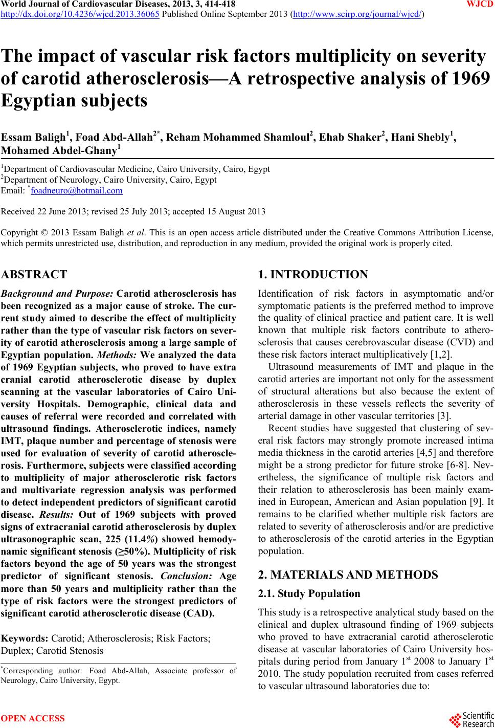 World Journal of Cardiovascular Diseases, 2013, 3, 414-418 WJCD http://dx.doi.org/10.4236/wjcd.2013.36065 Published Online September 2013 (http://www.scirp.org/journal/wjcd/) The impact of vascular risk factors multiplicity on severity of carotid atherosclerosis—A retrospective analysis of 1969 Egyptian subjects Essam Baligh1, Foad Abd-Allah2*, Reham Mohammed Shamloul2, Ehab Shaker2, Hani Shebly1, Mohamed Abdel-Ghany1 1Department of Cardiovascular Medicine, Cairo University, Cairo, Egypt 2Department of Neurology, Cairo University, Cairo, Egypt Email: *[email protected] Received 22 June 2013; revised 25 July 2013; accepted 15 August 2013 Copyright © 2013 Essam Baligh et al. This is an open access article distributed under the Creative Commons Attribution License, which permits unrestricted use, distribution, and reproduction in any medium, provided the original work is properly cited. ABSTRACT Background and Purpose: Carotid atherosclerosis has been recognized as a major cause of stroke. The cur- rent study aimed to describe the effect of multiplicity rather than the type of vascular risk factors on sever- ity of carotid atherosclerosis among a large sample of Egyptian population. Methods: We analyzed the data of 1969 Egyptian subjects, who proved to have extra cranial carotid atherosclerotic disease by duplex scanning at the vascular laboratories of Cairo Uni- versity Hospitals. Demographic, clinical data and causes of referral were recorded and correlated with ultrasound findings. Atherosclerotic indices, namely IMT, plaque number and percentage of stenosis were used for evaluation of severity of carotid atheroscle- rosis. Furthermore, subjects were classified according to multiplicity of major atherosclerotic risk factors and multivariate regression analysis was performed to detect independent predictors of significant carotid disease. Results: Out of 1969 subjects with proved signs of extracranial carotid atherosclerosis by duplex ultrasonographic scan, 225 (11.4%) showed hemody- namic significant stenosis (≥50%). Multiplicity of risk factors beyond the age of 50 years was the strongest predictor of significant stenosis. Conclusion: Age more than 50 years and multiplicity rather than the type of risk factors were the strongest predictors of significant carotid atherosclerotic disease (CAD). Keywords: Carotid; Atherosclerosis; Risk Factors; Duplex; Carotid Stenosis 1. INTRODUCTION Identification of risk factors in asymptomatic and/or symptomatic patients is the preferred method to improve the quality of clinical practice and patient care. It is well known that multiple risk factors contribute to athero- sclerosis that causes cerebrovascular disease (CVD) and these risk factors interact multiplicatively [1,2]. Ultrasound measurements of IMT and plaque in the carotid arteries are important not only for the assessment of structural alterations but also because the extent of atherosclerosis in these vessels reflects the severity of arterial damage in other vascular territories [3]. Recent studies have suggested that clustering of sev- eral risk factors may strongly promote increased intima media thickness in the carotid arteries [4,5] and therefore might be a strong predictor for future stroke [6-8]. Nev- ertheless, the significance of multiple risk factors and their relation to atherosclerosis has been mainly exam- ined in European, American and Asian population [9]. It remains to be clarified whether multiple risk factors are related to severity of atherosclerosis and/or are predictive to atherosclerosis of the carotid arteries in the Egyptian population. 2. MATERIALS AND METHODS 2.1. Study Population This study is a retrospective analytical study based on the clinical and duplex ultrasound finding of 1969 subjects who proved to have extracranial carotid atherosclerotic disease at vascular laboratories of Cairo University hos- pitals during period from January 1st 2008 to January 1st 2010. The study population recruited from cases referred to vascular ultrasound laboratories due to: *Corresponding author: Foad Abd-Allah, Associate professor o f N eurology, Cairo University, Egypt. OPEN ACCESS  E. Baligh et al. / World Journal of Cardiovascular Diseases 3 (2013) 414-418 415 1) Cerebrovascular symptoms either stroke or TIAs (sym- ptomatic group) 2) Checkup either routine or before coronary by- pass graft surgery (Asymptomatic group ) 2.2. Clinical Data and Cardiovascular Risk Factors The following data were collected from each individual patient prior to ultrasound examination: Age, Sex, Hy- pertension, Diabetes Mellitus, Dyslipidemia, Obesity, Smoking, and history of Ischemic heart disease (IHD). Hypertension was defined as blood pressure >140/90 mm Hg for ≥2 repeated measurements or if a subject was on antihypertensive medications [10]; diabetes mellitus as repeated fasting plasma glucose > 126 mg/dl or a patient on anti-diabetic measures [11]; dyslipidemia as fasting serum total cholesterol of ≥200 mg/dl, LDL cholesterol of ≥130 mg/dl, HDL cholesterol of <40 mg/dl for men and <50 mg/dl for women or triglyceride concentration of ≥150 mg/dl or if the subject was on lipid-lowering drugs or gave a history of established diagnosis of dys- lipidemia [12]. Obesity was defined as a body mass in- dex of ≥30 [13]. History of cigarette smoking was posi- tive if subjects smoked ≥10 cigarettes per day for >10 years and ischemic heart disease was defined as evi- dence of the disease based on ECG, echocardiography, stress test or coronary angiography or if the patient was known to have ischemic heart disease. 2.3. Ultrasonographic Data (Carotid Duplex Scan) Carotid duplex scanning was performed by qualified vas- cular operators using Philips HDI 5000 machines. A high-frequency (7- to 10-MHz) linear array transducer was employed to scan the carotid from the most proximal common carotid artery (CCA) to the internal carotid ar- tery (ICA) as far as the mandible permitted. The exami- nation starts by a B-mode transverse scanning of the vessels so as to examine the arterial wall morphology, detect intima media changes and presence of atheroma- tous plaques. 2.3.1. Intima Media Thickness Longitudinal scanning and quantification of the intima media thickness (IMT) at the distal far wall of the CCA was done, IMT ≥ 1.0 mm was considered abnormal [14]. 2.3.2. Assessment of Extra-Cranial Carotid Plaques Plaques were defined by the presence of focal, severe wall thickening (IMT > 2 mm). Echogenicity and surface characteristics were evaluated and number of plaques was recorded in each subject [15]. 2.3.3. Degree of Carotid Artery Stenosis Pulsed Doppler signals were routinely recorded from the CCA just proximal to the bifurcation, the origin of the external carotid artery, proximal, mid and if accessible distal ICA. They were recorded in the longitudinal view with less than 60˚ insonation angle. PSV and end-dia- stolic velocities were recorded at the proximal ICA, the distal CCA and the PSV ICA/CCA ratio was calculated. In case of a stenosis on B-mode and color Doppler im- aging, the Doppler signal was recorded from the site of maximum stenosis, i.e. from the area of maximum flow turbulence on color Doppler imaging. The degree of stenosis was primarily assessed according to the Doppler information. Hemodynamically insignificant plaque (< 50% stenosis) was diagnosed if plaque was detected on B-mode image and PSV was <125 cm/s. Moderate ICA stenosis (≥50%) was diagnosed by PSV ICA ≥ 125 cm/s and PSV ICA/CCA ratio ≥ 2, severe ICA stenosis (≥70%) according to NASCET criteria [16] by PSV ICA ≥ 230 cm/s and PSV ICA/CCA ratio ≥ 4, and critical ICA stenosis (>80%) by end-diastolic velocity ≥ 140 cm/s. ICA occlusion was diagnosed when occluding material was visualized in B-mode, color and pulsed Doppler signal was absent and diastolic velocity in the ipsilateral CCA was low or absent. Carotid arteries were considered normal if the intima-media thickness was <1.0 mm, no plaque detected and peak systolic velocity at the pro- ximal ICA (PSV ICA ) was less than 125 cm/s. Carotid atherosclerotic disease was considered present if the in- tima-media complex showed diffuse thickening (≥1.0 mm) or if carotid plaques were detected. 3. STATISTICAL ANALYSIS Data were described as mean ± standard deviation (SD), range, frequencies (number of cases) and relative fre- quencies (percentages). Categorical variables were ex- pressed as percentages and continuous variables were expressed as mean ± SD, with a 95% confidence interval (CI). Comparative statistics were performed with Stu- dent’s t test, Mann-Whitney U or Χ2 test as appropriate. Multivariate regression analysis was performed to detect independent predictors of carotid atherosclerosis and carotid stenosis. A probability value (p value) less than 0.05 was considered statistically significant. All statistic- cal calculations were performed using Microsoft Excel version 7 and SPSS version 15 for MS windows (Statis- tical Package for the Social Science, SPSS Inc., Chicago, Ill., USA). 4. RESULTS A total of 1969 subjects were included in the study. Age, gender and the number of risk factors are demonstrated in Table 1. Carotid Atherosclerotic Disease (CAD) was defined as: IMT ≥ 1 mm and/or presence of plaques. Increase IMT Copyright © 2013 SciRes. OPEN ACCESS 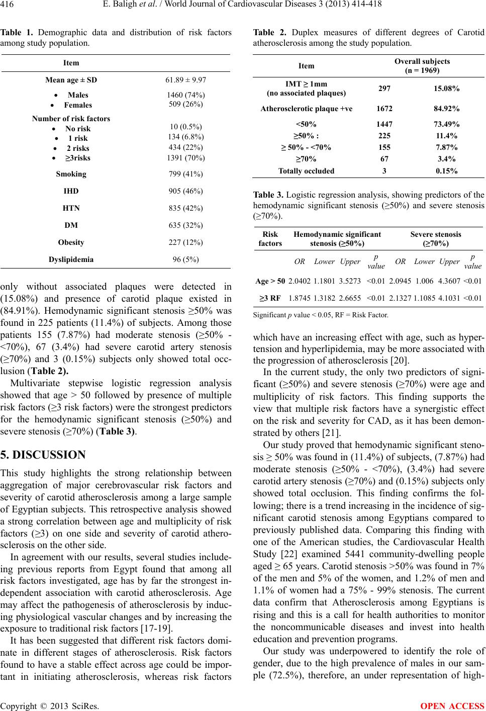 E. Baligh et al. / World Journal of Cardiovascular Diseases 3 (2013) 414-418 416 Table 1. Demographic data and distribution of risk factors among study population. Item Mean age ± SD 61.89 ± 9.97 Males Females 1460 (74%) 509 (26%) Number of risk factors No risk 1 risk 2 risks ≥3risks 10 (0.5%) 134 (6.8%) 434 (22%) 1391 (70%) Smoking 799 (41%) IHD 905 (46%) HTN 835 (42%) DM 635 (32%) Obesity 227 (12%) Dyslipidemia 96 (5%) only without associated plaques were detected in (15.08%) and presence of carotid plaque existed in (84.91%). Hemodynamic significant stenosis ≥50% was found in 225 patients (11.4%) of subjects. Among those patients 155 (7.87%) had moderate stenosis (≥50% - <70%), 67 (3.4%) had severe carotid artery stenosis (≥70%) and 3 (0.15%) subjects only showed total occ- lusion (Table 2). Multivariate stepwise logistic regression analysis showed that age > 50 followed by presence of multiple risk factors (≥3 risk factors) were the strongest predictors for the hemodynamic significant stenosis (≥50%) and severe stenosis (≥70%) (Table 3). 5. DISCUSSION This study highlights the strong relationship between aggregation of major cerebrovascular risk factors and severity of carotid atherosclerosis among a large sample of Egyptian subjects. This retrospective analysis showed a strong correlation between age and multiplicity of risk factors (≥3) on one side and severity of carotid athero- sclerosis on the other side. In agreement with our results, several studies include- ing previous reports from Egypt found that among all risk factors investigated, age has by far the strongest in- dependent association with carotid atherosclerosis. Age may affect the pathogenesis of atherosclerosis by induc- ing physiological vascular changes and by increasing the exposure to traditional risk factors [17-19]. It has been suggested that different risk factors domi- nate in different stages of atherosclerosis. Risk factors found to have a stable effect across age could be impor- tant in initiating atherosclerosis, whereas risk factors Table 2. Duplex measures of different degrees of Carotid atherosclerosis among the study population. Item Overall subjects (n = 1969) IMT ≥ 1mm (no associated plaques) 297 15.08% Atherosclerotic plaque +ve1672 84.92% <50% ≥50% : ≥ 50% - <70% ≥70% Totally occluded 1447 225 155 67 3 73.49% 11.4% 7.87% 3.4% 0.15% Table 3. Logistic regression analysis, showing predictors of the hemodynamic significant stenosis (≥50%) and severe stenosis (≥70%). Risk factors Hemodynamic significant stenosis (≥50%) Severe stenosis (≥70%) ORLower Upperp value OR Lower Upperp value Age > 502.04021.18013.5273<0.01 2.0945 1.006 4.3607<0.01 ≥3 RF1.87451.31822.6655<0.01 2.1327 1.1085 4.1031<0.01 Significant p value < 0.05, RF = Risk Factor. which have an increasing effect with age, such as hyper- tension and hyperlipidemia, may be more associated with the progression of atherosclerosis [20]. In the current study, the only two predictors of signi- ficant (≥50%) and severe stenosis (≥70%) were age and multiplicity of risk factors. This finding supports the view that multiple risk factors have a synergistic effect on the risk and severity for CAD, as it has been demon- strated by others [21]. Our study proved that hemodynamic significant steno- sis ≥ 50% was found in (11.4%) of subjects, (7.87%) had moderate stenosis (≥50% - <70%), (3.4%) had severe carotid artery stenosis (≥70%) and (0.15%) subjects only showed total occlusion. This finding confirms the fol- lowing; there is a trend increasing in the incidence of sig- nificant carotid stenosis among Egyptians compared to previously published data. Comparing this finding with one of the American studies, the Cardiovascular Health Study [22] examined 5441 community-dwelling people aged ≥ 65 years. Carotid stenosis >50% was found in 7% of the men and 5% of the women, and 1.2% of men and 1.1% of women had a 75% - 99% stenosis. The current data confirm that Atherosclerosis among Egyptians is rising and this is a call for health authorities to monitor the noncommunicable diseases and invest into health education and prevention programs. Our study was underpowered to identify the role of gender, due to the high prevalence of males in our sam- ple (72.5%), therefore, an under representation of high- Copyright © 2013 SciRes. OPEN ACCESS  E. Baligh et al. / World Journal of Cardiovascular Diseases 3 (2013) 414-418 417 risk women is also possible. However, the higher preva- lence of atherosclerotic lesions in males was in accor- dance with that found in other studies as the Cardiovas- cular Health Study which showed that among the elderly (≥65 years), women have less carotid atherosclerosis than men. The Atherosclerosis Risk in Communities (ARIC) Study [23] revealed that the carotid wall in- tima-media thickness is greater in men than in women aged 45 to 64 years. Caplan et al. [24] demonstrated that female may be more susceptible to intracranial arterial occlusive disease than extracranial carotid disease [25]. In conclusion, according to the current study, multiple risk factors are more predictive than a single risk factor for atherosclerosis in the carotid arteries. Since unfavor- able risk factor profile is associated with an increase in the severity of atherosclerosis and age is non modifiable, our study highlights the importance of controlling modi- fiable risk factors early in life which may retard athero- sclerosis development and hence delay the onset of pro- gression to clinical cerebrovascular insults. REFERENCES [1] Cardiovascular Diseases Prevention on Clinical Practice. Fourth Joint European Societies Task Force on Cardio- vascular Disease Prevention in Clinical Practice (2010) Chairperson: Graham I. In: Vahanian, A., Auricchio, A., Bax, J., Ceconi, C., Dean, V., Fillipatos, G., Funck-Bren- tano, C., Hobbs, R., Kearney, P., McDonagh, T., Popescu, B.A., Reiner, Z., Secthem, U., Sirnes, P.A., Tendera, M., Vardas, P. and Widimsky, P., Eds., Compendium of Abri- ged ESC Guidelines 2010, Springer Heatlhcare, London, 3-13. [2] Wilsgaard, T., Jacobsen, B.K., Schirmer, H., et al. (2001) Tracking of cardiovascular risk factors: The Tromso stu- dy. American Journal of Epidemiology, 154, 418-426. doi:10.1093/aje/154.5.418 [3] Benedetto, F.A., Mallamaci, F., Tripepi, G. and Zoccali, C. (2001) Prognostic value of ultrasonographic measure- ment of carotid intima-media thickness in dialysis pa- tients. Journal of the American Society of Nephrology, 12, 2458-2464. [4] Hulthe, J., Bokemark, L., Wikstrand, J. and Fagerberg, B. (2000) The metabolic syndrome, LDL particle size, and atherosclerosis: The atherosclerosis and insulin resistan- ce (AIR) study. Arteriosclerosis, Thrombosis, and Vas- cular Biology, 20, 2140-2147. doi:10.1161/01.ATV.20.9.2140 [5] Iglseder, B., Cip, P., Malaimare, L., Ladurner, G. and Paul- weber, B. (2005) The metabolic syndrome is a stronger risk factor for early carotid atherosclerosis in women than in men. Stroke, 36, 1212-1217. doi:10.1161/01.STR.0000166196.31227.91 [6] Koren-Morag, N., Goldbourt, U. and Tanne, D. (2005) Relation between the metabolic syndrome and ischemic stroke or transient ischemic attack. A prospective cohort study in patients with atherosclerotic cardiovascular dis- ease. Stroke, 36, 1366-1371. doi:10.1161/01.STR.0000169945.75911.33 [7] Milionis, H.J., Rizos, E., Goudevenos, J., Seferiadis, K., Mikhailidis, D.P. and Elisaf, M.S. (2005) Components of the metabolic syndrome and risk for first-ever acute is- chemic non-embolic stroke in elderly subjects. Stroke, 36, 1372-1376. doi:10.1161/01.STR.0000169935.35394.38 [8] Kurl, S., Laukkanen, J.A., Niskanen, L., et al. (2006) Metabolic syndrome and the risk of stroke in middle-aged men. Stroke, 37, 806-811. doi:10.1161/01.STR.0000204354.06965.44 [9] Takahashi, W., Ohnuki, T., Honma, K., Kawada, S. and Takagi, S. (2007) The significance of multiple risk fac- tors for early carotid atherosclerosis in Japanese sub- jects. Internal Medicine, 46, 1679-1684. doi:10.2169/internalmedicine.46.0240 [10] Chobanian, A.V., Bakris, G.L., Black, H.R., Cushman, W.C., Green, L.A., Izzo Jr., J.L., Jones, D.W., Materson, B.J., Oparil, S., Wright Jr., J.T. and Roccella, E.J. (2003) The seventh report of the joint national committee on prevention, detection, evaluation, and treatment of high blood pressure: national heart, lung, and blood institute joint national committee on prevention, detection, evalua- tion, and treatment of high blood pressure; national high blood pressure education program coordinating commit- tee. Journal of the American Medical Association, 289, 2560-2572. [11] Alberti, K.G. and Zimmet, P.Z. (1998) Definition, diag- nosis and classification of diabetes mellitus and its com- plications. Part 1: Diagnosis and classification of diabetes mellitus provisional report of a WHO consultation. Dia- betic Medicine, 15, 539-553. doi:10.1002/(SICI)1096-9136(199807)15:7<539::AID-DI A668>3.0.CO;2-S [12] Expert Panel on Detection, Evaluation, and Treatment of High Blood Cholesterol in Adults (2001) Executive sum- mary of the third report of the national cholesterol educa- tion program (NCEP) (adult treatment panel III). Journal of the American Medical Association, 285, 2486-2497. [13] World Health Organization (2000) Obesity: Preventing and managing the global epidemic. [14] Kanters, S.D., Algra, A., van Leeuwen, M.S. and Banga, J.D. (1997) Reproducibility of in vivo carotid intima-me- dia thickness measurements: A review. Stroke, 28, 665- 671. doi:10.1161/01.STR.28.3.665 [15] Rohde, L.E., Lee, R.T., Rivero, J., Jamacochian, M., Ar- royo, L.H., Briggs, W., Rifai, N., Libby, P., Creager, M.A. and Ridker, P.M. (1998) Circulating cell adhesion mo- lecules are correlated with ultrasound-based assessment of carotid atherosclerosis. Arteriosclerosis, Thrombosis, and Vascular Biology, 18, 1765-1770. doi:10.1161/01.ATV.18.11.1765 [16] North American Symptomatic Carotid Endarterectomy Trial Collaborators (1991) Beneficial effect of carotid en- darterectomy in symptomatic patients with high-grade carotid stenosis. New England Journal of Medicine, 325, 445-453. doi:10.1056/NEJM199108153250701 [17] Zimarino, M., Cappelletti, L., Venarucci, V., et al. (2001) Age dependence of risk factors for carotid stenosis: An observational study among candidates for coronary arte- Copyright © 2013 SciRes. OPEN ACCESS  E. Baligh et al. / World Journal of Cardiovascular Diseases 3 (2013) 414-418 Copyright © 2013 SciRes. 418 OPEN ACCESS riography. Atherosclerosis, 159, 165-173. doi:10.1016/S0021-9150(01)00477-4 [18] Stevens, J., Cai, J., Pamuk, E.R., et al. (1998) The effect of age on the association between body-mass index and mortality. New England Journal of Medicine, 338, 1-7. doi:10.1056/NEJM199801013380101 [19] Abd Allah, F., Baligh, E. and Ibrahim, M. (2010) Clinical relevance of carotid atherosclerosis among Egyptians: A 5-year retrospective analysis of 4733 subjects. Neuroepi- demiology, 35, 275-279. doi:10.1159/000319899 [20] Stensland-Bugge, E., Bønaa, K.H. and Joakimsen, O. (2001) Age and sex differences in the relationship between in- herited and lifestyle risk factors and subclinical carotid atherosclerosis: The Tromsø study. Atheroscle rosis, 154, 437-448. [21] Wilson, P.W., Hoeg, J.M., D'Agostino, R.B., Silbershatz, H., Belanger, A.M., Poehlmann, H., O’Leary, D. and Wolf, P.A. (1997) Cumulative effects of high cholesterol levels, high blood pressure, and cigarette smoking on ca- rotid stenosis. New England Journal of Medicine, 337, 516-522. doi:10.1056/NEJM199708213370802 [22] O’Leary, D.H., Polak, J.F., Kronmal, R.A., et al. (1992) Distribution and correlates of sonographically detected carotid artery disease in the cardiovascular health study. Stroke, 23, 1752-1760. doi:10.1161/01.STR.23.12.1752 [23] Howard, G., Sharrett, A.R., Heiss, G., et al. (1993) Ca- rotid artery intimal-medial thickness distribution in gen- eral populations as evaluated by B-mode ultrasound. ARIC investigators. Stroke, 24, 1297-1304. doi:10.1161/01.STR.24.9.1297 [24] Caplan, L.R., Gorelick, P.B. and Hier, D.B. (1986) Race, sex and occlusive cerebrovascular disease: A review. Stroke, 17, 648-655. doi:10.1161/01.STR.17.4.648 [25] Chang, C.H., Chang, Y.J., Lee, T.H., Hsu, K.C., Ryu, S.J. (2006) Risk factors of carotid stenosis in first-ever ische- mic stroke in Taiwan: A hospital-based study. Acta Neu- rol Taiwan, 15, 237-243. |