

Paper Menu >>
Journal Menu >>
 International Journal of Otolaryngology and Head & Neck Surgery, 2013, 2, 179-185 http://dx.doi.org/10.4236/ijohns.2013.25038 Published Online September 2013 (http://www.scirp.org/journal/ijohns) A Simple Combined Antegrade Radiological and Retrograde Endoscopic Procedure to Recanalise Fibrotic Hypopharyngo-Oesophageal Occlusions: Technical Description and Lessons from Clinical Outcome in Three Cases* Mohammed S. Miah1, Ian A. Zealley2, Afshin Alijani3, Barry McGuire4, Rodney E. Mountain1, Suresh Mahendran 1# 1Department of Otorhinolaryngology-Head & Neck Surgery, Ninewells Hospital and University of Dundee Medical School, Dundee, UK 2Department of Radiology, Ninewells Hospital and University of Dundee Medical School, Dundee, UK 3Department of Upper Gastrointestinal Surgery, Ninewells Hospital and University of Dundee Medical School, Dundee, UK 4Department of Anaesthesia, Ninewells Hospital and University of Dundee Medical School, Dundee, UK Email: #[email protected] Received June 2, 2013; revised June 29, 2013; accepted July 9, 2013 Copyright © 2013 Mohammed S. Miah et al. This is an open access article distributed under the Creative Commons Attribution Li- cense, which permits unrestricted use, distribution, and reproduction in any medium, provided the original work is properly cited. ABSTRACT Background: Complete hypophar yngo- o esoph ag eal o cclusion is a r ar e co mplication of h ead and n eck r ad ioth er apy an d a range of other conditions. Absolute dysphagia is acco mpanied by aspiration and depend ence on gastrostomy feeding. The condition presents a substantial management challenge. Surgical approaches to re-establish pharyngo-oesophageal continuity are varied, highly invasive and are associated with unpredictable outcomes. Minimally invasive techniques employing endoscopic and radiological techniques are emerging. This report describes a multidisciplinary approach which translates two interventional radiology techniques used in the management of central venous occlusions and bil- iary strictures to the management of three cases of complete hypopharyngo-oesophageal occlusion. Methods: Three cases with different underlying aetiologies had treatment initiated between 2009 and 2011. Antegrade pharyngoscopic access to the occlusions was accompanied by retrograde endoscopic access via a small gastrostomy. Luminal continuity was re-established by the interventional radiology technique of “sharp recanalisation” followed by passage of a wide bore nasogastric tube which was maintained in situ for 4 - 6 months, a duration of treatment analogous to that applied in the radiological management of fibrotic biliary strictures. After treatment a radiological contrast swallows examination was performed to gauge the calibre of the re-established lumen, assess functionality and to ru le out aspiration. Results: Pharyngo-oesophageal continuity was re-established in all three cases on the first attempt. No complications occurred as a result of the proced ures. In two cases, the excellent swallowing fun ction was re-established, although on e of these re- quired prolonged post-treatment adjuvant interventions. In one case no swallowing function resulted, despite apparently successful re-establishment of luminal continuity. Conclusions: Complete fibrotic o cclusion of the hypopharyngo-oeso- phageal lumen is rare and presents a substantial management challenge. A minimally invasive treatment combining antegrade radiological and retrograde endoscopic approaches resulted in successful re-establishment of luminal continu- ity in three cases of complete fibrotic occlusion of the hypopharyngo-oesophageal lumen. However variable responses to treatment suggest that both the underlyin g aetiology and the chronicity o f the occlusion may influence the likelihood of a successful functional outcome. Until definitiv e management guidelines are estab lished, we suggest that such cases are managed only by motivated multidisciplinary teams keen to develop their exp ertise in th is area. Keywords: Hypopharynx; Upper Oesophagus; Fibrotic Occlusion; Rendezvous Technique; Pharyngo-Oesophageal Continuity *Meetings: The work was presented at the Tri-Society of Head & Neck Oncology Meeting on 2nd Septem ber 20 11 in Sin gapore. #Corresponding a utho r . C opyright © 2013 SciRes. IJOHNS 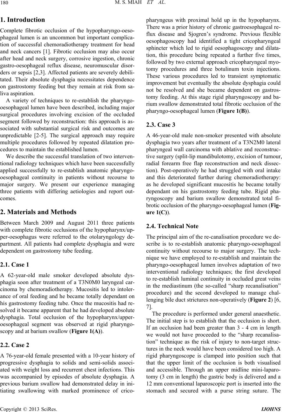 M. S. MIAH ET AL. Copyright © 2013 SciRes. IJOHNS 180 1. Introduction Complete fibrotic occlusion of the hypopharyngo-oeso- phageal lumen is an uncommon but important complica- tion of successful chemoradiotherapy treatment for head and neck cancers [1]. Fibrotic occlusion may also occur after head and neck surgery, corrosive ingestion, chronic gastro-oesophageal reflux disease, neuromuscular disor- ders or sepsis [2 ,3]. Affected patients are severely debili- tated. Their absolute dysphagia necessitates dependence on gastrostomy feeding but they remain at risk from sa- liva aspiration. A variety of techniques to re-establish the pharyngo- oesophageal lumen have been described, including major surgical procedures involving excision of the occluded segment followed by reconstruction: this approach is as- sociated with substantial surgical risk and outcomes are unpredictable [2-5]. The surgical approach may require multiple procedures followed by repeated dilatation pro- cedures to maintain the established lumen. We describe the successful translation of two interven- tional radiology tech niques which h ave been successfu lly applied successfully to re-establish anatomic pharyngo- oesophageal continuity in patients without recourse to major surgery. We present our experience managing three patients with differing aetiologies and report out- comes. 2. Materials and Methods Between March 2009 and August 2011 three patients with complete fibrotic occlusion s of the hypo pharynx /up- per-oesophagus were referred to the otolaryngology de- partment. All patients had complete dysphagia and were dependent on gastrostomy tub e feeding. 2.1. Case 1 A 62-year-old male smoker developed absolute dys- phagia soon after treatment of a T3N0M0 laryngeal car- cinoma by chemoradiotherapy. Mucositis led to intoler- ance of oral feeding and he became totally dependant on his gastrostomy feeding tube. Once the mucositis had re- solved it became apparent that he had developed absolute dysphagia. Total occlusion of the hypopharynx/upper- oesophageal segment was observed at rigid pharyngo- scopy and at barium swallow (Figure 1(A)). 2.2. Case 2 A 76-year-old female presented with a 10-year histor y of progressive dysphagia to solids and semi-solids associ- ated with weight loss and recurrent ch est infections. This was accompanied by episodes of absolute dysphagia. A previous barium swallow had demonstrated delay in ini- tiating swallowing with marked prominence of crico- pharyngeus with proximal hold up in the hypopharynx. There was a prior history of chronic gastroesophageal re- flux disease and Sjogren’s syndrome. Previous flexible oesophagoscopy had identified a tight cricopharyngeal sphincter which led to rigid oesophagoscopy and dilata- tion, this procedure being repeated a further five times, followed by two external approach cricopharyngeal myo- tomy procedures and three botulinum toxin injections. These various procedures led to transient symptomatic improvement but eventually the absolute dysphagia could not be resolved and she became dependent on gastros- tomy feeding. At this stage rigid pharyngoscopy and ba- rium swallow demonstrated total fibrotic occlusion of the pharyngo-oesop hageal lumen (Figure 1(B)). 2.3. Case 3 A 46-year-old male non-smoker presented with absolute dysphagia two years after treatment of a T3N2M0 lateral pharyngeal wall carcinoma with ablative and reconstruc- tive surgery (split-lip mandibu lotomy, excision of tumour, radial forearm free flap reconstruction and neck dissec- tion). Post-operatively he had struggled with oral intake and this deteriorated further during chemoradiotherapy: as he developed significant mucositis he became totally dependant on his gastrostomy feeding tube. Rigid pha- ryngoscopy and barium swallow demonstrated total fi- brotic occlusion of the pharyngo-oesophageal lumen (Fig- ure 1(C)). 2.4. Technical Note The principal aim of the re-canalisation procedure we de- scribe is to re-establish anatomic pharyngo-oesophageal continuity without recourse to major surgery. The tech- nique we have employed to re-establish and maintain th e pharyngo-oesophageal lumen involves adaptation of two interventional radiology techniques; the first developed to re-establish luminal co ntinuity in occluded great veins in the mediastinum (the so-called “sharp recanalisation” procedure) and the second developed to manage chal- lenging bile duct strictures non-op eratively (Figure 2) [6 , 7]. The procedure is performed under general anaesthetic. The initial step is to establish that the occlusion is short. If an occlusion had been greater than 3 - 4 cm in length we would not have proceeded to the “sharp recanalisa- tion” technique as the risk of injury to non-target struc- tures in the neck would have been considered too high. A rigid pharyngoscope is clamped into position such that that the upper limit of the occlusion is both visualised and accessible. Through an upper midline mini-laparo- tomy (3 cm in length) the gastric body is delivered and a 12 mm conventional laparoscopic port is inserted into the stomach and secured with a purse string suture. The 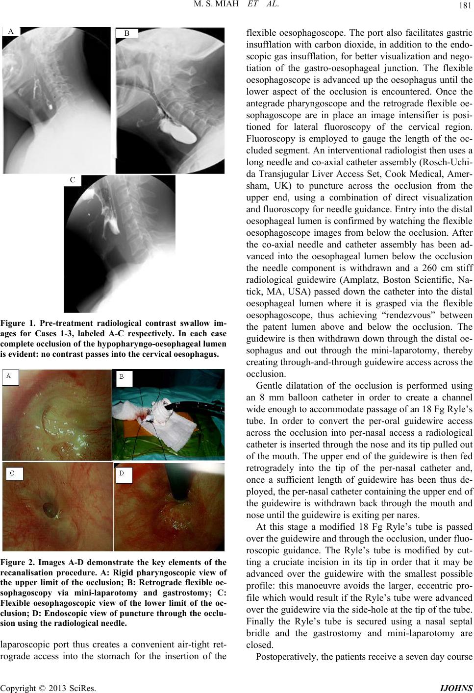 M. S. MIAH ET AL. 181 A B C Figure 1. Pre-treatment radiological contrast swallow im- ages for Cases 1-3, labeled A-C respectively. In each case complete occlusion of the hy popharyngo-oe sophageal lumen is evident: no contrast passes into the cervical oesophagus. Figure 2. Images A-D demonstrate the key elements of the recanalisation procedure. A: Rigid pharyngoscopic view of the upper limit of the occlusion; B: Retrograde flexible oe- sophagoscopy via mini-laparotomy and gastrostomy; C: Flexible oesophagoscopic view of the lower limit of the oc- clusion; D: Endoscopic view of puncture through the oc clu- sion using the radiological needle. laparoscopic port thus creates a convenient air-tight ret- rograde access into the stomach for the insertion of the flexible oesophagoscope. The port also facilitates gastric insufflation with carbon dioxide, in addition to the endo- scopic gas insufflation, for better visualization and nego- tiation of the gastro-oesophageal junction. The flexible oesophagoscope is advanced up the oesophagus until the lower aspect of the occlusion is encountered. Once the antegrade pharyngoscope and the retrograde flexible oe- sophagoscope are in place an image intensifier is posi- tioned for lateral fluoroscopy of the cervical region. Fluoroscopy is employed to gauge the length of the oc- cluded segment. An interventional radiologist then uses a long needle and co-axial catheter assembly (Rosch-Uchi- da Transjugular Liver Access Set, Cook Medical, Amer- sham, UK) to puncture across the occlusion from the upper end, using a combination of direct visualization and fluoroscopy for needle guid ance. Entry into th e distal oesophageal lumen is confirmed by watching the flex ible oesophagoscope images from below the occlusion. After the co-axial needle and catheter assembly has been ad- vanced into the oesophageal lumen below the occlusion the needle component is withdrawn and a 260 cm stiff radiological guidewire (Amplatz, Boston Scientific, Na- tick, MA, USA) passed down the catheter into the distal oesophageal lumen where it is grasped via the flexible oesophagoscope, thus achieving “rendezvous” between the patent lumen above and below the occlusion. The guidewire is then withdrawn down through the distal oe- sophagus and out through the mini-laparotomy, thereby creating through-and-through guidewire access across the occlusion. Gentle dilatation of the occlusion is performed using an 8 mm balloon catheter in order to create a channel wide enough to accommodate passage of an 18 Fg Ryle’s tube. In order to convert the per-oral guidewire access across the occlusion into per-nasal access a radiological catheter is inserted through the nose and its tip pulled out of the mouth. The upper end of the guidewire is then fed retrogradely into the tip of the per-nasal catheter and, once a sufficient length of guidewire has been thus de- ployed, the per-nasal catheter containing the upper end of the guidewire is withdrawn back through the mouth and nose until the guidewire is exiting per nares. At this stage a modified 18 Fg Ryle’s tube is passed over the guidewire and through the occlusion, under fluo- roscopic guidance. The Ryle’s tube is modified by cut- ting a cruciate incision in its tip in order that it may be advanced over the guidewire with the smallest possible profile: this manoeuvre avoids the larger, eccentric pro- file which would result if the Ryle’s tube were advanced over the guidewire via the side-hole at the tip of the tube. Finally the Ryle’s tube is secured using a nasal septal bridle and the gastrostomy and mini-laparotomy are closed. Postoperatively, the patients receive a seven day co urs e Copyright © 2013 SciRes. IJOHNS 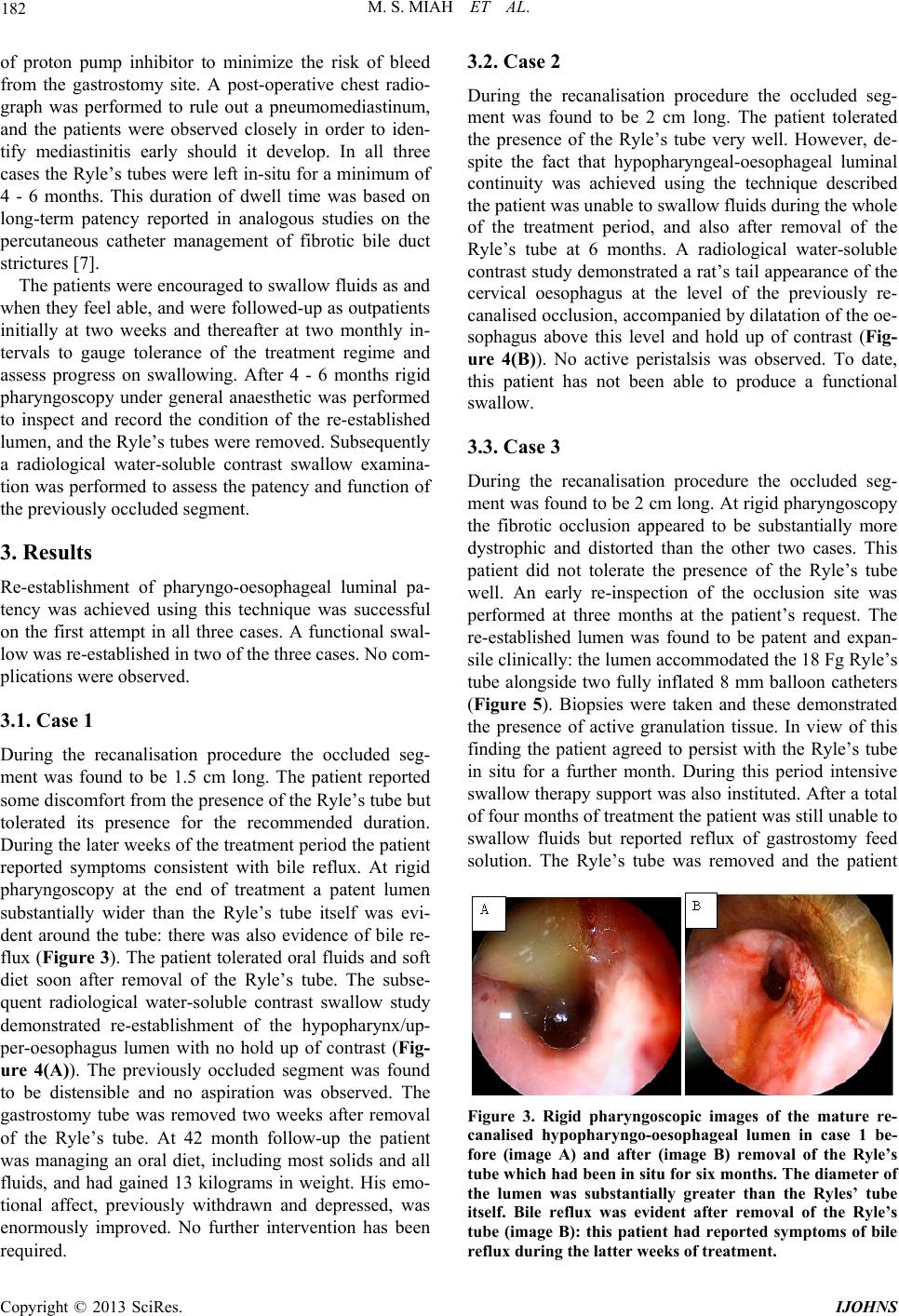 M. S. MIAH ET AL. 182 of proton pump inhibitor to minimize the risk of bleed from the gastrostomy site. A post-operative chest radio- graph was performed to rule out a pneumomediastinum, and the patients were observed closely in order to iden- tify mediastinitis early should it develop. In all three cases the Ryle’s tubes were left in-situ for a minimum of 4 - 6 months. This duration of dwell time was based on long-term patency reported in analogous studies on the percutaneous catheter management of fibrotic bile duct strictures [7]. The patients were encouraged to swallow fluids as and when they feel able, and were followed-up as outpatients initially at two weeks and thereafter at two monthly in- tervals to gauge tolerance of the treatment regime and assess progress on swallowing. After 4 - 6 months rigid pharyngoscopy under general anaesthetic was performed to inspect and record the condition of the re-established lumen, and the Ryle’s tubes were removed. Subsequently a radiological water-soluble contrast swallow examina- tion was performed to assess the patency and function of the previously occluded segment. 3. Results Re-establishment of pharyngo-oesophageal luminal pa- tency was achieved using this technique was successful on the first attempt in all three cases. A functional swal- low was re-established in two of the three cases. No com- plications were observed. 3.1. Case 1 During the recanalisation procedure the occluded seg- ment was found to be 1.5 cm long. The patient reported some discomfort from the presence of the Ryle’s tube but tolerated its presence for the recommended duration. During the later weeks of the treatment period the patient reported symptoms consistent with bile reflux. At rigid pharyngoscopy at the end of treatment a patent lumen substantially wider than the Ryle’s tube itself was evi- dent around the tube: there was also evidence of bile re- flux (Figure 3). The patient tolerated oral fluids and soft diet soon after removal of the Ryle’s tube. The subse- quent radiological water-soluble contrast swallow study demonstrated re-establishment of the hypopharynx/up- per-oesophagus lumen with no hold up of contrast (Fig- ure 4(A)). The previously occluded segment was found to be distensible and no aspiration was observed. The gastrostomy tube was removed two weeks after removal of the Ryle’s tube. At 42 month follow-up the patient was managing an oral diet, including most solids and all fluids, and had gained 13 kilograms in weight. His emo- tional affect, previously withdrawn and depressed, was enormously improved. No further intervention has been required. 3.2. Case 2 During the recanalisation procedure the occluded seg- ment was found to be 2 cm long. The patient tolerated the presence of the Ryle’s tube very well. However, de- spite the fact that hypopharyngeal-oesophageal luminal continuity was achieved using the technique described the patient was unable to swallow fluids during the whole of the treatment period, and also after removal of the Ryle’s tube at 6 months. A radiological water-soluble contrast study demonstrated a rat’s tail appearance of the cervical oesophagus at the level of the previously re- canalised occlusion, accompanied by dilatation of the oe- sophagus above this level and hold up of contrast (Fig- ure 4(B)). No active peristalsis was observed. To date, this patient has not been able to produce a functional swallow. 3.3. Case 3 During the recanalisation procedure the occluded seg- ment was found to be 2 cm long. At rigid pharyngo scopy the fibrotic occlusion appeared to be substantially more dystrophic and distorted than the other two cases. This patient did not tolerate the presence of the Ryle’s tube well. An early re-inspection of the occlusion site was performed at three months at the patient’s request. The re-established lumen was found to be patent and expan- sile clinically: the lumen accommodated the 18 Fg Ryle’s tube alongside two fully inflated 8 mm balloon catheters (Figure 5). Biopsies were taken and these demonstrated the presence of active granulation tissue. In view of this finding the patient agreed to persist with the Ryle’s tube in situ for a further month. During this period intensive swallow therapy support was also instituted. After a total of four months of treatment the patient was still unable to swallow fluids but reported reflux of gastrostomy feed solution. The Ryle’s tube was removed and the patient Figure 3. Rigid pharyngoscopic images of the mature re- canalised hypopharyngo-oesophageal lumen in case 1 be- fore (image A) and after (image B) removal of the Ryle’s tube which had been in situ for six months. The diameter of the lumen was substantially greater than the Ryles’ tube itself. Bile reflux was evident after removal of the Ryle’s tube (image B): this patient had reported symptoms of bile reflux during the latter weeks of treatment. Copyright © 2013 SciRes. IJOHNS 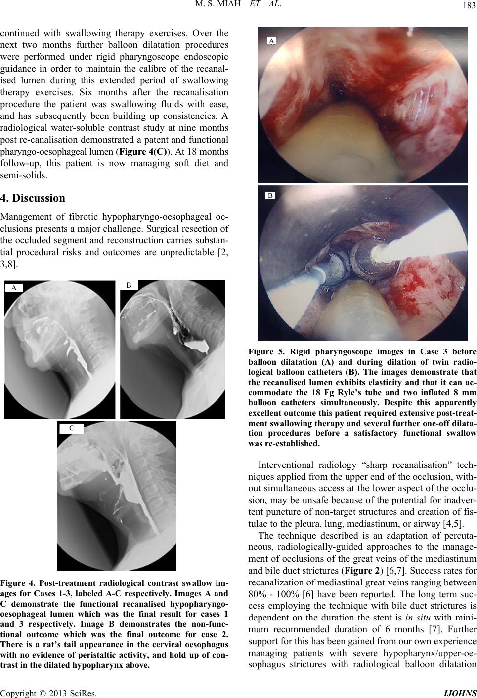 M. S. MIAH ET AL. 183 continued with swallowing therapy exercises. Over the next two months further balloon dilatation procedures were performed under rigid pharyngoscope endoscopic guidance in order to maintain the calibre of the recanal- ised lumen during this extended period of swallowing therapy exercises. Six months after the recanalisation procedure the patient was swallowing fluids with ease, and has subsequently been building up consistencies. A radiological water-soluble contrast study at nine months post re-canalisation demonstrated a patent and functional pharyngo-oesophageal lumen (Figure 4(C)). At 18 months follow-up, this patient is now managing soft diet and semi-solids. 4. Discussion Management of fibrotic hypopharyngo-oesophageal oc- clusions presents a major challenge. Surgical resection of the occluded segment and reconstruction carries substan- tial procedural risks and outcomes are unpredictable [2, 3,8]. A B C Figure 4. Post-treatment radiological contrast swallow im- ages for Cases 1-3, labeled A-C respectively. Images A and C demonstrate the functional recanalised hypopharyngo- oesophageal lumen which was the final result for cases 1 and 3 respectively. Image B demonstrates the non-func- tional outcome which was the final outcome for case 2. There is a rat’s tail appearance in the cervical oesophagus with no evidence of peristaltic activity, and hold up of con- trast in the dilated hypophary nx above. A B Figure 5. Rigid pharyngoscope images in Case 3 before balloon dilatation (A) and during dilation of twin radio- logical balloon catheters (B). The images demonstrate that the recanalised lumen exhibits elasticity and that it can ac- commodate the 18 Fg Ryle’s tube and two inflated 8 mm balloon catheters simultaneously. Despite this apparently excellent outcome this patient required extensive post-treat- ment swallowing therapy and several further one-off dilata- tion procedures before a satisfactory functional swallow was re-established. Interventional radiology “sharp recanalisation” tech- niques applied from the upper end of the occlusion, with- out simultaneous access at the lower aspect of the occlu- sion, may be unsafe because of the potential for inadver- tent puncture of non-target structures and creation of fis- tulae to the pleura, lung, mediastinum, or airway [4,5]. The technique described is an adaptation of percuta- neous, radiologically-guided approaches to the manage- ment of occlusions of the great veins of the mediastinum and bile duct strictures (Figure 2) [6,7]. Success rates for recanalization of mediastinal great veins ranging between 80% - 100% [6] have been reported. The long term suc- cess employing the technique with bile duct strictures is dependent on the duration the stent is in situ with mini- mum recommended duration of 6 months [7]. Further support for this has been gained from our own experience managing patients with severe hypopharynx/upper-oe- sophagus strictures with radiological balloon dilatation Copyright © 2013 SciRes. IJOHNS 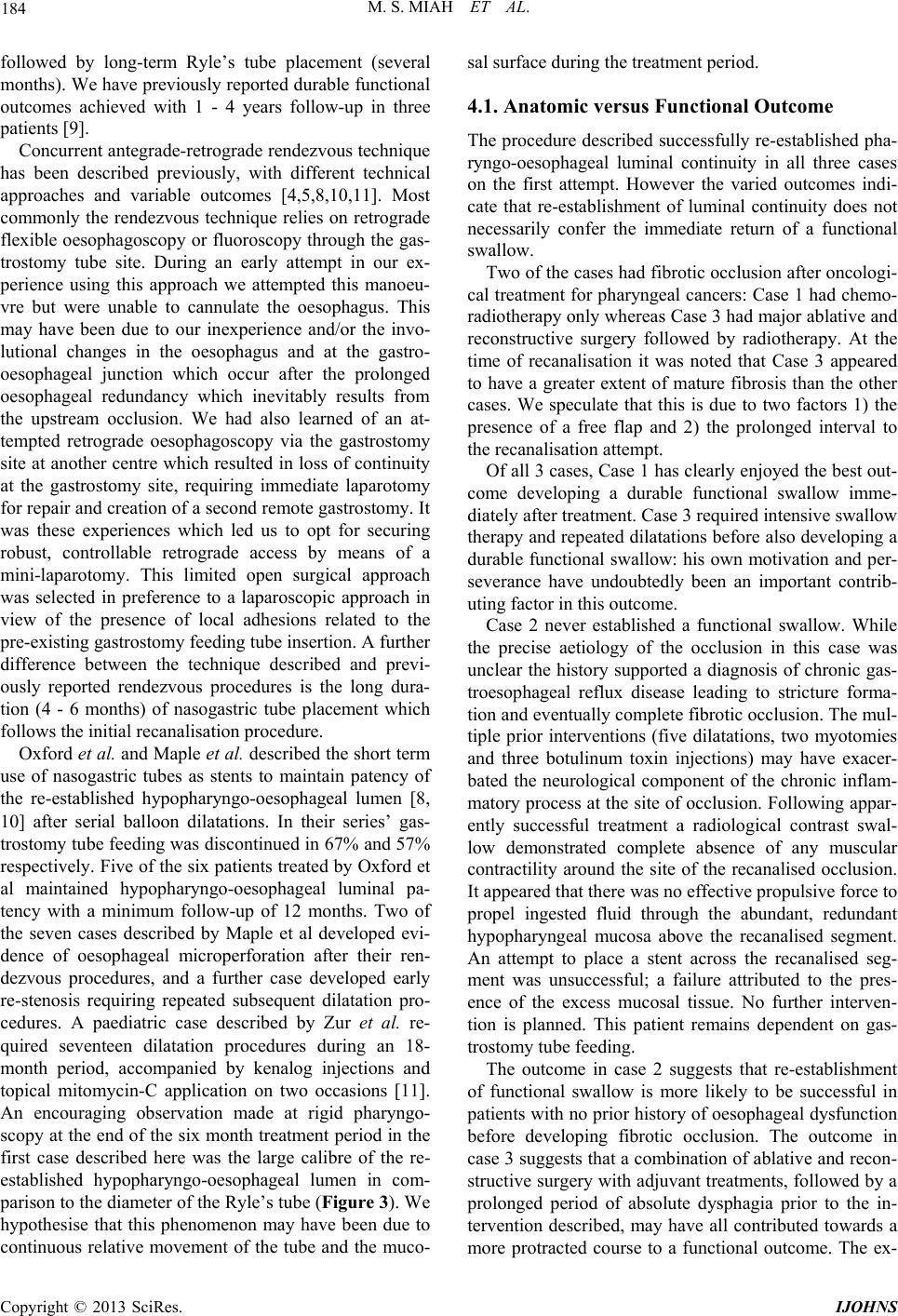 M. S. MIAH ET AL. 184 followed by long-term Ryle’s tube placement (several months). We have previously reported durable functional outcomes achieved with 1 - 4 years follow-up in three patients [9]. Concurrent antegr ade-retrograde r endezvous technique has been described previously, with different technical approaches and variable outcomes [4,5,8,10,11]. Most commonly the rendezvous technique relies on retrograde flexible oesophagoscopy or fluoroscopy through the gas- trostomy tube site. During an early attempt in our ex- perience using this approach we attempted this manoeu- vre but were unable to cannulate the oesophagus. This may have been due to our inexperience and/or the invo- lutional changes in the oesophagus and at the gastro- oesophageal junction which occur after the prolonged oesophageal redundancy which inevitably results from the upstream occlusion. We had also learned of an at- tempted retrograde oesophagoscopy via the gastrostomy site at another centre which resulted in loss of continuity at the gastrostomy site, requiring immediate laparotomy for repair and creatio n of a second remote gastrostomy. It was these experiences which led us to opt for securing robust, controllable retrograde access by means of a mini-laparotomy. This limited open surgical approach was selected in preference to a laparoscopic approach in view of the presence of local adhesions related to the pre-existing gastrostomy feeding tube insertion. A further difference between the technique described and previ- ously reported rendezvous procedures is the long dura- tion (4 - 6 months) of nasogastric tube placement which follows the initial recanalisation procedure. Oxford et al. and Maple et al. described the short term use of nasogastric tubes as stents to maintain patency of the re-established hypopharyngo-oesophageal lumen [8, 10] after serial balloon dilatations. In their series’ gas- trostomy tube feeding was discontinued in 67% and 57% respectively. Five of the six patients treated by Oxford et al maintained hypopharyngo-oesophageal luminal pa- tency with a minimum follow-up of 12 months. Two of the seven cases described by Maple et al developed evi- dence of oesophageal microperforation after their ren- dezvous procedures, and a further case developed early re-stenosis requiring repeated subsequent dilatation pro- cedures. A paediatric case described by Zur et al. re- quired seventeen dilatation procedures during an 18- month period, accompanied by kenalog injections and topical mitomycin-C application on two occasions [11]. An encouraging observation made at rigid pharyngo- scopy at the end of the six month treatment period in the first case described here was the large calibre of the re- established hypopharyngo-oesophageal lumen in com- parison to the diameter o f the Ryle’s tub e (Figure 3). We hypothesise that this phenomenon may have been due to continuous relative movement of the tube and the muco- sal surface during the treatment period. 4.1. Anatomic versus Functional Outcome The procedure described successfully re-established pha- ryngo-oesophageal luminal continuity in all three cases on the first attempt. However the varied outcomes indi- cate that re-establishment of luminal continuity does not necessarily confer the immediate return of a functional swallow. Two of the cases had fibrotic occlusion after oncologi- cal treatment for pharyngeal cancers: Case 1 had chemo- radiotherapy only whereas Case 3 had major ablative and reconstructive surgery followed by radiotherapy. At the time of recanalisation it was noted that Case 3 appeared to have a greater extent of mature fibrosis than the other cases. We speculate that this is due to two factors 1) the presence of a free flap and 2) the prolonged interval to the recanalisation attempt. Of all 3 cases, Case 1 has clearly enjoyed the best out- come developing a durable functional swallow imme- diately after treatment. Case 3 required intensive swallow therapy and repeated dilatations before also developing a durable functional swallow: his own motivation and per- severance have undoubtedly been an important contrib- uting factor in this outcome. Case 2 never established a functional swallow. While the precise aetiology of the occlusion in this case was unclear the history supported a diagnosis of chronic gas- troesophageal reflux disease leading to stricture forma- tion and even tually complete fibro tic occlusio n. The mul- tiple prior interventions (five dilatations, two myotomies and three botulinum toxin injections) may have exacer- bated the neurological component of the chronic inflam- matory process at the site of occlusion. Following appar- ently successful treatment a radiological contrast swal- low demonstrated complete absence of any muscular contractility around the site of the recanalised occlusion. It appeared that there was no effective propulsive force to propel ingested fluid through the abundant, redundant hypopharyngeal mucosa above the recanalised segment. An attempt to place a stent across the recanalised seg- ment was unsuccessful; a failure attributed to the pres- ence of the excess mucosal tissue. No further interven- tion is planned. This patient remains dependent on gas- trostomy tube feeding. The outcome in case 2 suggests that re-establishment of functional swallow is more likely to be successful in patients with no prior history of oesophageal dysfunction before developing fibrotic occlusion. The outcome in case 3 suggests that a combination of ablative and recon- structive surgery with adjuvant treatments, followed by a prolonged period of absolute dysphagia prior to the in- tervention described, may have all contributed towards a more protracted course to a functional outcome. The ex- Copyright © 2013 SciRes. IJOHNS 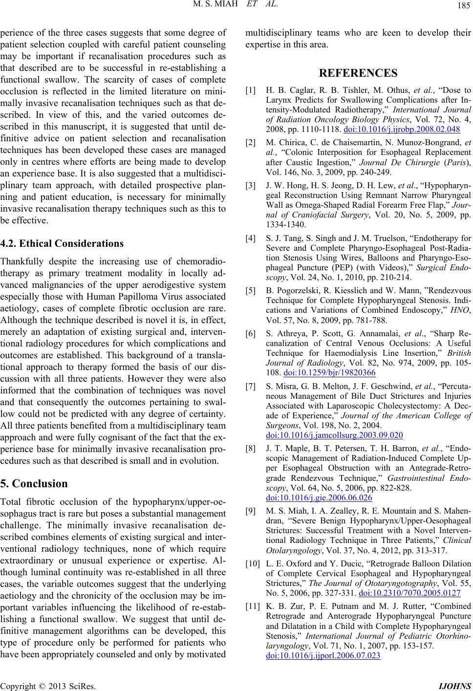 M. S. MIAH ET AL. Copyright © 2013 SciRes. IJOHNS 185 perience of the three cases suggests that some degree of patient selection coupled with careful patient counseling may be important if recanalisation procedures such as that described are to be successful in re-establishing a functional swallow. The scarcity of cases of complete occlusion is reflected in the limited literature on mini- mally invasive recanalisation techniques such as that de- scribed. In view of this, and the varied outcomes de- scribed in this manuscript, it is suggested that until de- finitive advice on patient selection and recanalisation techniques has been developed these cases are managed only in centres where efforts are being made to develop an experience base. It is also suggested that a multidisci- plinary team approach, with detailed prospective plan- ning and patient education, is necessary for minimally invasive recanalisation therapy techniques such as this to be effective. 4.2. Ethical Considerations Thankfully despite the increasing use of chemoradio- therapy as primary treatment modality in locally ad- vanced malignancies of the upper aerodigestive system especially those with Human Papilloma Virus associated aetiology, cases of complete fibrotic occlusion are rare. Although the technique described is novel it is, in effect, merely an adaptation of existing surgical and, interven- tional radiology procedures for which complications and outcomes are established. This background of a transla- tional approach to therapy formed the basis of our dis- cussion with all three patients. However they were also informed that the combination of techniques was novel and that consequently the outcomes pertaining to swal- low could not be predicted with any degree of certainty. All three patients benefited from a multidisciplinary team approach and w ere fully co gnisant of the fact that the ex- perience base for minimally invasive recanalisation pro- cedures such as that described is small and in evolution. 5. Conclusion Total fibrotic occlusion of the hypopharynx/upper-oe- sophagus tract is rare but poses a substantial man ag ement challenge. The minimally invasive recanalisation de- scribed combines elements of existing surgical and inter- ventional radiology techniques, none of which require extraordinary or unusual experience or expertise. Al- though luminal continuity was re-established in all three cases, the variable outcomes suggest that the underlying aetiology and the chronicity of the occlusion may be im- portant variables influencing the likelihood of re-estab- lishing a functional swallow. We suggest that until de- finitive management algorithms can be developed, this type of procedure only be performed for patients who ave been appropriately counseled and only by motivated multidisciplinary teams who are keen to develop their expertise in this area. h REFERENCES [1] H. B. Caglar, R. B. Tishler, M. Othus, et al., “Dose to Larynx Predicts for Swallowing Complications after In- tensity-Modulated Radiotherapy,” International Journal of Radiation Oncology Biology Physics, Vol. 72, No. 4, 2008, pp. 1110-1118. doi:10.1016/j.ijrobp.2008.02.048 [2] M. Chirica, C. de Chaisemartin, N. Munoz-Bongrand, et al., “Colonic Interposition for Esophageal Replacement after Caustic Ingestion,” Journal De Chirurgie (Paris), Vol. 146, No. 3, 2009, pp. 240-249. [3] J. W. Hong, H. S. Jeong, D. H. Lew, et al., “Hypopharyn- geal Reconstruction Using Remnant Narrow Pharyngeal Wall as Omega-Shaped Radial Forearm Free Flap,” Jour- nal of Craniofacial Surgery, Vol. 20, No. 5, 2009, pp. 1334-1340. [4] S. J. Tang, S. Singh and J. M. Truelson, “Endotherapy for Severe and Complete Pharyngo-Esophageal Post-Radia- tion Stenosis Using Wires, Balloons and Pharyngo-Eso- phageal Puncture (PEP) (with Videos),” Surgical Endo- scopy, Vol. 24, No. 1, 2010, pp. 210-214. [5] B. Pogorzelski, R. Kiesslich and W. Mann, ”Rendezvous Technique for Complete Hypopharyngeal Stenosis. Indi- cations and Variations of Combined Endoscopy,” HNO, Vol. 57, No. 8, 2009, pp. 781-788. [6] S. Athreya, P. Scott, G. Annamalai, et al., “Sharp Re- canalization of Central Venous Occlusions: A Useful Technique for Haemodialysis Line Insertion,” British Journal of Radiology, Vol. 82, No. 974, 2009, pp. 105- 108. doi:10.1259/bjr/19820366 [7] S. Misra, G. B. Melton, J. F. Geschwind, et al., “Percuta- neous Management of Bile Duct Strictures and Injuries Associated with Laparoscopic Cholecystectomy: A Dec- ade of Experience,” Journal of the American College of Surgeons, Vol. 198, No. 2, 2004. doi:10.1016/j.jamcollsurg.2003.09.020 [8] J. T. Maple, B. T. Petersen, T. H. Barron, et al., “Endo- scopic Management of Radiation-Induced Complete Up- per Esophageal Obstruction with an Antegrade-Retro- grade Rendezvous Technique,” Gastrointestinal Endo- scopy, Vol. 64, No. 5, 2006, pp. 822-828. doi:10.1016/j.gie.2006.06.026 [9] M. S. Miah, I. A. Zealley, R. E. Mountain and S. Mahen- dran, “Severe Benign Hypopharynx/Upper-Oesophageal Strictures: Successful Treatment with a Novel Interven- tional Radiology Technique in Three Patients,” Clinical Otolaryngology, Vol. 37, No. 4, 2012, pp. 313-317. [10] L. E. Oxford and Y. Ducic, “Retrograde Balloon Dilation of Complete Cervical Esophageal and Hypopharyngeal Strictures,” The Journal of Ototaryngotography, Vol. 55, No. 5, 2006, pp. 327-331. doi:10.2310/7070.2005.0127 [11] K. B. Zur, P. E. Putnam and M. J. Rutter, “Combined Retrograde and Anterograde Hypopharyngeal Puncture and Dilatation in a Child with Complete Hypopharyngeal Stenosis,” International Journal of Pediatric Otorhino- laryngology, Vol. 71, No. 1, 2007, pp. 153-157. doi:10.1016/j.ijporl.2006.07.023 |