

Paper Menu >>
Journal Menu >>
 Neuroscience & Medicine, 2013, 4, 145-149 http://dx.doi.org/10.4236/nm.2013.43023 Published Online September 2013 (http://www.scirp.org/journal/nm) 145 Course of Atypical Manifestations of a Case of Charcot-Marie-Tooth Disease over 35 Years of Clinical Observation Giuseppe Lanza1*, Luisa Vinciguerra1, Valentina Puglisi1, Daniela Modica1, Alfio Catalano1, Giuseppe Zelante1, Riccardo Ricceri1, Lorenzo Lupo2, Salvatore Giuffrida1, Manuela Pennisi3 1Department “G. F. Ingrassia”, Section of Neurosciences, University of Catania, Catania, Italy; 2Department “G. F. Ingrassia”, MED 01, University of Catania, Catania, Italy; 3Department of Chemistry, University of Catania, Catania, Italy. Email: *[email protected] Received April 16th, 2013; revised May 20th, 2013; accepted June 5th, 2013 Copyright © 2013 Giuseppe Lanza et al. This is an open access article distributed under the Creative Commons Attribution License, which permits unrestricted use, distribution, and reproduction in any medium, provided the original work is properly cited. ABSTRACT Introduction: Atypical manifestations of peripheral neuropathy are not rare, challenging the differential diagnosis. In the past, the diagnosis of hereditary neuropathy was mainly based on the clinical and electromyographic (EMG) findings and, occasionally, biopsy. Nowadays, the genetic tests allow us to identify more than 40 different genes/loci associated with Charcot-Marie-Tooth (CMT) disease, although some subtypes are clinically indistinguishable. We have followed a patient with a clinical diagnosis of apparent sporadic and atypical CMT and recently diagnosed genetically as distal hereditary motor neuropathy, type V (dHMN-V). Case Report: Thirty-five years ago, a 16 years old patient complained muscular weakness and wasting at the hands small muscles. Sporadic fasciculations were observed whereas deep ten- don reflexes and sensation were normal. EMG examination revealed neurogenic muscular denervation in the distribu- tion of C7, C8 and T1 segments bilaterally. Muscular biopsy of the left Biceps Brachii showed rare atrophic fibers and some cellular atypia. The disease has undergone a clinical and EMG progression and diffusion over the years, involving the lower limbs and leading to a bilateral steppage. A more slight diffuse axonal motor neuropathy was also identified in the proband’s son and second cousin. The genetic study found a known missense mutation in BSCL2 gene related to a dHMN-V. Interestingly, there was a remarkable intra-familiar phenotypic variability, especially in the clinical onset and severity. Discussion: Atypical manifestations of hereditary neuropathies often overlap with other conditions. The pre- sent case highlights how a comprehensive clinical evaluation and a careful follow-up have led to a correct diagnosis even 35 years later and have allowed to identify other affected family members. The apparent lack of familiarity was probably due to the very soft presentation in the proband’s relatives. Although the genetic study was not available at that time, the first clinical diagnosis was not disavowed. The main differential diagnoses and a brief review of similar reported cases are discussed. Keywords: Hereditary Neuropathies; Clinical Overlap; Genetic Analysis; Phenotypic Variability 1. Introduction Charcot-Marie-Tooth (CMT) neuropathy refers to a group of hereditary disorders characterized by a chronic motor and sensory polyneuropathy. The affected individual typically has distal muscle weakness and atrophy often associated with mild to moderate distal sensory loss, de- pressed tendon reflexes, high-arched feet and weak ankle dorsiflexion. However, atypical clinical manifestations are not rare, often making the differential diagnosis with other neurological disorders challenging. For instance, some Hereditary Motor Neuropathies (HMN) presents with distal weakness [1], thus, suggesting a clinical pic- ture resembling CMT disease, although the HMN does not usually show sensory disturbances. In the past, classification of CMT spectrum disorders was mainly based on the neurophysiological testing and, occasionally, sural nerve or muscle biopsy. Nowadays, genetic testing is available depending on inheritance pat- terns and molecular genetics and more than 40 different genes/loci are associated with CMT. However, there are areas of overlap between different types of CMT and some subtypes are clinically indistinguishable and are *Corresponding author. Copyright © 2013 SciRes. NM 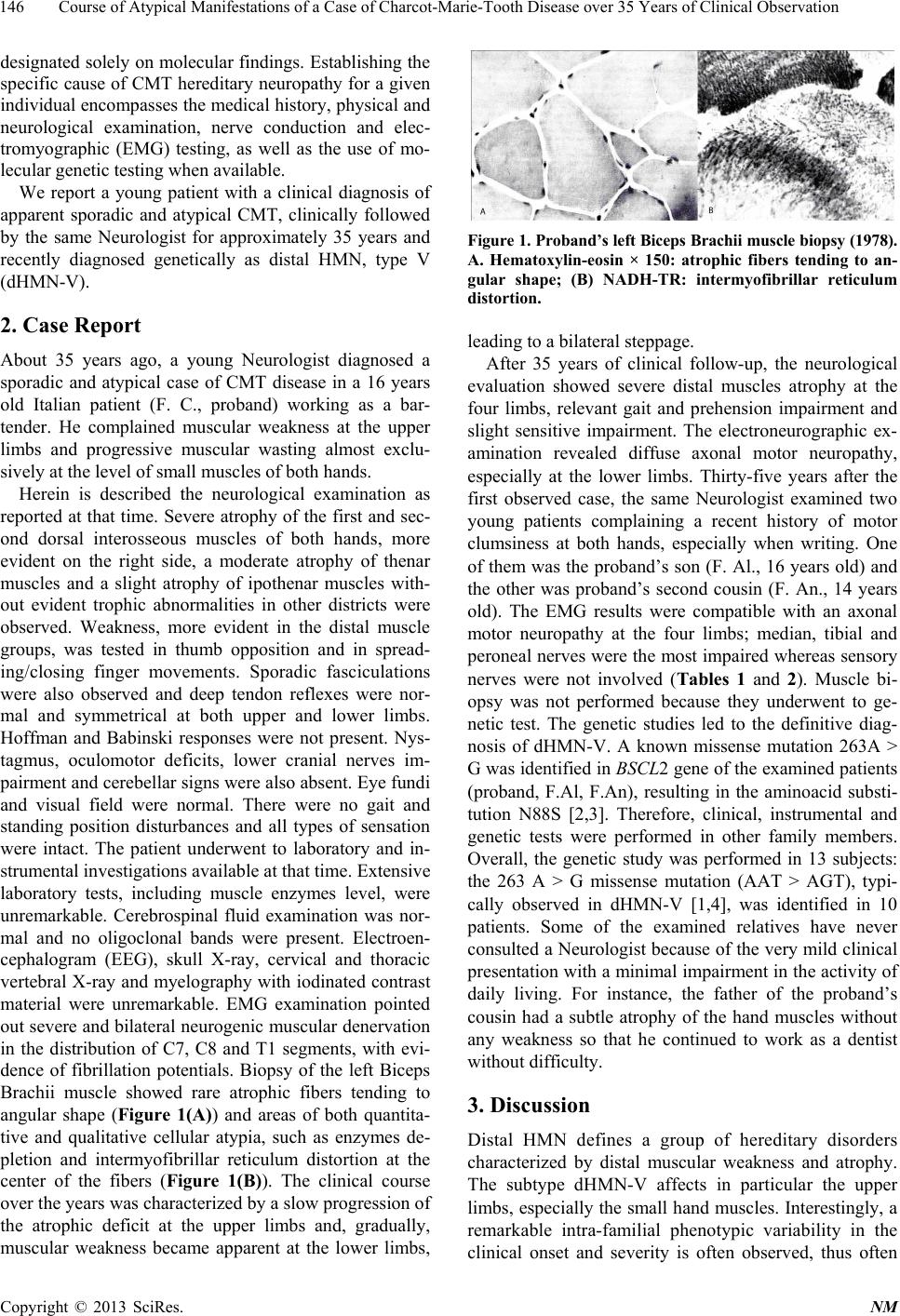 Course of Atypical Manifestations of a Case of Charcot-Marie-Tooth Disease over 35 Years of Clinical Observation 146 designated solely on molecular findings. Establishing the specific cause of CMT hereditary neuropathy for a given individual encompasses the medical history, physical and neurological examination, nerve conduction and elec- tromyographic (EMG) testing, as well as the use of mo- lecular genetic testing when available. We report a young patient with a clinical diagnosis of apparent sporadic and atypical CMT, clinically followed by the same Neurologist for approximately 35 years and recently diagnosed genetically as distal HMN, type V (dHMN-V). 2. Case Report About 35 years ago, a young Neurologist diagnosed a sporadic and atypical case of CMT disease in a 16 years old Italian patient (F. C., proband) working as a bar- tender. He complained muscular weakness at the upper limbs and progressive muscular wasting almost exclu- sively at the level of small muscles of both hands. Herein is described the neurological examination as reported at that time. Severe atrophy of the first and sec- ond dorsal interosseous muscles of both hands, more evident on the right side, a moderate atrophy of thenar muscles and a slight atrophy of ipothenar muscles with- out evident trophic abnormalities in other districts were observed. Weakness, more evident in the distal muscle groups, was tested in thumb opposition and in spread- ing/closing finger movements. Sporadic fasciculations were also observed and deep tendon reflexes were nor- mal and symmetrical at both upper and lower limbs. Hoffman and Babinski responses were not present. Nys- tagmus, oculomotor deficits, lower cranial nerves im- pairment and cerebellar signs were also absent. Eye fundi and visual field were normal. There were no gait and standing position disturbances and all types of sensation were intact. The patient underwent to laboratory and in- strumental investigations available at that time. Extensive laboratory tests, including muscle enzymes level, were unremarkable. Cerebrospinal fluid examination was nor- mal and no oligoclonal bands were present. Electroen- cephalogram (EEG), skull X-ray, cervical and thoracic vertebral X-ray and myelography with iodinated contrast material were unremarkable. EMG examination pointed out severe and bilateral neurogenic muscular denervation in the distribution of C7, C8 and T1 segments, with evi- dence of fibrillation potentials. Biopsy of the left Biceps Brachii muscle showed rare atrophic fibers tending to angular shape (Figure 1(A)) and areas of both quantita- tive and qualitative cellular atypia, such as enzymes de- pletion and intermyofibrillar reticulum distortion at the center of the fibers (Figure 1(B)). The clinical course over the years was characterized by a slow progression of the atrophic deficit at the upper limbs and, gradually, muscular weakness became apparent at the lower limbs, Figure 1. Proband’s le ft Biceps Brachii muscle biop sy (1978). A. Hematoxylin-eosin × 150: atrophic fibers tending to an- gular shape; (B) NADH-TR: intermyofibrillar reticulum distortion. leading to a bilateral steppage. After 35 years of clinical follow-up, the neurological evaluation showed severe distal muscles atrophy at the four limbs, relevant gait and prehension impairment and slight sensitive impairment. The electroneurographic ex- amination revealed diffuse axonal motor neuropathy, especially at the lower limbs. Thirty-five years after the first observed case, the same Neurologist examined two young patients complaining a recent history of motor clumsiness at both hands, especially when writing. One of them was the proband’s son (F. Al., 16 years old) and the other was proband’s second cousin (F. An., 14 years old). The EMG results were compatible with an axonal motor neuropathy at the four limbs; median, tibial and peroneal nerves were the most impaired whereas sensory nerves were not involved (Tables 1 and 2). Muscle bi- opsy was not performed because they underwent to ge- netic test. The genetic studies led to the definitive diag- nosis of dHMN-V. A known missense mutation 263A > G was identified in BSCL2 gene of the examined patients (proband, F.Al, F.An), resulting in the aminoacid substi- tution N88S [2,3]. Therefore, clinical, instrumental and genetic tests were performed in other family members. Overall, the genetic study was performed in 13 subjects: the 263 A > G missense mutation (AAT > AGT), typi- cally observed in dHMN-V [1,4], was identified in 10 patients. Some of the examined relatives have never consulted a Neurologist because of the very mild clinical presentation with a minimal impairment in the activity of daily living. For instance, the father of the proband’s cousin had a subtle atrophy of the hand muscles without any weakness so that he continued to work as a dentist without difficulty. 3. Discussion Distal HMN defines a group of hereditary disorders characterized by distal muscular weakness and atrophy. The subtype dHMN-V affects in particular the upper limbs, especially the small hand muscles. Interestingly, a remarkable intra-familial phenotypic variability in the clinical onset and severity is often observed, thus often Copyright © 2013 SciRes. NM 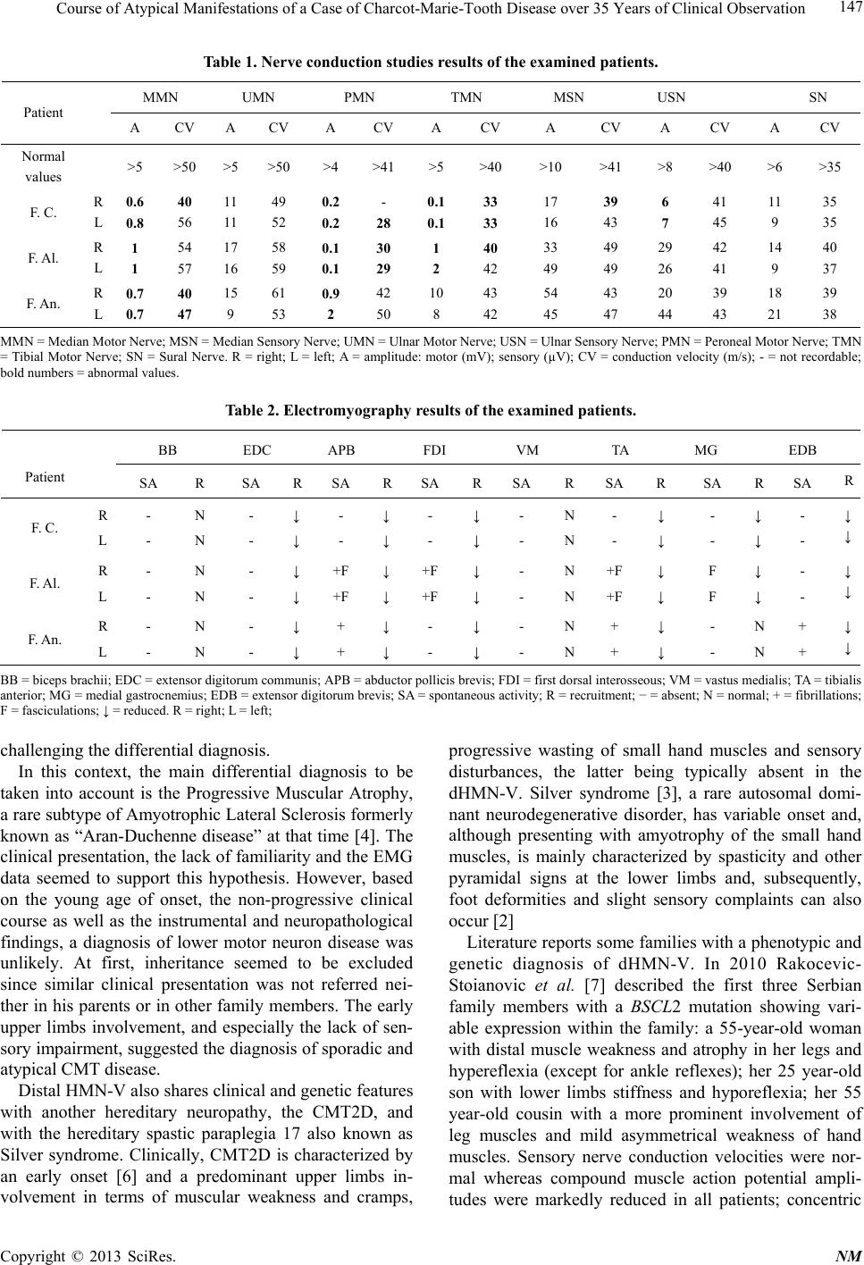 Course of Atypical Manifestations of a Case of Charcot-Marie-Tooth Disease over 35 Years of Clinical Observation Copyright © 2013 SciRes. NM 147 Table 1. Nerve conduction studies results of the examined patients. MMN UMN PMN TMN MSN USN SN Patient A CV A CV A CV A CV A CV A CV A CV Normal values >5 >50 >5 >50 >4 >41 >5 >40 >10 >41 >8 >40 >6 >35 F. C. R L 0.6 0.8 40 56 11 11 49 52 0.2 0.2 - 28 0.1 0.1 33 33 17 16 39 43 6 7 41 45 11 9 35 35 F. Al. R L 1 1 54 57 17 16 58 59 0.1 0.1 30 29 1 2 40 42 33 49 49 49 29 26 42 41 14 9 40 37 F. An. R L 0.7 0.7 40 47 15 9 61 53 0.9 2 42 50 10 8 43 42 54 45 43 47 20 44 39 43 18 21 39 38 MMN = Median Motor Nerve; MSN = Median Sensory Nerve; UMN = Ulnar Motor Nerve; USN = Ulnar Sensory Nerve; PMN = Peroneal Motor Nerve; TMN = Tibial Motor Nerve; SN = Sural Nerve. R = right; L = left; A = amplitude: motor (mV); sensory (µV); CV = conduction velocity (m/s); - = not recordable; bold numbers = abnormal values. Table 2. Electromyography results of the examined patients. BB EDC APB FDI VM TA MG EDB Patient SA R SA R SA RSA RSA RSA R SA R SA R F. C. R L - - N N - - ↓ ↓ - - ↓ ↓ - - ↓ ↓ - - N N - - ↓ ↓ - - ↓ ↓ - - ↓ ↓ F. Al. R L - - N N - - ↓ ↓ +F +F ↓ ↓ +F +F ↓ ↓ - - N N +F +F ↓ ↓ F F ↓ ↓ - - ↓ ↓ F. An. R L - - N N - - ↓ ↓ + + ↓ ↓ - - ↓ ↓ - - N N + + ↓ ↓ - - N N + + ↓ ↓ BB = biceps brachii; EDC = extensor digitorum communis; APB = abductor pollicis brevis; FDI = first dorsal interosseous; VM = vastus medialis; TA = tibialis anterior; MG = medial gastrocnemius; EDB = extensor digitorum brevis; SA = spontaneous activity; R = recruitment; − = absent; N = normal; + = fibrillations; F = fasciculations; ↓ = reduced. R = right; L = left; challenging the differential diagnosis. In this context, the main differential diagnosis to be taken into account is the Progressive Muscular Atrophy, a rare subtype of Amyotrophic Lateral Sclerosis formerly known as “Aran-Duchenne disease” at that time [4]. The clinical presentation, the lack of familiarity and the EMG data seemed to support this hypothesis. However, based on the young age of onset, the non-progressive clinical course as well as the instrumental and neuropathological findings, a diagnosis of lower motor neuron disease was unlikely. At first, inheritance seemed to be excluded since similar clinical presentation was not referred nei- ther in his parents or in other family members. The early upper limbs involvement, and especially the lack of sen- sory impairment, suggested the diagnosis of sporadic and atypical CMT disease. Distal HMN-V also shares clinical and genetic features with another hereditary neuropathy, the CMT2D, and with the hereditary spastic paraplegia 17 also known as Silver syndrome. Clinically, CMT2D is characterized by an early onset [6] and a predominant upper limbs in- volvement in terms of muscular weakness and cramps, progressive wasting of small hand muscles and sensory disturbances, the latter being typically absent in the dHMN-V. Silver syndrome [3], a rare autosomal domi- nant neurodegenerative disorder, has variable onset and, although presenting with amyotrophy of the small hand muscles, is mainly characterized by spasticity and other pyramidal signs at the lower limbs and, subsequently, foot deformities and slight sensory complaints can also occur [2] Literature reports some families with a phenotypic and genetic diagnosis of dHMN-V. In 2010 Rakocevic- Stoianovic et al. [7] described the first three Serbian family members with a BS CL 2 mutation showing vari- able expression within the family: a 55-year-old woman with distal muscle weakness and atrophy in her legs and hypereflexia (except for ankle reflexes); her 25 year-old son with lower limbs stiffness and hyporeflexia; her 55 year-old cousin with a more prominent involvement of leg muscles and mild asymmetrical weakness of hand muscles. Sensory nerve conduction velocities were nor- mal whereas compound muscle action potential ampli- tudes were markedly reduced in all patients; concentric  Course of Atypical Manifestations of a Case of Charcot-Marie-Tooth Disease over 35 Years of Clinical Observation 148 needle EMG showed chronic denervation in distal mus- cles. In 2006, van de Warrenburg et al. [8] reported the first two Dutch families with BSCL2 mutations and un- derscored the phenotypic spectrum of BSCL2 gene muta- tions with an overlap between Silver syndrome, dHMN- V and dHMN-II, highlighting the variable phenotypic patterns between and within the families. Despite a rather early onset in the second or first decade, disease progres- sion was usually slow and mildly impacts the daily living functions. The heterogeneous clinical presentations in- cluded: foot muscle weakness and atrophy, with or with- out gradually evolving pyramidal tract signs or hand muscle involvement; predominant involvement of hand muscles, with minimal or no lower limb involvement and the early or late presence of Babinski’s sign; predomi- nant spastic paraparesis with minimal distal lower limb involvement. Therefore, although the phenol-type may eventually become compatible with Silver syndrome va- riant (with predominant foot rather than hand muscle involvement), the dHMN-II or dHMN-V resembling fea- tures are often encountered in early stages and pyramidal tract signs might even remain absent. In a report of 90 patients with the N88S mutation [9], the following distribution of clinical subtypes was ob- served: 4.4% (still) unaffected; 20% subclinically/very mildly affected; 31.1% dHMN-type V; 14.5% classical Silver syndrome; 20% CMT like phenotype (either spinal form or hereditary motor sensory neuropathies-II de- pending on the absence or presence of sensory abnor- malities, respectively); 10% a pure spastic paraparesis. More recently, a Chinese family over four generations with seipinopathy was studied [10]. Six family members were dHMN-II with predominant weakness of lower ex- tremities, whereas one was accompanied by pyramidal signs. Other three women were dHMN-V with predomi- nant hands atrophy. Taken together, these findings interestingly highlight the view that the phenotype of motor neuropathies asso- ciated with BSCL2 mutations is broader than Silver syn- drome and dHMN-V [1]. In this context, Irobi et al. [1] reported two families, one with members affected by marked spasticity in the lower limbs and very striking distal amyotrophy that always started in the legs and the other with distal amyotrophy sometimes starting and pre- dominating in the legs, but no pyramidal tract signs. Fur- ther evidence for genetic heterogeneity of dHMN-V, CMT2 with predominant hand involvement and Silver syndrome came from Rohkamm et al. [11], confirming that most likely only two mutations (N88S, S90L) in exon 3 of BSCL2 may lead to dHMN-V or Silver syn- drome phenotypes whereas mutations in other genes (such as GARS, HSPB1 and HSPB8) are not a common cause of dHMN-V, Silver syndrome and CMT2D. 4. Conclusion Some atypical manifestations of CMT disease, such as the apparent lack of familiarity, the predominant upper limbs involvement at the onset, together with no tendon reflexes or sensation abnormalities, intrigued a young Neurologist 35 years ago. Although the genetic study was not available at that time, a comprehensive diagnos- tic work-up and a continuous clinical observation, in ad- dition to modern advanced diagnostic procedures such as genetic analysis, allowed us to delineate a definitive diag- nosis and to identify other family members affected by a rare hereditary neuropathy. The apparent lack of famili- arity that led to the diagnosis of atypical and sporadic CMT in the past was probably due to the very soft pres- entation and course of the disease in the proband’s rela- tives, highlighting the impressive intra-familiar variabil- ity that might characterize these conditions. In conclu- sion, 35 years later, the same Neurologist has not dis- avowed his first diagnosis. REFERENCES [1] J. Irobi, P. De Jonghe and V. Timmerman, “Molecular Genetics of Distal Hereditary Motor Neuropathies,” Hu- man Molecular Genetics, Vol. 13, No. 2, 2004, pp. R195- R202. [2] C. Windpassinger, M. Auer-Grumbach, J. Irobi, H. Patel, E. Petek, G. Horl, et al., “Heterozygous Missense Muta- tions in BSCL2 Are Associated with Distal Hereditary Motor Neuropathy and Silver Syndrome,” Nature Genet- ics, Vol. 36, No. 3, 2004, pp. 271-276. doi:10.1038/ng1313 [3] D. Ito and N. Suzuki, “Seipinopathy: A Novel Endoplas- mic Reticulum Stress-Associated Disease,” Brain, Vol. 132, 2009, pp. 8-15. [4] M. Bonduelle, “Aran-Duchenne? Duchenne-Aran? The Quarrel around Progressive Muscular Atrophy,” Revue Neurologique Société de Neurologie de Paris, Vol. 146, No. 2, 1990, pp. 97-106. [5] J. Magrè, M. Delépine, E. Khallout, T. Gedde-Dahl, L. Van Maldergem, E. Sobel, et al., “Identification of the Gene Altered in Berardinelli-Seip Congenital Lipodystro- phy on Chromosome 11q13,” Nature Genetics, Vol. 28, No. 4, 2001, pp. 365-370. doi:10.1038/ng585 [6] K. Sivakumar, T. Kyriakides, I. Puls, G. A. Nicholson, B. Funalot, A. Antonellis, et al., “Phenotypic Spectrum of Disorders Associated with Glycyl-tRNA Synthetase Mu- tations,” Brain, Vol. 128, No. 10, 2005, pp. 2304-2314. doi:10.1093/brain/awh590 [7] V. Rakocević-Stojanović, V. Milić-Rasić, S. Perić, J. Ba- ets, V. Timmerman, I. Dierick, et al., “N88S Mutation in the BSCL2 Gene in a Serbian Family with Distal Heredi- tary Motor Neuropathy Type V or Silver Syndrome,” Journal of the Neurological Sciences, Vol. 296, No. 1-2, 2010, pp. 107-109. doi:10.1016/j.jns.2010.06.015 [8] B. P. van de Warrenburg, H. Scheffer, J. J. van Eijk, M. H. Copyright © 2013 SciRes. NM  Course of Atypical Manifestations of a Case of Charcot-Marie-Tooth Disease over 35 Years of Clinical Observation Copyright © 2013 SciRes. NM 149 Versteeg, H. Kremer, M. J. Zwarts, et al., “BSCL2 Muta- tions in Two Dutch Families with Overlapping Silver Syndrome-Distal Hereditary Motor Neuropathy,” Neuro- muscular Disorders, Vol. 16, No. 2, 2006, pp. 122-125. doi:10.1016/j.nmd.2005.11.003 [9] M. Auer-Grumbach, B. Schlotter-Weigel, H. Lochmüller, G. Strobl-Wildemann, P. Auer-Grumbach, R. Fischer, et al., “Phenotypes of the N88S Berardinelli-Seip Congeni- tal Lipodystrophy 2 Mutation,” Annals of Neurology, Vol. 57, No. 3, 2005, pp. 415-424. doi:10.1002/ana.20410 [10] B. Chen, R. Zheng, X. Luan, W. Zhang, Z. Wang and Y. Yuan, “Clinical and Pathological Study of Distal Motor Neuropathy with N88S Mutation in BSCL2,” Neuropa- thology, Vol. 29, No. 5, 2009, pp. 543-547. doi:10.1111/j.1440-1789.2009.01011.x [11] B. Rohkamm, M. M. Reilly, H. Lochmüller, B. Schlotter- Weigel, N. Barisic, L. Schöls L, et al., “Further Evidence for Genetic Heterogeneity of Distal HMN Type V, CMT2 with Predominant Hand Involvement and Silver Syn- drome,” Neurological Sciences, Vol. 263, No. 1-2, 2007, pp. 100-106. doi:10.1016/j.jns.2007.06.047 |