

Paper Menu >>
Journal Menu >>
 Modern Research in Catalysis, 2013, 2, 13-18 http://dx.doi.org/10.4236/mrc.2013.23A003 Published Online September 2013 (http://www.scirp.org/journal/mrc) Synthesis, Characterization, and Activity of Tin Oxide Nanoparticles: Influence of Solvothermal Time on Photocatalytic Degradation of Rhodamine B Zuoli He1*, Jiaqi Zhou2 1Electronic Materials Research Laboratory, School of Electronic and Information Engineering, Xi’an Jiaotong University, Xi’an, China 2School of Materials Science and Engineering, Shaanxi University of Science and Technology, Xi’an, China Email: *[email protected], *[email protected] Received April 26, 2013; revised June 9, 2013; accepted July 8, 2013 Copyright © 2013 Zuoli He, Jiaqi Zhou. This is an open access article distributed under the Creative Commons Attribution License, which permits unrestricted use, distribution, and reproduction in any medium, provided the original work is properly cited. ABSTRACT The SnO2 spheres-like nanoparticles have been successfully synthesized by a microwave solvothermal method, in which SnCl2·2H2O, poly(vinylpyrrolidone) PVP, H2O2 and NaOH as raw materials. The as-synthesized products have been characterized by X-ray diffraction, scanning electron microscope, and UV/Vis/NIR spectrophotometer. Photocata- lytic activities of the samples have been evaluated by the degradation of rhodamine B (RhB) under UV-light illumina- tion. Results showed that these products with diameter about 1 - 2 µm, and when the reaction time prolong, the surface of the SnO2 spheres will change to rough and then smooth when the time even longer. The product with nanorods on its surface shows the higher photocatalytic activity and red shift in the UV-vis absorption, which are relative to the unique structure. At last we studied the electron transfer reactions during photo-oxidation of RhB. Keywords: SnO2; Nanoparticles; Photocatalytic; Solvothermal 1. Introduction Environmental problems, especially, the sustained pollu- tions of water by various organic and metallic ion con- taminants have been one of the most serious problems. And many efforts are dedicated to the remediation of en- vironmental pollution [1-3]. For instance, photodegrada- tion of organic compounds provides an available way to deal with the water pollution. Nanostructured semicon- ductors (such as TiO2, ZnO, SnO2 and so on) are proved to be an excellent photocatalyst which can degrade many kinds of persistent organic pollution [4-10]. Tin oxide (SnO2), as one of the most important semi- conductor oxides, has been used as photocatalyst for pho- todegradation of organic compounds. The results indicate that SnO2 has exhibited photoactivity toward degradation of dye and other organic compounds [11,12]. However, just like other transition metal oxides photocatalysts such as TiO2 and ZnO, SnO2 suffer from low photocatalytic efficiency because of its wide-bandgap (energy of the band gap is about 3.6 eV) [13] and high recombination rates of photogenerated electron-hole pairs. This defects hinder SnO2 photocatalyst using widely and practically in the environmental application [14]. To overcome this pro- blem, the fabrication of nanostructures provides an ef- fective way. The synthesis of TiO2-based photocatalysts has been reported in our previous papers [15-18]. Recently, SnO2- based photocatalysts were also synthesized by various approaches and have exhibited attractive performances [19-22]. In this work, we describe the synthesis of SnO2 nanoparticles via a microwave solvothermal method and discuss the effect of solvothermal time on the morpholo- gies and nanostructures. The as-synthesized SnO2 nano- particles were well characterised and their photocatalytic activities were evaluated by the photodegradation of Rho- damine B (RhB). 2. Experimental 2.1. Preparation All the reagents used in this experiment were analytical grade and were used without further purification. In a typical preparation procedure, first, 1.353 g of SnCl2·2H 2O was dissolved into 40 mL distilled water under conti- *Corresponding author. C opyright © 2013 SciRes. MRC 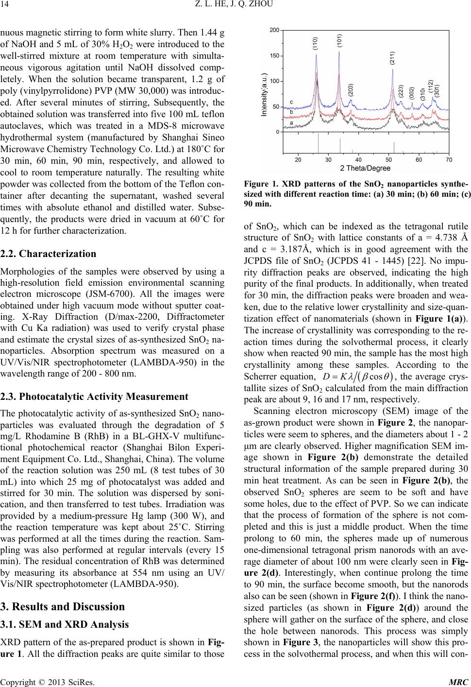 Z. L. HE, J. Q. ZHOU 14 nuous magnetic stirring to form white slurry. Then 1.44 g of NaOH and 5 mL of 30% H2O2 were introduced to the well-stirred mixture at room temperature with simulta- neous vigorous agitation until NaOH dissolved comp- letely. When the solution became transparent, 1.2 g of poly (vinylpyrrolidone) PVP (MW 30,000) was introduc- ed. After several minutes of stirring, Subsequently, the obtained solution was transferred into five 100 mL teflon autoclaves, which was treated in a MDS-8 microwave hydrothermal system (manufactured by Shanghai Sineo Microwave Chemistry Technology Co. Ltd.) at 180˚C for 30 min, 60 min, 90 min, respectively, and allowed to cool to room temperature naturally. The resulting white powder was collected from the bottom of the Teflon con- tainer after decanting the supernatant, washed several times with absolute ethanol and distilled water. Subse- quently, the products were dried in vacuum at 60˚C for 12 h for further characterization. 2.2. Characterization Morphologies of the samples were observed by using a high-resolution field emission environmental scanning electron microscope (JSM-6700). All the images were obtained under high vacuum mode without sputter coat- ing. X-Ray Diffraction (D/max-2200, Diffractometer with Cu Ka radiation) was used to verify crystal phase and estimate the crystal sizes of as-synthesized SnO2 na- noparticles. Absorption spectrum was measured on a UV/Vis/NIR spectrophotometer (LAMBDA-950) in the wavelength range of 200 - 800 nm. 2.3. Photocatalytic Activity Measurement The photocatalytic activity of as-synthesized SnO2 nano- particles was evaluated through the degradation of 5 mg/L Rhodamine B (RhB) in a BL-GHX-V multifunc- tional photochemical reactor (Shanghai Bilon Experi- ment Equipment Co. Ltd., Shanghai, China). The volume of the reaction solution was 250 mL (8 test tubes of 30 mL) into which 25 mg of photocatalyst was added and stirred for 30 min. The solution was dispersed by soni- cation, and then transferred to test tubes. Irradiation was provided by a medium-pressure Hg lamp (300 W), and the reaction temperature was kept about 25˚C. Stirring was performed at all the times during the reaction. Sam- pling was also performed at regular intervals (every 15 min). The residual concentration of RhB was determined by measuring its absorbance at 554 nm using an UV/ Vis/NIR spectrophotometer (LAMBDA-950). 3. Results and Discussion 3.1. SEM and XRD Analysis XRD pattern of the as-prepared product is shown in Fig- ure 1. All the diffraction peaks are quite similar to those Figure 1. XRD patterns of the SnO2 nanoparticles synthe- sized with different reaction time: (a) 30 min; (b) 60 min; (c) 90 min. of SnO2, which can be indexed as the tetragonal rutile structure of SnO2 with lattice constants of a = 4.738 Å and c = 3.187Å, which is in good agreement with the JCPDS file of SnO2 (JCPDS 41 - 1445) [22]. No impu- rity diffraction peaks are observed, indicating the high purity of the final products. In additionally, when treated for 30 min, the diffraction peaks were broaden and wea- ken, due to the relative lower crystallinity and size-quan- tization effect of nanomaterials (shown in Figure 1(a)). The increase of crystallinity was corresponding to the re- action times during the solvothermal process, it clearly show when reacted 90 min, the sample has the most high crystallinity among these samples. According to the Scherrer equation, cosDK , the average crys- tallite sizes of SnO2 calculated from the main diffraction peak are about 9, 16 and 17 nm, respectively. Scanning electron microscopy (SEM) image of the as-grown product were shown in Figure 2, the nanopar- ticles were seem to spheres, and the diameters about 1 - 2 μm are clearly observed. Higher magnification SEM im- age shown in Figure 2(b) demonstrate the detailed structural information of the sample prepared during 30 min heat treatment. As can be seen in Figure 2(b), the observed SnO2 spheres are seem to be soft and have some holes, due to the effect of PVP. So we can indicate that the process of formation of the sphere is not com- pleted and this is just a middle product. When the time prolong to 60 min, the spheres made up of numerous one-dimensional tetragonal prism nanorods with an ave- rage diameter of about 100 nm were clearly seen in Fig- ure 2(d). Interestingly, when continue prolong the time to 90 min, the surface become smooth, but the nanorods also can be seen (shown in Figure 2(f)). I think the nano- sized particles (as shown in Figure 2(d)) around the sphere will gather on the surface of the sphere, and close the hole between nanorods. This process was simply shown in Figure 3, the nanoparticles will show this pro- cess in the solvothermal process, and when this will con- Copyright © 2013 SciRes. MRC 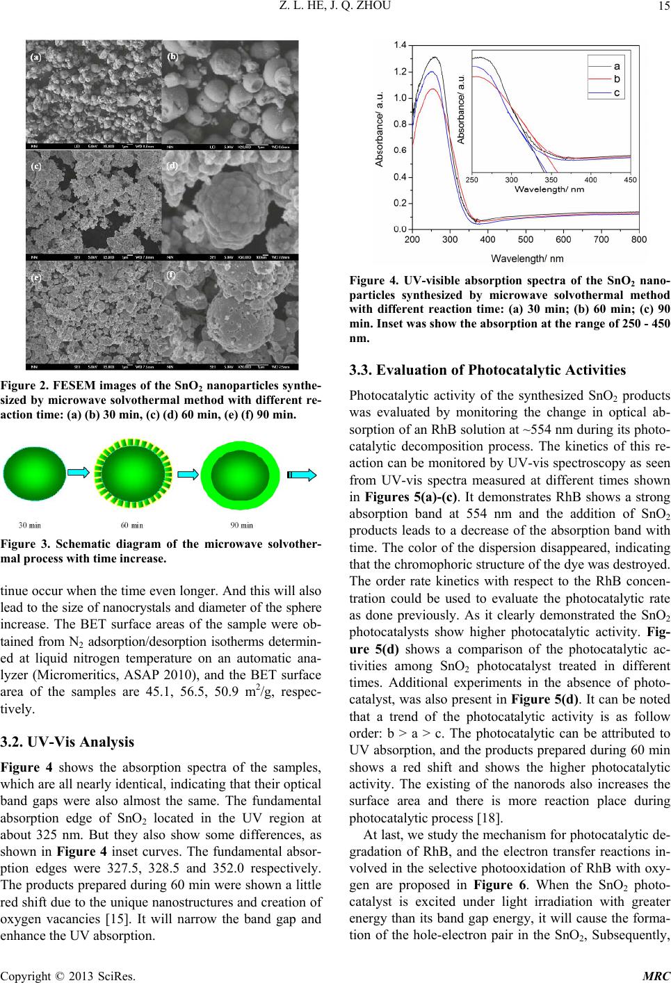 Z. L. HE, J. Q. ZHOU 15 Figure 2. FESEM images of the SnO2 nanoparticles synthe- sized by microwave solvothermal method with different re- action time: (a) (b) 30 min, (c) (d) 60 min, (e) (f) 90 min. Figure 3. Schematic diagram of the microwave solvother- mal process with time increase. tinue occur when the time even longer. And this will also lead to the size of nanocrystals and diameter of the sphere increase. The BET surface areas of the sample were ob- tained from N2 adsorption/desorption isotherms determin- ed at liquid nitrogen temperature on an automatic ana- lyzer (Micromeritics, ASAP 2010), and the BET surface area of the samples are 45.1, 56.5, 50.9 m2/g, respec- tively. 3.2. UV-Vis Analysis Figure 4 shows the absorption spectra of the samples, which are all nearly identical, indicating that their optical band gaps were also almost the same. The fundamental absorption edge of SnO2 located in the UV region at about 325 nm. But they also show some differences, as shown in Figure 4 inset curves. The fundamental absor- ption edges were 327.5, 328.5 and 352.0 respectively. The products prepared during 60 min were shown a little red shift due to the unique nanostructures and creation of oxygen vacancies [15]. It will narrow the band gap and enhance the UV absorption. Figure 4. UV-visible absorption spectra of the SnO2 nano- particles synthesized by microwave solvothermal method with different reaction time: (a) 30 min; (b) 60 min; (c) 90 min. Inset was show the absorption at the range of 250 - 450 nm. 3.3. Evaluation of Photocatalytic Activities Photocatalytic activity of the synthesized SnO2 products was evaluated by monitoring the change in optical ab- sorption of an RhB solution at ~554 nm during its photo- catalytic decomposition process. The kinetics of this re- action can be monitored by UV-vis spectroscopy as seen from UV-vis spectra measured at different times shown in Figures 5(a)-(c). It demonstrates RhB shows a strong absorption band at 554 nm and the addition of SnO2 products leads to a decrease of the absorption band with time. The color of the dispersion disappeared, indicating that the chromophoric structure of the dye was destroyed. The order rate kinetics with respect to the RhB concen- tration could be used to evaluate the photocatalytic rate as done previously. As it clearly demonstrated the SnO2 photocatalysts show higher photocatalytic activity. Fig- ure 5(d) shows a comparison of the photocatalytic ac- tivities among SnO2 photocatalyst treated in different times. Additional experiments in the absence of photo- catalyst, was also present in Figure 5(d). It can be noted that a trend of the photocatalytic activity is as follow order: b > a > c. The photocatalytic can be attributed to UV absorption, and the products prepared during 60 min shows a red shift and shows the higher photocatalytic activity. The existing of the nanorods also increases the surface area and there is more reaction place during photocatalytic process [18]. At last, we study the mechanism for photocatalytic de- gradation of RhB, and the electron transfer reactions in- volved in the selective photooxidation of RhB with oxy- gen are proposed in Figure 6. When the SnO2 photo- catalyst is excited under light irradiation with greater energy than its band gap energy, it will cause the forma- ion of the hole-electron pair in the SnO2, Subsequently, t Copyright © 2013 SciRes. MRC 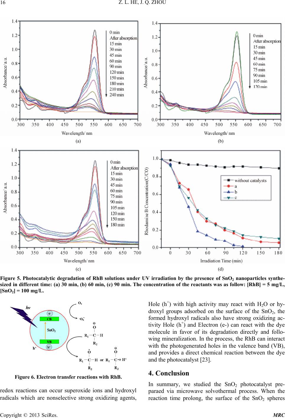 Z. L. HE, J. Q. ZHOU Copyright © 2013 SciRes. MRC 16 (a) (b) (c) (d) Figure 5. Photocatalytic degradation of RhB solutions under UV irradiation by the presence of SnO2 nanoparticles synthe- sized in different time: (a) 30 min, (b) 60 min, (c) 90 min. The concentration of the reactants was as follow: [RhB] = 5 mg/L, [SnO2] = 100 mg/L. Hole (h+) with high activity may react with H2O or hy- droxyl groups adsorbed on the surface of the SnO2, the formed hydroxyl radicals also have strong oxidizing ac- tivity Hole (h+) and Electron (e-) can react with the dye molecule in favor of its degradation directly and follo- wing mineralization. In the process, the RhB can interact with the photogenerated holes in the valence band (VB), and provides a direct chemical reaction between the dye and the photocatalyst [23]. 4. Conclusion Figure 6. Electron transfer reactions with RhB. In summary, we studied the SnO2 photocatalyst pre- paraed via microwave solvothermal process. When the reaction time prolong, the surface of the SnO2 spheres redox reactions can occur superoxide ions and hydroxyl radicals which are nonselective strong oxidizing agents,  Z. L. HE, J. Q. ZHOU 17 will change to rough and then smooth when the time even longer. We also propose the modeling to explain this interesting phenomenon. The product with nanorods on its surface shows the higher photocatalytic activity and red shift in the Uv-vis absorption, which are relative to the unique structure. 5. Acknowledgements This work was supported by the Research Fund for the Doctoral Program of Higher Education of China under grant 20120201130004, the Science and Technology De- veloping Project of Shaanxi Province (2012KW-11), and the Fundamental Research Funds for the Central Univer- sities. REFERENCES [1] C. Burda, X. Chen, R. Narayanan and M. A. El-Sayed, “Chemistry and Properties of Nanocrystals of Different Shapes,” Chemical Reviews, Vol. 105, No. 4, 2005, pp, 1025-1102. doi:10.1021/cr030063a [2] X. Chen and S. S. Mao, “Titanium Dioxide Nanomateri- als: Synthesis, Properties, Modifications, and Applica- tions,” Chemical Reviews, Vol. 107, No. 7, 2007, pp, 2891-2959. doi:10.1021/cr0500535 [3] Z. He, W. Que, J. Chen, X. Yin, Y. He, J. Ren, “Photoca- talytic Degradation of Methyl Orange over Nitrogen— Fluorine Codoped TiO2 Nanobelts Prepared by Solvother- mal Synthesis,” ACS Applied Materials & Interfaces, Vol. 4, No. 12, 2012, pp. 6816-6826. doi:10.1021/am3019965 [4] J. C. Yu, J. G. Yu, W. K. Ho, Z. T. Jiang and L. Z. Zhang, “Effects of F Doping on the Photocatalytic Activity and Microstructures of Nanocrystalline TiO2 Powders,” Che- mistry of Materials, Vol. 14, No. 9, 2002, pp. 3808-3816. doi:10.1021/cm020027c [5] J. Ng, J. H. Pan and D. D. Sun, “Hierarchical Assembly of Anatase Nanowhiskers and Evaluation of Their Photo- catalytic Efficiency in Comparison to Various One-Di- mensional TiO2 Nanostructures,” Journal of Materials Chemistry, Vol. 21, No. 32, 2011, pp. 11844-11853. doi:10.1039/c1jm11088h [6] Z. He, W. Que, Y. He, J. Chen, H. Xie and G. Wang, “Na- nosphere Assembled Mesoporous Titanium Dioxide with Advanced Photocatalystic Activity Using Absorbent Cot- ton as Template,” Journal of Materials Science, Vol. 47, No. 20, 2012, pp. 7210-7216. doi:10.1007/s10853-012-6667-9 [7] C. A. Martinez-Huitle and E. Brillas, “Decontamination of Wastewaters Containing Synthetic Organic Dyes by Electrochemical Methods: A General Review,” Applied Catalysis B: Environmental, Vol. 87, No. 4-5, 2009, pp. 105-145. doi:10.1016/j.apcatb.2008.09.017 [8] H. J. Zhang, G. H. Chen and D. W. Bahnemann, “Photo- electrocatalytic Materials for Environmental Applica- tions,” Journal of Materials Chemistry, Vol. 19, No. 29, 2009, pp. 5089-5121. doi:10.1039/b821991e [9] H. J. Wang, F. Q. Sun, Y. Zhang, K. Y. Gu, W. Chen and W. S. Li, “Photochemical Construction of Free-Standing Sn-Filled SnO2 Nanotube Arrayon a Solution Surface for Flexible Use in Photocatalysis,” Journal of Materials Chemistry, Vol. 21, No. 33, 2011, pp. 12407-12413. doi:10.1039/c1jm10887e [10] Z. He and W. Que, “Enhanced Photocatalytic Activity of N-Cetyl-N,N,N-Trimethyl Ammonium Bromide-Assisted Solvothermal Grown Fluff-Like ZnO Nanoparticles,” Jour- nal of Nanoengineering and Nanomanufacturing, Vol. 2, No. 1, 2012, pp. 17-21. doi:10.1166/jnan.2012.1043 [11] M. Dimitrov, T. Tsoncheva, S. Shao and R. KÖhn, “No- vel Preparation of Nanosized Mesoporous SnO2 Powders: Physicochemical and Catalytic Properties,” Applied Ca- talysis B: Environmental, Vol. 94, No. 1-2, 2010, pp, 158- 165. doi:10.1016/j.apcatb.2009.11.004 [12] S. S. Wu, H. Q. Cao, S. F. Yin, X. W. Liu and X. R. Zhang, “Amino Acid-Assisted Hydrothermal Synthesis and Pho- tocatalysis of SnO2 Nanocrystals,” The Journal of Physi- cal Chemistry C, Vol. 113, No. 41, 2009, pp. 17893- 17898. doi:10.1021/jp9068762 [13] Y. T. Han, X. Wu, Y. L. Ma, L. H. Gong, F. Y. Qu and H. J. Fan, “Porous SnO2 Nanowire Bundles for Photocatalyst and Li Ion Battery Applications,” CrystEngComm, Vol. 13, No. 10, 2011, pp. 3506-3510. doi:10.1039/c1ce05171g [14] Z. Wen, G. Wang, W. Lu, Q. Wang, Q. Zhang and J. Li, “Enhanced Photocatalytic Properties of Mesoporous SnO2 Induced by Low Concentration ZnO Doping,” Crystal Growth & Design, Vol. 7, No. 9, 2007, pp. 1722-1725. doi:10.1021/cg060801z [15] Z. He, W. Que, H. Xie, J. Chen, Y. Yuan and P. Sun, “Fa- cile Synthesis of Self-Sensitized TiO2 Photocataslysts and Their Higher Photocatalytic Activity,” Journal of The American Ceramic Society, Vol. 95, No. 12, 2012, pp, 3941-3946. doi:10.1111/j.1551-2916.2012.05426.x [16] Z. He, W. Que, Y. He, J. Chen, J. Hu, H. Javed, Y. Ji, X. Li and D. Fei, “Electrochemical Behavior and Photocata- lytic Performance of Nitrogen-Doped TiO2 Nanotubes Arrays Powders Prepared by Combining Anodization with Solvothermal Process,” Ceramics International, Vol. 39, No. 5, 2013, pp. 5545-5552. doi:10.1016/j.ceramint.2012.12.068 [17] Z. He, W. Que, J. Chen, Y. He and G. Wang, “Surface Che- mical Analysis on the Carbon-Doped Mesoporous TiO2 Nanoparticles after Post-thermal Treatment: XPS and FTIR Characterization,” Journal of Physics and Chemistry of Solids, Vol. 74, No. 7, 2013, pp. 924-928. doi:10.1016/j.jpcs.2013.02.001 [18] Z. He, Z. Zhu, J. Li, N. Wei and J. Zhou, “Characteriza- tion and Activity of Mesoporous Titanium Dioxide beads with High Surface Areas and Controllable Pore Sizes,” Journal of Hazardous Materials, Vol. 190, No. 1-3, 2011, pp. 133-139. doi:10.1016/j.jhazmat.2011.03.011 [19] E. M. Seftel, E. Popovici, M. Mertens, E. A. Stefaniak, R. V. Grieken, P. Cool and E. F. Vansant, “SnIV-Containing Layered Double Hydroxides as Precursors for Nano-Sized ZnO/SnO2 Photocatalysts,” Applied Catalysis B: Environ- mental, Vol. 84, No. 3-4, 2008, pp. 699-705. doi:10.1016/j.apcatb.2008.06.006 Copyright © 2013 SciRes. MRC  Z. L. HE, J. Q. ZHOU Copyright © 2013 SciRes. MRC 18 [20] C. Wang, J. C. Zhao, X. M. Wang, B. X. Mai, G. Y. Sheng, P. A. Peng and J. M. Fu, “Preparation, Characterization and Photocatalytic Activity of Nano-Sized ZnO/SnO2 Coupl- ed Photocatalysts,” Applied Catalysis B: Environmental, Vol. 39, No. 3, 2002, pp. 269-279. doi:10.1016/S0926-3373(02)00115-7 [21] N. Wang, J. X. Xu and L. H. Guan, “Synthesis and En- hanced Photocatalytic Activity of Tin Oxide Nanoparti- cles Coated on Multi-Walled Carbon Nanotube,” Materi- als Research Bulletin, Vol. 46, No. 9, 2011, pp, 1372- 1376. doi:10.1016/j.materresbull.2011.05.014 [22] L. R. Zheng, Y. H. Zheng, C. Q. Chen, Y. Y. Zhan, X. Y. Lin, Q. Zheng, K. M. Wei and J. F. Zhu, “Network Struc- tured SnO2/ZnO Heterojunction Nanocatalyst with High Photocatalytic Activity,” Inorganic Chemistry, Vol. 48, No. 5, 2009, pp. 1819-1825. doi:10.1021/ic802293p [23] Z. Zhu, J. Zhou and Z. He, “Effects of Heat Treatment Scheme on Luminescence Properties and Photocatalytic Activity of Mo2O3/TiO2 Nanowire Prepared via Solvo- thermal Method,” Journal of Nanoengineering and Nano- manufacturing, Vol. 2, No. 1, 2012, pp. 80-84. doi:10.1166/jnan.2012.1055 |