

Paper Menu >>
Journal Menu >>
 Surgical Science, 2013, 4, 401-404 http://dx.doi.org/10.4236/ss.2013.49078 Published Online September 2013 (http://www.scirp.org/journal/ss) Influence of Peritoneal Suture on the Formation of Abdominal Adhesions in Wistar Rats: Is Suturing Worthwhile? Nathália Andrade1, Michel Vinagre1, Luciana Can a ba r r o2, Willy Marcus França3* 1Faculty of Medical Science and Health, Catholic University of São Paulo, São Paulo, Brazil 2Faculty of Medical Science and Health, Pontifical Catholic University of São Paulo, São Paulo, Brazil 3Laboratory of Experimental Surgery, FCMS, PUC/SP, São Paulo, Brazil Email: *[email protected] Received March 24, 2013; revised April 25, 2013; accepted May 2, 2013 Copyright © 2013 Nathália Andrade et al. This is an open access article distributed under the Creative Commons Attribution License, which permits unrestricted use, distribution, and reproduction in any medium, provided the original work is properly cited. ABSTRACT Purpose: The purpose of the present study was to determine the effect of peritoneal closure on the formation of ab- dominal adhesions by verifying their degree of damage on intestinal portions and the omentum with the abdominal wall. Given the different reports found in the literature concerning peritoneal closure mostly in obstetrics and gynecology, any objective information based on statistically tested results may be of great value in the everyday surgery practice. Material and Method: This is an experimental model on which a laparotomy is performed on the free cavity of the abdominal wall in growing rats. Young Wistar rats (approximately 1 month old) were operated through a long median xipho-umbilical abdominal incision. The animals were divided in 3 groups with fifteen rats each: in Group I, only the peritoneum was left open and all the other layers of the abdominal wall were closed; the rats in Group II had their peri- toneums closed with unabsorbable cord (Prolene 4-0, Ethicon®). The abdominal wall of the rats in Group III (control) was only opened up to the musculature. The peritoneum, which remained intact and closed, was carefully prodded with the grip of tweezers to avoid lesions and/or perforations in the peritoneum. Results: There were no deaths nor incisional dehiscence and/or hernias among the animals. Nine animals of Group I presented adhesions (60%), whereas there were adhesions in all the fifteen animals of Group II (100%). In Group III adhesions were found in two animals (13%). The percentage of adhesions in Group II was significantly higher than those observed in Groups I and III (p < 0.0001). Ad- hesions were mostly formed by the abdominal omentum. It was not observed any effect of the procedure on viscera. Conclusion: The experimental model that was suggested is appropriate for the establishment and study of peritoneal adhesions. The rate of adhesions found in the Group II was significantly higher (p < 0.0001) than the rate observed in the Group I and Group III, suggesting that peritoneum suture can play a important role in the adhesion processes. Keywords: Abdominal Adherence; Incision Closure; Peritoneal Suture 1. Introduction The need for peritoneum closure has been discussed for many years. Increase of operative time and formation of abdominal adhesions raise questions concerning the per- formance of this procedure [1]. Besides being one of the main causes of intestinal obstruction, affecting particu- larly the small intestine [2], which is related to the type of surgery and the extension of peritoneal damage, adhe- sions may also affect fertility and cause chronic abdomi- nal pain [3]. Reoperations are more complicated when abdominal adhesions are present, and increase surgery time and the risk of iatrogenic intestinal lesion. Moreover, the chances are that it may be impossible to perform a laparoscopy on the patient in the future [1]. The ongoing discussion about peritoneal closure has engaged gynecologists, obstetricians and surgeons for years [4,5]. Despite the assertion in several studies that there is no statistical difference between peritoneal clo- sure and non-closure in lower abdominal incisions (cae- sarean sections) [4,5], some authors have shown signifi- cant results in favor of peritoneal non-closure, such as intraoperative time and hospitalization period (p < 0.01) [6]. *Corresponding author. According to McNally and Curtain [7] the incidence of C opyright © 2013 SciRes. SS 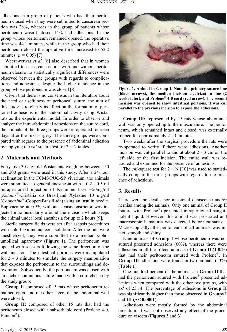 N. ANDRADE ET AL. 402 adhesions in a group of patients who had their perito- neum closed when they were submitted to caesarean sec- tion was 28%, whereas in the group of patients whose peritoneum wasn’t closed 14% had adhesions. In the group whose peritoneum remained opened, the operative time was 44.1 minutes, while in the group who had their peritoneum closed the operative time increased to 52.2 minutes (p = 0.05) [7]. Weerawetwat et al. [8] also described that in women submitted to caesarean section with and without perito- neum closure no statistically significant differences were observed between the groups with regards to complica- tions and adhesions, despite the higher incidence in the group whose peritoneum was closed [8]. Given that there is no consensus in the literature about the need or usefulness of peritoneal suture, the aim of this study is to clarify its effect on the formation of peri- toneal adhesions in the abdominal cavity using Wistar rats as the experimental model. In order to observe and analyze the intra-abdominal adhesions on the suture cord, the animals of the three groups were re-operated fourteen days after the first surgery. The three groups were com- pared with regards to the presence of abdominal adhesion by applying the chi-square test for 2 × N tables. 2. Materials and Methods Forty five 30-day-old Wistar rats weighing between 150 and 200 grams were used in this study. After a 24-hour acclimation in the FCMS/PUC-SP vivarium, the animals were submitted to general anesthesia with a 0.2 - 0.5 ml intraperitoneal injection of Ketamine base −50mg/ml (Ketalar®-Cristália do Brasil)and Xylazine 10 mg/ml (Coopazine®-CoopersBrasilLtda) using an insulin needle. Bupivacaine at 0.5% without a vasoconstrictor was in- jected intramuscularly around the incision which keeps the animal under local anesthesia for up to 2 hours [9]. Sterile surgery fields were set after asepsis procedures with chlorhexidine aqueous solution. After the rats were anesthetized, they were submitted to a median xipho- umbilical laparotomy (Figure 1). The peritoneum was opened with scissors following the same direction of the wall incision. The intestinal portions were manipulated for 2 - 3 minutes to simulate the surgery manipulation that exposes the peritoneum to the surroundings and de- hydration. Subsequently, the peritoneum was closed with an anchor continuous suture made with a cord chosen by the study group: Group I: composed of 15 rats whose peritoneum re- mained open, and the other layers of the abdominal wall were closed; Group II: composed of other 15 rats that had the peritoneum closed with unabsorbable cord (Prolene 4-0, Ethicon®). Figure 1. Animal in Group I. Note the primary suture line (black arrows), the median incision cicatrization line (2 weeks later), and Prolene® 4-0 cord (red arrow). The second incision was opened to show intestinal portions, it was cut parallel to the previous incision to expose the adhesions. Group III: represented by 15 rats whose abdominal wall was only opened up to the musculature. The perito- neum, which remained intact and closed, was externally rubbed for approximately 2 - 3 minutes. Two weeks after the surgical procedure the rats were re-operated to verify if there were adhesions. Another incision was cut parallel to and at about 2 - 3 cm on the left side of the first incision. The entire wall was re- tracted and examined for the presence of adhesions. The chi-square test for 2 × N [10] was used to statisti- cally compare the three groups with regards to the pres- ence of adhesions. 3. Results There were no deaths nor incisional dehiscence and/or hernias among the animals. Only one animal of Group II (suture with Prolene®) presented intraperitoneal sangui- nolent liquid. However, this animal was prostrated and macroscopic hematuria was detected before euthanasia. Macroscopically, the peritoneum of all animals was in- tact, smooth and shiny. Nine animals of Group I whose peritoneum was not sutured presented adhesions (60%), whereas there were adhesions in all the fifteen animals of Group II (100%) that had their peritoneum sutured with Prolene®. In Group III adhesions were found in two animals (13%) (Table 1). One hundred percent of the animals in Group II that had the peritoneum sutured with Prolene® presented ad- hesions when compared with the other two groups, with ax2 of 23.14. The percentage of adhesions in Group II was significantly higher than those observed in Groups I and III (p < 0.0001). Adhesions were mostly formed by the abdominal omentum. It was not observed any effect of the proce- dure on viscera (Figures 2 and 3). Copyright © 2013 SciRes. SS  N. ANDRADE ET AL. 403 Figure 2. Animal of Group I. The image shows the unsu- tured peritoneum, only muscular layers of the abdominal wall were sutured. Note Prolene® blue stiches through transparency. Figure 3. Animal in Group II. Peritonium was sutured with an unabsorbable cord (Prolene®). Note Prolene® blue stich- esthroughthetransparency (arrows). Table 1. Group I is composed of animals whose peritoneum was opened, but was not sutured; the peritonium of the animals in Group II was sutured with Prolene® 4-0 and the peritoneum Group III animals w as not ope ne d. Percentage of adhesions in the 3 groups. ADHESIONS GROUP YES NO TOTAL % ADHESIONS I (No suture) 9 6 15 60 II (Prolene) 15 0 15 100 III (Control) 2 13 15 13 TOTAL 26 19 45 62 4. Discussion Having a vast impact on the patients’ quality of life and health care costs 1, postoperative adhesions are the main cause of intestinal obstruction and complicated reopera- tions [1], accounting for more than 40% of all cases and 60% to 70% of those involving the small bowel [2]. Pa- tients submitted to laparotomy have 90% risk of devel- oping intra-peritoneal adhesions and 5% - 20% incidence of re-hospitalization due to adhesion formation 1and also with effects on fertility and chronic pain [3]. This is the main reason for the ongoing discussion about peritoneal closure; yet, research is mostly focused on obstetric patients [4,5]. The reconstitution of the ab- dominal wall anatomy, tissue approximation for better cicatrization, decreased risk of infection by restoring the peritoneal barrier, decreased risk of dehiscence and inci- sional hernia, and also decreased abdominal adhesions are some of the positive outcomes attributed to peritoneal closure [7,11-13]. On the other hand, faster cicatrization of the abdominal wall and decrease of operative time are described as the advantages of the no-suture approach [7,11,13]. Microscopically, the postoperative peritoneal adhe- sions were the result of fibrin effusion after a lesion or tissue inflammation with an initial phagocyte wave cleaning up the traumatic debris, and a secondary fibro- blast wave healing the lesion and differentiating into new mesothelium cells [14,15]. Most of the fibrotic tissue formed is resorbed within five days. However, in some cases organization with fibroblast and capillary growth may develop, resulting in fibrotic adhesion especially if physiological fibrinolysis doesn’t occur in five to seven days [11,12]. The main anatomo-pathological aspect of peritoneal adhesion is the presence of tissue ischemia [11,12], which would be the result of adhesion formation for revascularization of the areas where blood supply was impaired by the surgical act, given that tissue ischemia decreases plasminogen activation [16]. The establish- ment of adhesion may also occur due to other reasons, such as: trauma, infection, thrombosis and presence of foreign bodies [17]. Other possible risk factors with re- gards to the development of adhesions include patient’s age (under 60 years old), previous laparoscopy in less than five years, peritonitis, multiple laparotomies, emer- gency surgeries, omental resection and penetrating ab- dominal trauma, particularly through stab [18]. Nagele et al. [19] presented different conclusions and suggest that routine peritoneal closure should be dis- carded, despite its low morbidity with regards to fever and infection [15,19]. In a prospective randomized study performed by Grundsell et al. [6], significant results were found that support the peritoneal non-closure approach concerning intraoperative time and hospitalization period (p < 0.01) [6], however, the authors do not mention peritoneal adhesions. McNally M. et al. [7], concluded that closing the peri- toneum in caesarean section is not necessary, and that the procedure can even result in adhesion formation [7]. Na- Copyright © 2013 SciRes. SS  N. ANDRADE ET AL. Copyright © 2013 SciRes. SS 404 gele et al. [19] also suggested that routine peritoneal closure should be discarded, despite its association with low morbidity related to fever and infection [19]. On the other hand, Cheong et al. [4] concluded in an extensive literature review that there are no differences between peritoneal closure and non-closure in lower ab- dominal incisions with regards to comorbidities such as infections, fever and peritoneal adhesion formation, among others [3]. Weerawetwat et al. [8] also described that in women submitted to caesarean section with and without peritoneum closure there are no statistically sig- nificant differences, despite their higher incidence in the group whose peritoneum was closed [8]. In the present study, it was observed a higher inci- dence of peritoneal adhesions in animals whose perito- neum was sutured (Group II, 100%) when compared to the group whose peritoneum was not sutured (Group I, 60%). These results contrast with Cheong et al. [4] con- clusion, who asserted that there are no significant differ- ences between suturing and not suturing the peritoneum. The animals in Group III (Control group), were only opened up to the musculature and the peritoneum re- mained intact and had only 13% of adhesions. The experimental model that was suggested is appro- priate for the establishment and study of peritoneal adhe- sions. The rate of adhesions found in the Group II was sig- nificantly higher (p < 0.0001) than the rate observed in the Group I and Group III. 5. Acknowledgements Special thanks to the FCMS/PUC-SP Vivarium team. To the Biologist Luciana Canabarro, assistant at the Labora- tory of Surgery Technique & Experimental Surgery for her invaluable work with the animals. This study was performed with the support of a PIBIC-CNPq scholarship, N. 122.775/2010-4. This project was sponsored with a scholarship by PIBIC-CNPq (122.775/2010-4). REFERENCES [1] B. Schnüriger, et al., “Prevention of Postoperative Peri- toneal Adhesions: A Review of the Literature,” The American Journal of Surgery, Vol. 201, No. 1, 2011, pp. 111-121. doi:10.1016/j.amjsurg.2010.02.008 [2] H. Ellis, “The Clinical Significance of Adhesions: Focus on Intestinal Obstruction,” European Journal of Surgery, Vol. 577, 1997, pp. 5-9. [3] H. Ellis, et al., “The Cause and Prevention of Postopera- tive Intraperitoneal Adhesions,” Surgery, Gynecology & Obstetrics, Vol. 133, No. 3, 1971, pp. 497-511. [4] Y. C. Cheong, et al., “Peritoneal Closure—To Close or Not to Close,” Human Reproduction, Vol. 16, No. 8, 2001, pp. 1548-1552. doi:10.1093/humrep/16.8.1548 [5] G. H. Limpscomb, et al., “Peritoneal Closure at Vaginal Hysterectomy: A Reassessment,” Obstetrics & Gynecol- ogy, Vol. 87, No. 1, 1996, pp. 40-43. doi:10.1016/0029-7844(95)00362-2 [6] H. S. Grundsell, et al., “Randomized Study of Non-Clo- sure of Peritoneum in Lower Segment Cesarean Section,” Acta Obstetricia et Gynecologica Scandinavica, Vol. 77, No. 1, 1998, pp. 110-115. doi:10.1080/00016349808565823 [7] M. McNally, et al., “Does Closure of the Peritoneum during Caesarian Section Influence Postoperative Mor- bidity and Subsequent Bladder Adhesion Formation?” Journal of Obstetrics & Gynaecology, Vol. 17, No. 3, 1997, pp. 239-241. doi:10.1080/01443619750113131 [8] W. Weerawetwat, et al., “Closure versus Non-Closure of Visceral and Parietal Peritoneum at Caesarean Delivery: 16 Years Study,” Journal of the Medical Association of Thailand, Vol. 87, No. 9, 2004, pp. 1007-1011. [9] W. França, et al., “Maturity of the Myenteric Plexus Is Decreased in the Gastroschisis Rat Model,” Fetal Diag- nosis and Therapy, Vol. 23, No. 1, 2008, pp. 60-68. doi:10.1159/000109228 [10] W. G. Cochran, Some Methods for Strengthening the Commun x² Test,” Biometrics, Vol. 10, No. 4, 1954, pp. 417-451. doi:10.2307/3001616 [11] C. J. Leon, et al., “Comparison of Closure vs Non-Closure of the Peritoneal Layer in the Formation of Postoperative Adhesions to the Abdominal Wall,” Ar- quivos Brasileiros de Cirurgia Digestiva, Vol. 9, No. 4, 1994, pp. 102-107. [12] R. C. Glasmeyer, et al., “The Importance of Closing the Peritoneum at Laparotomy: Experimental Study,” Revista Médica do Paraná, Vol. 43, No. 2, 1985, pp. 26-27. [13] D. M. Duffy, et al., “Is Peritoneal Closure Necessary?” Surgery Gynecology & Obstetrics, Vol. 49, No. 12, 1994, pp. 817-822. doi:10.1097/00006254-199412000-00005 [14] H. Ellis, “The Scientific Basis of the Management of Injury, Wounds, and Ulcers in the Twentieth Century,” Annals of the Royal College of Surgeons of England, Vol. 60, 1978, pp. 219-221. [15] H. Ellis, “The Cause and Prevention of Post Operative Intraperitoneal Adhesions,” Surgery Gynecology & Obs- tetrics, Vol. 133, No. 3, 1971, pp. 497-511. [16] H. Ellis, “The Aetiology of Post-Operative Abdominal Adhesion: An Experimental Study,” British Journal of Surgery, Vol. 50, No. 219, 1962 pp. 10-16. doi:10.1002/bjs.18005021904 [17] M. L. P. B. Simões, et al., “Closure x Non-Closure of the Peritoneum and the Adhesions Formation: Experimental Study in Rats,” Revista Brasileira de Cirurgia, Vol. 86, No. 6, 1996, pp. 303-305. [18] C. K. Sung and K. H. Kim, “Missed Injuries in Abdomi- nal Trauma,” Journal of Trauma, Vol. 41, No. 2, 1996, pp. 276-282. [19] F. Nagele, et al., “Closure or Nonclosure of the Visceral Peritoneum at Cesarean Delivery,” American Journal of Obstetrics & Gynecology, Vol. 174, No. 4, 1996, pp. 1366-1370. doi:10.1016/S0002-9378(96)70686-5 |