

Paper Menu >>
Journal Menu >>
 Open Journal of Orthopedics, 2013, 3, 243-247 http://dx.doi.org/10.4236/ojo.2013.35045 Published Online September 2013 (http://www.scirp.org/journal/ojo) 243 Initial Experience with BoneBac PressTM: A Novel Autologous Bone Graft Harvesting and Collecting Device Mick Perez-Cruet, Evan M. Begun, Robert Collins, Daniel Fahim Department of Neurosurgery, William Beaumont Hospital, Royal Oak, USA. Email: [email protected] Received July 12th, 2013; revised August 15th, 2013; accepted August 29th, 2013 Copyright © 2013 Mick Perez-Cruet et al. This is an open access article distributed under the Creative Commons Attribution License, which permits unrestricted use, distribution, and reproduction in any medium, provided the original work is properly cited. ABSTRACT Object: The objective of this study is to analyze the utility of a novel autologous bone graft harvesting and collecting device in spinal fusion surgeries. Methods: 35 patients underwent fusion procedures in the cervical or lumbar spine using the BoneBac PressTM. Procedures included anterior cervical discectomy with fusion (ACDF), lumbar laminectomy and posterolateral arthrodesis, and transforaminal lumbar interbody fusion (TLIF). The amount of bone graft collected from each level was determined as well as the need for additional bone graft extenders. Fusion rates were determined based on an independent radiographic evaluation performed 5 to 12 months postoperatively. Results: 54 total levels were operated upon, collecting a total of 176.0 cc of autograft. The average amount of bone collected was 3.26 cc/level. In the cervical and lumbar spine the average amount of bone collected per level was 2.30 cc and 6.77 cc respectively. The fusion rate was 94.3% at 10 months postoperatively. In most cases no additional bone graft extender was needed. Autologous bone collected had excellent handling characteristics and was easily packed into cages or placed poster- olaterally. Conclusions: The use of autograft bone material collected using the BoneBac PressTM is cost-effective, sig- nificantly reduces bone graft cost, and eliminates donor graft site morbidity while promoting successful fusion. Keywords: Minimally Invasive; Autograft; Fusion; Transforaminal Lumbar Interbody Fusion 1. Introduction Spinal fusion surgery has become more common as in- dications have expanded and clinical data regarding sus- tained outcomes improvements are published. Spine fu- sion is used to treat traumatic fractures, degenerative disease, and pain from abnormal motion. For successful fusion, there must be growth of new bone, which pro- vides a more ideal modulus of stiffness than instrumenta- tion alone. Even though there has been tremendous growth in the number of bone graft extenders, biologically active agents, and other materials to replace or regenerate bone, autograft remains the gold standard for fusion surgery. Several investigations have reported fusion rates >95% for procedures utilizing autograft bone [1-4]. Autologous bone exhibits properties that are ideal for grafting in- cluding osteoinduction (growth factors), osteoconduction (scaffold), osteogenesis (osteoprogenitor cells), and a lack of immunogenicity [5,6]. Spinal fusion procedures have traditionally been performed with the use of iliac crest autograft. This requires an additional incision, and can result in significant donor site morbidity including chronic pain, infection, hematoma, and fracture [7]. This technique has largely been abandoned secondary to a high rate of complications, ranging from 8% - 34% [7- 11]. Autograft material can also be harvested from bone at the surgical site, which eliminates morbidity resulting from an additional donor site. The piecemeal resection of bone involving rongeurs and kerrisons for the acquisition of bone graft can be time-consuming and cumbersome when compared to the utilization of a high-speed drill. Furthermore, the yield of this piecemeal technique is largely dependent on the surgical technologist’s ability to retrieve the various bone fragments from a gauze sponge. All these factors make the local acquisition of bone graft inefficient. Ideally, the surgeon would be able to gather autolo- gous bone graft consistently and efficiently while per- forming the decompression portion of the procedure. Using the drill to decompress while simultaneously col- lecting the drilled bone fragments would be optimal. Several additional factors are necessary to promote fu- Copyright © 2013 SciRes. OJO 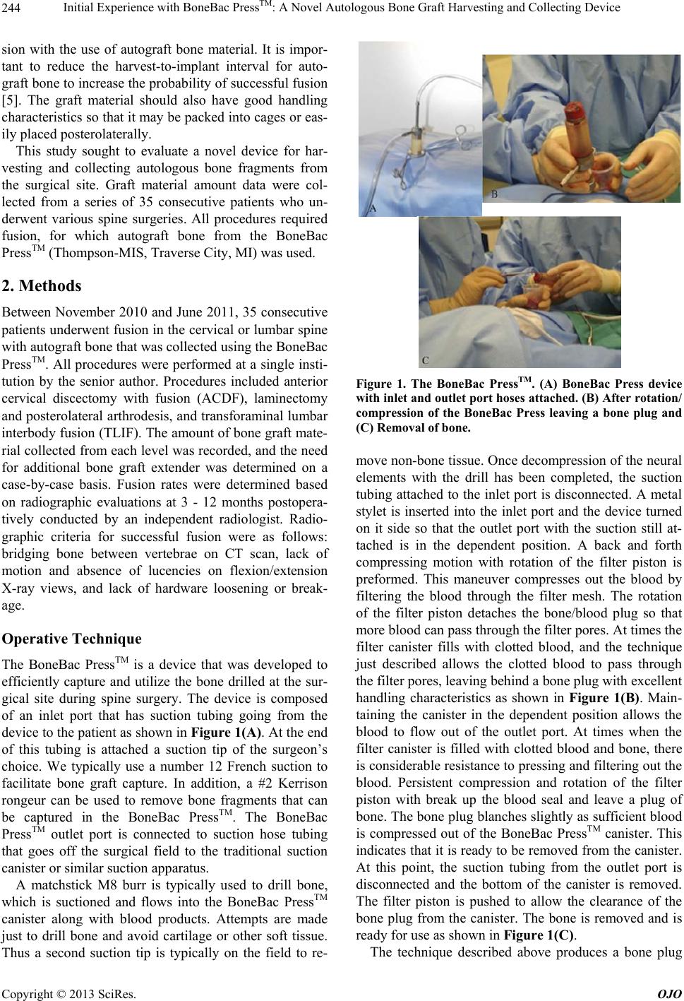 Initial Experience with BoneBac PressTM: A Novel Autologous Bone Graft Harvesting and Collecting Device 244 sion with the use of autograft bone material. It is impor- tant to reduce the harvest-to-implant interval for auto- graft bone to increase the probability o f successful fusion [5]. The graft material should also have good handling characteristics so that it may be packed into cages or eas- ily placed posterolaterally. This study sought to evaluate a novel device for har- vesting and collecting autologous bone fragments from the surgical site. Graft material amount data were col- lected from a series of 35 consecutive patients who un- derwent various spine surgeries. All procedures required fusion, for which autograft bone from the BoneBac PressTM (Thompson-MIS, Traverse City, MI) was used. 2. Methods Between November 2010 and June 2011, 35 consecutive patients underwent fusion in the cervical or lumbar spine with autogr aft bone that was collected us ing the BoneBac PressTM. All procedures were performed at a single insti- tution by the senior author. Procedures included anterior cervical discectomy with fusion (ACDF), laminectomy and posterolateral arthrodesis, and transforaminal lumbar interbody fusion (TLIF). The amount of bone graft mate- rial collected from each level was recorded, and the need for additional bone graft extender was determined on a case-by-case basis. Fusion rates were determined based on radiographic evaluations at 3 - 12 months postopera- tively conducted by an independent radiologist. Radio- graphic criteria for successful fusion were as follows: bridging bone between vertebrae on CT scan, lack of motion and absence of lucencies on flexion/extension X-ray views, and lack of hardware loosening or break- age. Operative Technique The BoneBac PressTM is a device that was developed to efficiently capture and utilize the bone drilled at the sur- gical site during spine surgery. The device is composed of an inlet port that has suction tubing going from the device to the patient as shown in Figure 1(A). At the end of this tubing is attached a suction tip of the surgeon’s choice. We typically use a number 12 French suction to facilitate bone graft capture. In addition, a #2 Kerrison rongeur can be used to remove bone fragments that can be captured in the BoneBac PressTM. The BoneBac PressTM outlet port is connected to suction hose tubing that goes off the surgical field to the traditional suction canister or similar suction apparatus. A matchstick M8 burr is typically used to drill bone, which is suctioned and flows into the BoneBac PressTM canister along with blood products. Attempts are made just to drill bone and avoid cartilage or other soft tissue. Thus a second suction tip is typically on the field to re- Figure 1. The BoneBac PressTM. (A) BoneBac Press device with inlet and outlet port hoses attached. (B) After rotation/ compression of the BoneBac Press leaving a bone plug and (C) Removal of bone. move non-bone tissue. Once decompression of the neural elements with the drill has been completed, the suction tubing attached to the inlet port is disconnected. A metal stylet is inserted into the inlet port and the device turned on it side so that the outlet port with the suction still at- tached is in the dependent position. A back and forth compressing motion with rotation of the filter piston is preformed. This maneuver compresses out the blood by filtering the blood through the filter mesh. The rotation of the filter piston detaches the bone/blood plug so that more blood can pass through the filter pores. At times the filter canister fills with clotted blood, and the technique just described allows the clotted blood to pass through the filter pores, leaving behind a bone plug with excellen t handling characteristics as shown in Figure 1(B). Main- taining the canister in the dependent position allows the blood to flow out of the outlet port. At times when the filter canister is filled with clotted blood and bone, there is considerable resistance to pressing and filtering out the blood. Persistent compression and rotation of the filter piston with break up the blood seal and leave a plug of bone. The bone p lug blanches slightly as suf ficient blood is compressed out of the BoneBac PressTM canister. This indicates that it is ready to be removed from the canister. At this point, the suction tubing from the outlet port is disconnected and the bottom of the canister is removed. The filter piston is pushed to allow the clearance of the bone plug from the canister. The bone is removed and is ready for use as shown in Figure 1(C). The technique described above produces a bone plug Copyright © 2013 SciRes. OJO  Initial Experience with BoneBac PressTM: A Novel Autologous Bone Graft Harvesting and Collecting Device 245 with excellent handling characteristics. It can be packed into cages, placed postero laterally, or used to fill the disc space directly. If additional bone graft is needed, the autograft bone can be mixed in a 1:1 ratio with bone graft extenders such as demineralized bone matrix. 3. Results A total of 54 levels were operated upon in the series of 35 patients, which included 21 females and 14 males with a mean age of 62 years old (range, 19 - 84). Patient disease pathology included degenerative disc disease with bilateral foraminal stenosis (n = 7), disc herniation with spinal cord compression (n = 7), spondylolisthesis (grade I or II) with bilateral foraminal stenosis and/or spinal cord compression (n = 13), traumatic spine injury causing spinal cord compression (n = 3), large osteophyte causing foraminal stenosis and/or spinal cord compres- sion (n = 2), metastatic tumor causing spinal cord com- pression (n = 1), lumbar spondylolysis of the bilateral pars (n = 1), and ossification of the longitudinal ligament causing spinal cord compression (n = 1). The number of patients undergoing procedures in the cervical and lumbar spine was 13 and 22, respectively. Cervical levels fused included C3-4 (n = 2), C4-5 (n = 5), C5-6 (n = 11), and C6-7 (n = 5). Lumbar levels fused included L1- 2 (n = 1), L2-3 (n = 4) , L3-4 (n = 7), L4 -5 (n = 12), and L5-S1 (n = 7). A total of 176.0 cc of autogr aft bone was collected, and the average amount of bone col- lected was 3.26 cc/level (range 1 - 17 cc/level). In the cervical and lumbar spine the average amount of bone collected per level was 2.30 cc and 6.77 cc, respectively. No additional bone graft extender was needed in any of the cases. Thirty three patients had evidence of stable fusion at 5 months to 12 months postoperatively as shown in Fig- ures 2(A) and (B), for an overall fusion rate of 94.3%. Two patients experienced resorption of bone graft mate- Figure 2. L4-5 Interbody Fusion using the BoneBac PressTM. (A) Coronal and (B) Saggital computer tomography recon- struction showing L4-5 interbody fusion after using Bone- Bac press bone. rial, both at the C3-4 level. The Average time to fusion was 6.1 month s. 4. Discussion The history of spine fusion has progressed as the overall number of surgeries has increased and novel materials to extend bone graft and regenerate bone are discovered. In the past, harvesting bone from the iliac crest was a popu- lar method of obtaining autograft bone for fusion proce- dures. Autograft bone is ideal for fusion and can be packed into cages or placed posterolaterally to regrow bone and fuse unstable spinal segments. The placement of graft material into the interbody space can improve fusion rates, restore sagittal alignment, and increase fo- raminal height and canal diameter [12]. Allograft bone and bone extenders have frequently been used in spine fusion. Allograft is obtained from ca- daveric sources and is the most widely used graft mate- rial other than autogenous bone [13]. Allograft bone has several downsides including a lack of osteogenicity and osteoinductivity, greater tendency to undergo resorption, and less tensile strength than autograft [14]. In addition, allograft is costly, and has been occasionally associated with disease transmission [5,14]. This combination of factors may lead physicians and patients to seek more attractive alternatives such as autograft. Autograft bone possesses qualities that allow for re- growth of bone and complete arthrodesis between verte- brae [6]. This includes osteoinduction, or the presence of growth factors that stimulate osteoblast production, os- teoconduction, or the graft material’s ability to act as a scaffold for new bone growth, and osteogenesis, which involves osteoblasts from the graft material producing new bone [5,6]. Autograft bone has the added advantage of not provoking an immunogenic response or transmit- ting disease [14]. One of the most popular bone graft extenders is bone morphogenetic protein (BMP). BMP is an osteoinductive morphogen capable of inducing de novo bone formation and stimulating bony healing [15]. In a clinical and eco- nomic review of recombinant human bone morphoge- netic protein-2 (rhBMP-2) use in the United Kingdom, Song et al. reported a 6.4% probability of rhBMP-2 be- ing cost effective, and an estimated total cost for using rh-BMP in the UK of about 4.2 million pounds per year [16]. Although there have been a wide range of opinions on the viability of BMP presented in the literature, more economically feasible strategies of implanting BMP should be develop ed before it is widely accepted [14]. Iliac crest bone graft (ICBG) is the most commonly used autologous bone graft as compared to other donor sites, and can produce larg e quantities of cancellous, co r- tico-cancellous, or vascularized bone graft [9]. However, ICBG harvesting has been widely reported to be associ- Copyright © 2013 SciRes. OJO  Initial Experience with BoneBac PressTM: A Novel Autologous Bone Graft Harvesting and Collecting Device 246 ated with significant morbidity. A 6449 patient study re- ported a 19.37% morbidity rate with ICBG for maxillo- facial surgery, including infection, postoperative hema- toma, iliac crest fracture, and chronic donor site pain [9]. A 170 patient study by Schwartz et al. reported that 20% of patients undergoing ICBG for cervical and lumbar fusion experienced substantial pain and disability at the harvest site for up to three years post surgery [11 ]. Other studies have reported neurological or vascular injury, cosmetic defects, seromas, and avulsion fracture of the anterior iliac crest (in rare cases) as a result of ICBG [7]. The only way to eliminate donor site morbidity is to avoid autologous bone harvesting from the iliac crest altogether. Although many bone graft extenders and synthetic substitutes have been developed to help avoid ICBG, the high cost of these materials fuels the search for an effective and affordable alternative. Using autolo- gous bone drilled directly from the surgical site elimi- nates morbidity related to ICBG and the high cost asso- ciated with bone graft extenders. Transforaminal lumbar interbody fusion (TLIF) is per- formed to treat lower back pain caused by degenerative disc disease, spondylolisth esis, and stenosis. Bo ne drilled from the lamina and articular processes in TLIF can be excised and collected for bone graft material, rather than simply disposing of it. This graft material can then be placed into an interbody cage for vertebral fusion [4,14]. A number of studies have utilized autologous bone col- lected from laminectomy, facetectomy, or drilling the articular processes for fusion in open and minimally in- vasive TLIF procedures. These procedures resulted in high fusion rates based on previously published methods of determining vertebral fusion (92% - 100% postopera- tively), as well as improved postoperative clinical out- comes scores [1-4,17,18]. It is clear that autograft bone drilled directly from the surgical site can result in out- comes that are as good or better than those of procedu res utilizing ICBG and bone graft extenders. We utilized the BoneBac PressTM in a variety of spinal fusion procedures to correct pain resulting from degen- erative disc disease, traumatic fractures, and spondylo- listhesis. The bone chips and dust suctioned into the de- vice’s canister were easily compressed into a circular disc of morselized bone. This graft material had excellent handling characteristics an d was easily packed into cages or placed posterolaterally for successful fusion. The har- vest-to-implant interval was kept to a minimum, as the graft material was almost immediately utilized for ar- throdesis. The BoneBac PressTM was also easy to set up and connect to the operating room wall suction. These procedures were cost-effective and significantly reduced our bone graft expenditures . We found our 94.3% fusion rate to be comparable to other studies on TLIF fusion rates as previously men- Figure 3. Cost Savings Analysis. Cost comparison Chart for 176 cc of common allograft bone products. Costs are based the following: BMP ($3500/case), OsteoCell® ($472/cc), De- mineralized Bone Matrix ($180/cc). tioned. The average time for successful fusion of 6.06 months postoperatively is also similar to these studies [1-4,17,18]. Use of the BoneBac PressTM resulted in suc- cessful fusion except in two cases. Both patients under- went ACDF at the C3-4 level. This resulted in resorption of the bone graft material, which was not incorporated into anterior arthro desis between the vertebrae. Cost Savings Prior to incorporating the BoneBac PressTM, our cost for 176.0 cc of cellular bon e matrix was $63,360 ($5400 per 15 cc of material). We also conducted a cost-savings- analysis for other allograft bone products based on cur- rent (2012) market cost as seen in Figure 3. The equiva- lent cost for 176 cc of Bone Morphogenic Protein (BMP) ($3500/case), OsteoCell® ($472/cc), and demineralized bone matrix ($180/cc) would be $122,500, $83,000, and $31,680, respectively. The BoneBac PressTM resulted in complete elimination of th is cost for the 176.0 cc of bon e graft that was harvested from the present series of 35 patients. 5. Conclusion We have found the BoneBac PressTM to be a safe, effec- tive, and cost-efficient means of collecting autograft during spine fusions. Bone graft material collected from the device resulted in high rates of fusion and low bone graft costs. We will continue to follow our patients over the coming years in order to evaluate fusion results, fur- ther cost-analysis, and outcomes related to patient satis- faction, health, and morbidity. REFERENCES [1] Z. Chen, J. Zhao, A. Liu, J. Yuan and Z. Li, “Surgical Copyright © 2013 SciRes. OJO  Initial Experience with BoneBac PressTM: A Novel Autologous Bone Graft Harvesting and Collecting Device Copyright © 2013 SciRes. OJO 247 Treatment of Recurrent Lumbar Disc Herniation by Transforaminal Lumbar Interbody Fusion,” International Orthopaedics, Vol. 33, No. 1, 2009, pp. 197-201. doi:10.1007/s00264-008-0531-1 [2] Z. Ito, Y. Matsuyama, Y. Sakai, S. Imagama, N. Wakao, K. Ando, et al., “Bone Union Rate with Autologous Iliac Bone versus Local Bone Graft in Posterior Lumbar In- terbody Fusion,” Spine, Vol. 35, No. 21, 2010, pp. E1101-E1105. doi:10.1097/BRS.0b013e3181de4f2e [3] M. C. Kim, H. T. Chung, D. J. Kim, S. H. Kim and S. H. Jeon, “The Clinical and Radiological Outcomes of Mini- mally Invasive Transforaminal Lumbar Interbody Single Level Fusion,” Asian Spine Journal, Vol. 5, No. 2, 2011, pp. 111-116. doi:10.4184/asj.2011.5.2.111 [4] Y. Xiao, F. Li and Q. Chen, “Transforaminal Lumbar In- terbody Fusion with One Cage and Excised Local Bone,” Archives of Orthopaedic and Trauma Surgery, Vol. 130, No. 5, 2010, pp. 591-597. doi:10.1007/s00402-009-0917-6 [5] E. Guerado and C. H. Fuerstenberg, “What Bone Graft Substitutes Should We Use in Post-Traumatic Spinal Fu- sion?” Injury: International Journal of the Care of the Injured, Vol. 42, Suppl. 2, 2011, pp. S64-S71. doi:10.1016/j.injury.2011.06.200 [6] T. Ichiyanagi, K. Anabuki, Y. Nishijima and H. Ono, “Isolation of Mesenchymal Stem Cells from Bone Mar- row Wastes of Spinal Fusion Procedure (TLIF) for Low Back Pain Patients and Preparation of Bone Dusts for Transplantable Autologous Bone Graft with a Serum Glue,” BioScience Trends, Vol. 4, No. 3, 2010, pp. 110- 118. [7] A. Ozdemir Ovalioglu, C. Kilincer, T. C. Ovalioglu and O. Simsek, “Avulsion Fracture of the Anterior Iliac Crest after Bone Graft Harvest: Case Report and Review of Techniques, Risk Factors and Treatment,” Turkish Neu- rosurgery, Vol. 21, No. 3, 2011, pp. 423-426. [8] E. Ahlmann, M. Patzakis, N. Roidis, L. Shepherd and P. Holtom, “Comparison of Anterior and Posterior Iliac Crest Bone Grafts in Terms of Harvest-Site Morbidity and Functional Outcomes,” Journal of Bone & Joint Sur- gery, Vol. 84, No. 5, 2002, pp. 716-720. [9] R. Dimitriou, G. I. Mataliotakis, A. G. Angoules, N. K. Kanakaris and P. V. Giannoudis, “Complications Follow- ing Autologous Bone Graft Harvesting from the Iliac Crest and Using the RIA: A Systematic Review,” Injury: International Journal of the Care of the Injured, Vol. 42, Suppl. 2, 2011, pp. S3-S15. doi:10.1016/j.injury.2011.06.015 [10] R. F. Heary, R. P. Schlenk, T. A. Sacchieri, D. Barone and C. Brotea, “Persistent Iliac Crest Donor Site Pain: Independent Outcome Assessment,” Neurosurgery, Vol. 50, No. 3, 2002, pp. 510-517. [11] C. E. Schwartz, J. F. Ma rtha, P. Kowalski, D. A. Wang, R. Bode, L. Li, et al., “Prospective Evaluation of Chronic Pain Associated with Posterior Autologous Iliac Crest Bone Graft Harvest and Its Effect on Postoperative Out- come,” Health and Quality of Life Outcomes, Vol. 7, 2009, p. 49. doi:10.1186/1477-7525-7-49 [12] M. Perez-Cruet, “Minimally Invasive Spine Fusion: Te- chniques and Nuances,” Quality Medical Publishing, St. Louis, 2011, pp. 346-367. [13] S. Berven, B. K. Tay, F. S. Kleinstueck and D. S. Brad- ford, “Clinical Applications of Bone Graft Substitutes in Spine Surgery: Consideration of Mineralized and Demin- eralized Preparations and Growth Factor Supplementa- tion,” European Spine Journal, Vol. 10, No. 2, 2001, pp. S169-S177. doi:10.1007/s005860100270 [14] M. Perez-Cruet, “Minimally Invasive Spine Fusion: Te- chniques and Nuances,” Quality Medical Publishing, St. Louis, 2011, pp. 96-120. [15] J. M. Toth, S. D. Boden, J. K. Burkus, J. M. Badura, S. M. Peckham and W. F. McKay, “Short-Term Osteoclastic Activity Induced by Locally High Concentrations of Re- combinant Human Bone Morphogenetic Protein-2 in a Cancellous Bone Environment,” Spine, Vol. 34, No. 6, 2009, pp. 539-550. doi:10.1097/BRS.0b013e3181952695 [16] K. R. Garrison, S. Donell, J. Ryder, I. Shemilt, M. Mug- ford, I. Harvey, et al., “Clinical Effectiveness and Cost- Effectiveness of Bone Morphogenetic Proteins in the Non-Healing of Fractures and Spinal Fusion: A System- atic Review,” Health Technology Assessment, Vol. 11, No. 30, 2007, pp. 1-150. [17] D. J. Jo, J. K. Jun, K. T. Kim and S. M. Kim, “Lumbar Interbody Fusion Outcomes in Degenerative Lumbar Dis- ease: Comparison of Results between Patients over and under 65 Years of Age,” Journal of Korean Neurosurgi- cal Society, Vol. 48, No. 5, 2010, pp. 412-418. doi:10.3340/jkns.2010.48.5.412 [18] C. Schizas, N. Tzinieris, E. Tsiridis and V. Kosmopolous, “Minimally Invasive versus Open Transforaminal Lum- bar Interbody Fusion: Evaluating Initial Experience,” In- ternational Orthopaedics, Vol. 33, No. 6, 2009, pp. 1683- 1688. |