

Paper Menu >>
Journal Menu >>
 Open Journal of Orthopedics, 2013, 3, 227-233 http://dx.doi.org/10.4236/ojo.2013.35043 Published Online September 2013 (http://www.scirp.org/journal/ojo) 227 Comparison between Distractor Application on Both Radial & Ulnar Side and Radial Side Only for Fracture Distal Radius with Ulnar Styloid Fracture Ujjal Bhakat1, Arindam Mukherjee2, Ranadeb Bandyopadhyay3 1B. S. Medical College, Bankura, India; 2The Mission Hospital, Durgapur, India; 3Department of Orthopaedics, B. S. Medical Col- lege, Bankura, India. Email: [email protected] Received June 28th, 2013; revised July 29th, 2013; accepted August 14th, 2013 Copyright © 2013 Ujjal Bhakat et al. This is an open access article distributed under the Creative Commons Attribution License, which permits unrestricted use, distribution, and reproduction in any medium, provided the original work is properly cited. ABSTRACT Background: Fracture of distal radius with involvement of the ulnar styloid process is a common clinical problem. It can be treated conservatively, u sually involving wrist immob ilization in plaster cast or surgically. A key method of sur- gical fixation is external fixation by distractor. Distractor can be applied either only on the radial side or on both ulnar and radial sides. Materials and Methods: A prospective randomized and comparative study of 1 year duration was conducted on 32 patients admitted in the Department of Orthopaedics of BSMC & H in the age group of 20 to 75 years old with AO types B and C distal radius fracture along with involvement of the ulnar styloid process. The parameters studied were resto ration of radial len gth, restoration of rad ial angle, intracarpal step-off an d palmar tilt which were sta- tistically evaluated and Fisher’s exact test was performed. The two tailed P-value was calculated and both the groups were statistically compared. Results: In our study, 37.5% patients in Group A and 81.25% in Group B had a radial dif- ference <3 mm which was statistically sig nificant ( Tab le 1 , Chart 1). 43.75% patients in Group A and 87 .5% in Group B had radial angle <5’ which was significant (Table 2, Chart 2). 31.25% in Group A and 75% had intra carpal step off <2 mm which was again statistically significant (Table 3, Chart 3). 62.5% had an abnormal palmar tilt in Group A while only 6.25% had an abnormal palmar tilt in Group B which is extremely statistically sign ificant. On an average, 2 mm of distraction was required in 75% patients of Group A while only 30% patients in Group B required distraction (Table 4, Chart 4). Conclusion: In our study, the radial difference, radial angle, intra carpal step off and palmar tilt returned significantly to normal in the patients treated with distractor on radial side only when compared with distractor application on both radial and ulnar sides for distal radius fracture with ulnar styloid process involvement. Also post-operative distraction required under image intensifier was higher in the group treated with distractor on either side than those with distractor only on rad ial side. Keywords: Fracture Distal Radius; Plaster Cast; Distractor; Radial Length; Radial Angle; Intra Carpal Step Off; Palmar Tilt; Fischer’s Test; P-Value 1. Introduction The incidence of distal radius fracture with ulnar styloid fracture is increasing together with average age of popu- lation. Intra-articular incongruity is the most probable cause of unsatisfactory outcome of these fractures in younger and most active adults. Thus the main goal in the treatment should be restoration of articular congru- ence. Persistent intra-articular incongruity has been shown to cause a 9.9 fold increased risk of radiological osteoarthritis and restriction of range of motion [1,2]. New implants have been designed to provide stable and enough fixations for early mobilization after surgery and to lower complication rates, such as ex ternal fixation by distractor and internal fixation by Allie’s plate. Distal radius fracture along with involvement of the ulnar sty- loid, closed or open, can be treated by distractor applica- tion on the radial side only. It can also be treated by ap- plication of distractor on both the radial and ulnar sides of the affected forearm [3]. Hence we conducted a study in our rural set up to compare the results of treatment of these fractures by external fixation with a distractor by either of the two methods. Copyright © 2013 SciRes. OJO 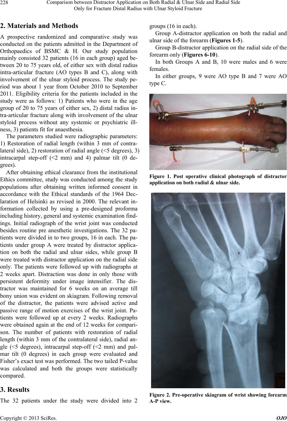 Comparison between Distractor Application on Both Radial & Ulnar Side and Radial Side Only for Fracture Distal Radius with Ulnar Styloid Fracture 228 2. Materials and Methods A prospective randomized and comparative study was conducted on the patients admitted in the Department of Orthopaedics of BSMC & H. Our study population mainly consisted 32 patients (16 in each group) aged be- tween 20 to 75 years old, of either sex with distal radius intra-articular fracture (AO types B and C), along with involvement of the ulnar styloid process. The study pe- riod was about 1 year from October 2010 to September 2011. Eligibility criteria for the patients included in the study were as follows: 1) Patients who were in the age group of 20 to 75 years of either sex, 2) distal radius in- tra-articular fracture along with involvement of the ulnar styloid process without any systemic or psychiatric ill- ness, 3) patients fit for anaesthesia. The parameters studied were radiographic parameters: 1) Restoration of radial length (within 3 mm of contra- lateral side), 2) restoration of radial angle (<5 degrees), 3) intracarpal step-off (<2 mm) and 4) palmar tilt (0 de- grees). After obtaining ethical clearance from the institutional Ethics committee, study was conducted among the study populations after obtaining written informed consent in accordance with the Ethical standards of the 1964 Dec- laration of Helsinki as revised in 2000. The relevant in- formation collected by using a pre-designed proforma including history, general and systemic examination find- ings. Initial radiograph of the wrist joint was conducted besides routine pre anesthetic investigations. The 32 pa- tients were divided in to two groups, 16 in each. The pa- tients under group A were treated by distractor applica- tion on both the radial and ulnar sides, while group B were treated with distractor application on the radial side only. The patients were followed up with radiographs at 2 weeks apart. Distraction was done in only those with persistent deformity under image intensifier. The dis- tractor was maintained for 6 weeks on an average till bony union was eviden t on skiagram. Following removal of the distractor, the patients were advised active and passive range of motion exercises of the wrist joint. Pa- tients were followed up at every 2 weeks. Radiographs were obtained again at the end of 12 weeks for compari- son. The number of patients with restoration of radial length (within 3 mm of the contralateral side), radial an- gle (<5 degrees), intracarpal step-off (<2 mm) and pal- mar tilt (0 degrees) in each group were evaluated and Fisher’s exact test was performed. The two tailed P-value was calculated and both the groups were statistically compared. 3. Results The 32 patients under the study were divided into 2 groups (16 in each). Group A-distractor application on both the radial and ulnar side of the forearm (Figures 1- 5). Group B-distractor application on the radial side of the forearm only (Figures 6-10). In both Groups A and B, 10 were males and 6 were females. In either groups, 9 were AO type B and 7 were AO type C. Figure 1. Post operative clinical photograph of distractor application on both radial & ulnar side. Figure 2. Pre-operative skiagram of wrist showing forearm A-P view. Copyright © 2013 SciRes. OJO 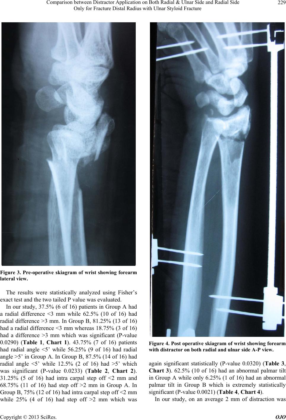 Comparison between Distractor Application on Both Radial & Ulnar Side and Radial Side Only for Fracture Distal Radius with Ulnar Styloid Fracture 229 Figure 3. Pre-operative skiagram of wrist showing forearm lateral view. The results were statistically analyzed using Fisher’s exact test and the two tailed P valu e was evaluated. In our study, 37 .5% (6 of 16) patien ts in Group A had a radial difference <3 mm while 62.5% (10 of 16) had radial difference >3 mm. In Group B, 81.25% (13 of 16) had a radial difference <3 mm whereas 18.75% (3 of 16) had a difference >3 mm which was significant (P-value 0.0290) (Table 1, Chart 1). 43.75% (7 of 16) patients had radial angle <5’ while 56.25% (9 of 16) had radial angle >5 ’ in Group A. I n Group B, 87.5 % (14 of 16) h ad radial angle <5’ while 12.5% (2 of 16) had >5’ which was significant (P-value 0.0233) (Table 2, Chart 2). 31.25% (5 of 16) had intra carpal step off <2 mm and 68.75% (11 of 16) had step off >2 mm in Group A. In Group B, 75% (12 of 16) had intra carpal step off <2 mm while 25% (4 of 16) had step off >2 mm which was Figure 4. Post operative skiagram of wrist showing forearm with distractor on both radial and ulnar side A-P view. again significant statistically (P-value 0.0320) (Table 3, Chart 3). 62.5% (10 of 16) had an abnormal palmar tilt in Group A while only 6 .25% (1 of 16) had an abnormal palmar tilt in Group B which is extremely statistically significant (P-value 0.0021) (Table 4, Chart 4). In our study, on an average 2 mm of distraction was Copyright © 2013 SciRes. OJO  Comparison between Distractor Application on Both Radial & Ulnar Side and Radial Side Only for Fracture Distal Radius with Ulnar Styloid Fracture 230 Figure 5. Post operative skiagram of wrist showing forearm with distractor on both radial and ulnar side lateral view. Figure 6. Post operative clinical photograph of distractor application on radial side. Figure 7. Pre-operative skiagram of wrist showing forearm A-P view. Figure 8. Pre-operative skiagram of wrist showing forearm lateral view. Copyright © 2013 SciRes. OJO 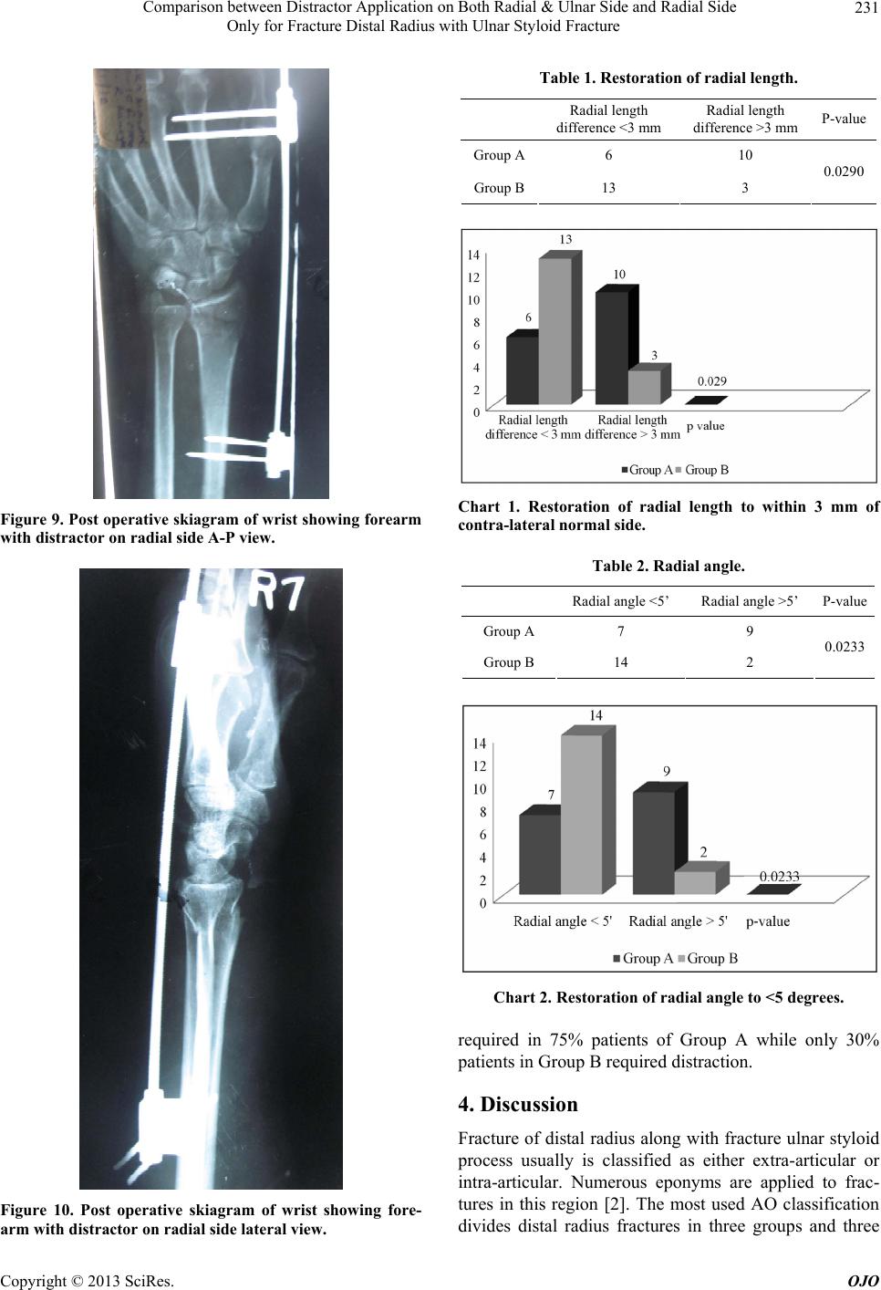 Comparison between Distractor Application on Both Radial & Ulnar Side and Radial Side Only for Fracture Distal Radius with Ulnar Styloid Fracture 231 Figure 9. Post operative skiagram of wrist showing forearm with distractor on radial side A-P view. Figure 10. Post operative skiagram of wrist showing fore- arm with distractor on radial side lateral view. Table 1. Restoration of radial length. Radial length difference <3 mm Radial length difference >3 mm P-value Group A6 10 Group B13 3 0.0290 Chart 1. Restoration of radial length to within 3 mm of contra-lateral normal side. Table 2. Radial angle. Radial angle <5’ Radial angle >5’ P-value Group A 7 9 Group B 14 2 0.0233 Chart 2. Restoration of radial angle to <5 degrees. required in 75% patients of Group A while only 30% patients in Group B required distraction. 4. Discussion Fracture of distal radius along with fracture ulnar styloid process usually is classified as either extra-articular or intra-articular. Numerous eponyms are applied to frac- tures in this region [2]. The most used AO classification divides distal radius fractures in three groups and three Copyright © 2013 SciRes. OJO 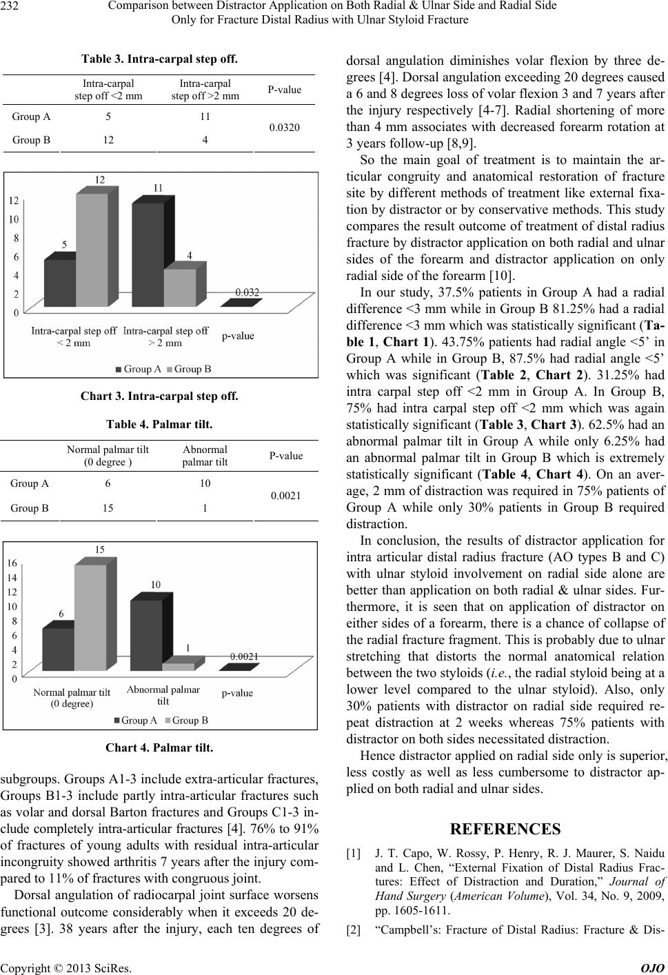 Comparison between Distractor Application on Both Radial & Ulnar Side and Radial Side Only for Fracture Distal Radius with Ulnar Styloid Fracture 232 Table 3. Intra-carpal step off. Intra-carpal step off <2 mm Intra-carpal step off >2 mm P-value Group A 5 11 Group B 12 4 0.0320 Chart 3. Intra-carpal step off. Table 4. Palmar tilt. Normal palmar tilt (0 degree ) Abnormal palmar tilt P-value Group A 6 10 Group B 15 1 0.0021 Chart 4. Palmar tilt. subgroups. Groups A1-3 include extra-articular fractures, Groups B1-3 include partly intra-articular fractures such as volar and dorsal Barton fractures and Groups C1-3 in- clude completely intra-articular fractures [4]. 76% to 91% of fractures of young adults with residual intra-articular incongruity showed arthritis 7 years after the injury com- pared to 11% of fractures with congruous joint. Dorsal angulation of radiocarpal joint surface worsens functional outcome considerably when it exceeds 20 de- grees [3]. 38 years after the injury, each ten degrees of dorsal angulation diminishes volar flexion by three de- grees [4]. Dorsal angulation exceeding 20 degrees caused a 6 and 8 degrees loss of volar flexion 3 and 7 years after the injury respectively [4-7]. Radial shortening of more than 4 mm associates with decreased forearm rotation at 3 years follow-up [8,9]. So the main goal of treatment is to maintain the ar- ticular congruity and anatomical restoration of fracture site by different methods of treatment like external fixa- tion by distractor or by conservative methods. This study compares the result outcome of treatment of distal radius fracture by distractor application on both radial and ulnar sides of the forearm and distractor application on only radial side of the forearm [10]. In our study, 37.5% patients in Group A had a radial difference <3 mm wh ile in Group B 81.25% had a radial difference <3 mm which was statistically significant (Ta- ble 1, Chart 1). 43.75% patients had radial angle <5’ in Group A while in Group B, 87.5% had radial angle <5’ which was significant (Table 2, Chart 2). 31.25% had intra carpal step off <2 mm in Group A. In Group B, 75% had intra carpal step off <2 mm which was again statistically significant (Tab le 3, Chart 3). 62.5% had an abnormal palmar tilt in Group A while only 6.25% had an abnormal palmar tilt in Group B which is extremely statistically significant (Table 4, Chart 4). On an aver- age, 2 mm of distraction was required in 75% patients of Group A while only 30% patients in Group B required distraction. In conclusion, the results of distractor application for intra articular distal radius fracture (AO types B and C) with ulnar styloid involvement on radial side alone are better than application on both radial & ulnar sides. Fur- thermore, it is seen that on application of distractor on either sides of a forearm, there is a chance of collapse of the radial fracture fragment. This is probably due to ulnar stretching that distorts the normal anatomical relation between the two styloids (i.e., the radial styloid being at a lower level compared to the ulnar styloid). Also, only 30% patients with distractor on radial side required re- peat distraction at 2 weeks whereas 75% patients with distractor on both sides necessitated distraction. Hence distractor applied on radial side only is superior, less costly as well as less cumbersome to distractor ap- plied on both radial and ulnar sides. REFERENCES [1] J. T. Capo, W. Rossy, P. Henry, R. J. Maurer, S. Naidu and L. Chen, “External Fixation of Distal Radius Frac- tures: Effect of Distraction and Duration,” Journal of Hand Surgery (American Volume), Vol. 34, No. 9, 2009, pp. 1605-1611. [2] “Campbell’s: Fracture of Distal Radius: Fracture & Dis- Copyright © 2013 SciRes. OJO 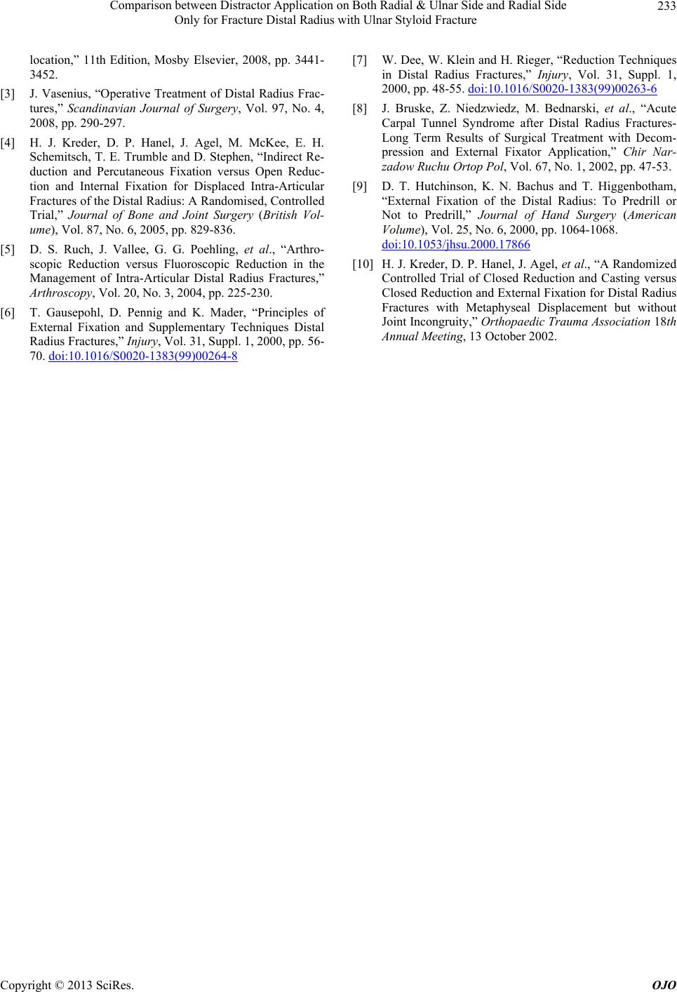 Comparison between Distractor Application on Both Radial & Ulnar Side and Radial Side Only for Fracture Distal Radius with Ulnar Styloid Fracture Copyright © 2013 SciRes. OJO 233 location,” 11th Edition, Mosby Elsevier, 2008, pp. 3441- 3452. [3] J. Vasenius, “Operative Treatment of Distal Radius Frac- tures,” Scandinavian Journal of Surgery, Vol. 97, No. 4, 2008, pp. 290-297. [4] H. J. Kreder, D. P. Hanel, J. Agel, M. McKee, E. H. Schemitsch, T. E. Trumble and D. Stephen, “Indirect Re- duction and Percutaneous Fixation versus Open Reduc- tion and Internal Fixation for Displaced Intra-Articular Fractures of the Distal Radius: A Randomised, Controlled Trial,” Journal of Bone and Joint Surgery (British Vol- ume), Vol. 87, No. 6, 2005, pp. 829-836. [5] D. S. Ruch, J. Vallee, G. G. Poehling, et al., “Arthro- scopic Reduction versus Fluoroscopic Reduction in the Management of Intra-Articular Distal Radius Fractures,” Arthroscopy, Vol. 20, No. 3, 2004, pp. 225-230. [6] T. Gausepohl, D. Pennig and K. Mader, “Principles of External Fixation and Supplementary Techniques Distal Radius Fractures,” Injury, Vol. 31, Suppl. 1, 2000, pp. 56- 70. doi:10.1016/S0020-1383(99)00264-8 [7] W. Dee, W. Klei n and H. Rieger, “Reduction Techniques in Distal Radius Fractures,” Injury, Vol. 31, Suppl. 1, 2000, pp. 48-55. doi:10.1016/S0020-1383(99)00263-6 [8] J. Bruske, Z. Niedzwiedz, M. Bednarski, et al., “Acute Carpal Tunnel Syndrome after Distal Radius Fractures- Long Term Results of Surgical Treatment with Decom- pression and External Fixator Application,” Chir Nar- zadow Ruchu Ortop Pol, Vol. 67, No. 1, 2002, pp. 47-53. [9] D. T. Hutchinson, K. N. Bachus and T. Higgenbotham, “External Fixation of the Distal Radius: To Predrill or Not to Predrill,” Journal of Hand Surgery (American Volume), Vol. 25, No. 6, 2000, pp. 1064-1068. doi:10.1053/jhsu.2000.17866 [10] H. J. Kreder, D. P. Hanel, J. Agel, et al., “A Randomized Controlled Trial of Closed Reduction and Casting versus Closed Reduction and External Fixation for Distal Radius Fractures with Metaphyseal Displacement but without Joint Incongruity,” Orthopaedic Trauma Association 18th Annual Meeting, 13 October 2002. |