

Paper Menu >>
Journal Menu >>
 Open Journal of Gastroenterology, 2013, 3, 281-287 OJGas http://dx.doi.org/10.4236/ojgas.2013.35048 Published Online September 2013 (http://www.scirp.org/journal/ojgas/) Management of ac ute esophageal variceal b le ed i ng by endoscopic sclerotherapy in technically difficult endoscopic band ligation cases—A population based cohort study Gamal E. Esmat1, Iman M. Hamza1*, Bahaa E. Abbas2, Ahmed M. Hashem1, Hossam S. Ghoneim3 1Endemic Medicine Department, Division of Endoscopy, Cairo University, Giza, Egypt 2Division of Endoscopy, Air Force Hospital, Cairo, Egypt 3Tropical Medicine Department, Beni Sweif University, Cairo, Egypt Email: *[email protected] Received 14 June 2013; revised 15 July 2013; accepted 29 July 2013 Copyright © 2013 Gamal E. Esmat et al. This is an open access article distributed under the Creative Commons Attribution License, which permits unrestricted use, distribution, and reproduction in any medium, provided the original work is properly cited. ABSTRACT Endoscopic band ligation is regarded as the main therapeutic option for acute esophageal variceal bleed- ing, while sclerotherapy may be used in the acute set- ting if ligation is technically difficult. The incidence of difficult-to-perform band ligation in acute esophageal variceal bleeding, as well as the outcome of patients subjected to injection sclerotherapy as an alternative treatment, has not been clearly investigated. Our aim is to study the outcome of patients subjected to injec- tion sclerotherapy in the acute setting of esophageal variceal bleeding when endoscopic band ligation is technically difficult to perform. We included 151 pa- tients with acute esophageal variceal bleeding origin- nating from medium or large sized varices. All pa- tients were planned for EBL as the 1st treatment op- tion (EBL group 61.6%), meanwhile, EIS using 5% ethanolamine oleate was reserved as the 2nd treat- ment option when EBL was technically difficult (EIS group 38.4%). The mean time to restore hemody- namic stability was significantly prolonged in the EIS group (11.5 ± 6.5 hrs versus 9.5 ± 5.0 hrs, p 0.05). Ini- tial control of bleeding was significantly higher in the EBL group versus the EIS group (96.7% vs 84.5%, p 0.021). Re-bleeding was more among the EIS group (42.9% vs 24.2%, p 0.04). There were no significant differences as regarding mortality and duration of hospital stay. So, a considerable proportion of cases presenting with acute variceal bleeding will have dif- ficulty in performing EBL. In these patients, sclero- therapy is not a waning procedure with an accepted success rate, without much additional complications and without dera nging mortality. Keywords: Esophageal Varices; Band Ligation; Injection Sclerotherapy 1. INTRODUCTION The management of acute variceal bleeding remains a clinical challenge. Regarding the best endoscopic thera- peutic option, a meta-analysis of 10 randomized con- trolled trials including 404 patients showed an almost significant benefit of endoscopic band ligation in the initial control of bleeding compared to sclerotherapy [1]. At the same time, Baveno IV and V consensuses had re- peatedly confirmed that ligation is the recommended me- thod of endoscopic therapy for acute esophageal variceal bleeding although sclerotherapy may be used in the acute setting if ligation is technically difficult [2,3]. The incidence of difficulty to perform band ligation in acute esophageal variceal bleeding, as well as the out- come of patients subjected to injection sclerotherapy as an alternative treatment, has not been clearly investi- gated. 2. AIM OF THE WORK Our aim is to study the outcome of patients subjected to injection sclerotherapy in the setting of acute esophageal variceal bleeding in case of technical difficulty to per- form band ligation. 3. PATIENTS AND METHODS This study was approved by the Hepatogastroenterology Department and the Research Board of Cairo University. It included 151 patients with acute bleeding from eso- phageal varices originating from medium and large sized varices. They were admitted to Gastroenterology unit of *Corresponding author. OPEN ACCESS  G. E. Esmat et al. / Open Journal of Gastroenterology 3 (2013) 281-287 282 Air force Hospital, Cairo; during the period from 15 January 2010 till 15 July same year. Patients with bleed- ing gastric varices, bleeding small sized oesophageal varices, bleeding portal hypertensive gastropathy, hepatic coma or hepatocellular carcinoma were not included in the study. Patients were resuscitated after relevant history taking and clinical examination. Stabilization of hemodynamics was firstly performed. Blood transfusion was initiated depending upon the severity of bleeding and hemo-dy- namic status with a transfusion target hematocrit of 24% and/or hemoglobin of 8 g/dl. I.V antibiotics were rou- tinely given. Octreotide was started whenever indicated in a dose of 50 μg IV bolus, followed by continuous in- fusion 50 μg/hr for 2 - 5 days. Further assessment was done including laboratory investigations and abdominal ultrasonography. The severity of bleeding was consid- ered as: mild when pulse rate <100/min, orthostatic hy- potension or cold extremities, moderate when pulse rate 100 - 120 beats/min with restlessness or severe when pulse rate >120 beats/min, systolic blood pressure <60 mmHg, severe pallor or oliguria [4]. Endoscopy session was scheduled once the patient’s hemodynamics permit- ted after an informed and written consent. Emergency endotherapy was performed by a single endoscopist, all patients were planned for endoscopic band ligation (EBL) using the Saeed multi-band ligators manufactured by the Wilson-Cook Medical GI endoscopy company as the 1st treatment option to control the acute bleeding episode (EBL group) (Figure 1), meanwhile, endoscopic injec- tion sclerotherapy (EIS) using ethanolamine oleate was reserved as a 2nd treatment option when EBL was tech- nically difficult (only when the view of the bleeding source was obscured) (sclerotherapy group) (Figure 2). Varices were assigned grades according to their shape and size: grade F1, small straight veins; grade F2, Figure 1. EVL to bleeding varices. Figure 2. Sclerotherapy for bleeding varices. slightly enlarged tortuous veins occupying less than one- third of the esophageal lumen; and grade F3, large coil- shaped varices that occupied more than one-third of the esophageal lumen [5]. The assessment for treatment side-effects was per- formed during hospital stay and included monitoring of symptoms, physical examination and laboratory data. Complications were defined as untoward events related to treatment and require active therapy or prolonged hos- pitalization. All available patients in both groups were followed up for 42 days for clinical assessment, variceal re-bleeding, and hepatic de-compensation. Failure to control active bleeding was considered according to the UK guidelines [6] as transfusion requirement of 4 units or more and inability to achieve an increase in systolic blood pressure by 20 mm Hg or to 70 mm Hg or more, and/or inability to achieve a pulse rate reduction to less than 100 beat/min or a reduction of 20 beat/min from baseline pulse rate (within the first six hours). Or the occurrence of haematemesis from the six hour point, reduction in blood pressure of more than 20 mm Hg from the six hour point and/or increase in pulse rate of more than 20 beat/min from the six hour point on two con- secutive readings an hour apart, transfusion of 2 units of blood or more (over and above the previous transfusions) required to increase the haematocrit to above 27%, or haemoglobin to above 9 g/l. Re-bleeding: was considered as the occurrence of new hematemesis or melena after a period of 24 hours or more of stable vital signs and he- matocrit/hemoglobin following an episode of acute bleed- ing up to 42 days from the acute bleeding episode. Stop- page of bleeding: is considered when there is no he- matemesis, stable hemoglobin concentration without blood transfusions, stable hemodynamic conditions at the be- ginning of the first 24-hour interval from time zero [2]. Copyright © 2013 SciRes. OPEN ACCESS  G. E. Esmat et al. / Open Journal of Gastroenterology 3 (2013) 281-287 Copyright © 2013 SciRes. 283 OPEN ACCESS Patients’ data were tabulated and processed using SPSS (10.0) statistical package. Quantitative variables were expressed by means and standard deviation. While qualitative data were expressed by frequency and percent. Qualitative variables were analyzed using Chi-square or Ficher’s exact test when appropriate. Quantitative vari- ables were analyzed using student’s T-test or Fried- mann’s test when appropriate. In all tests p value was significant when <0.05. 4. RESULTS This study included 151 patients with acute esophageal variceal bleeding. They were sub-grouped based on the type of endoscopic intervention into EBL (band ligation) group (93 patients 61.6%) and EIS (sclerotherapy group) (58 patients 38.4%). The majority of patients in the EBL group had a mild bleeding episode (64.5%) versus none in the sclerother- apy group. While the majority of those in the scerother- apy group had a moderate bleeding episode (67.2%, p 0.38). Time from admission to patient stabilization was significantly prolonged in the EIS group 11.5 ± 6.5 hrs versus 9.5 ± 5.0 hrs (p 0.05) (Tab le 1). The post endo- scopy outcome is demonstrated in (Table 2). 5. DISCUSSION Bleeding from esophageal varices is a life-threatening complication of portal hypertension which accounts for most of cirrhosis-related mortalities [7,8]. Endoscopic therapy is an integral component of the management of acute variceal bleeding as well as prevention of recurrent bleeding [8,9]. Since it first evolved as a new therapeutic option, EBL has been regarded as a more effective mo- dality than endoscopic injection sclerotherapy in the con- trol of acute hemorrhage with less reported adverse events [10-12]. On the other hand, some studies still ad- vocate the use of sclerotherapy as a better treatment op- tion up to regarding it as the gold-standard treatment [13,14]. Despite the routine standard of care given to patients presenting with hematemesis such as pharmacotherapy and gastric lavage, still up to one third of cases are ac- tively bleeding during endoscopy, and it is this group of patients that poses a challenge to endoscopists and en- Table 1. Base-line characteristics of EIS and EVL groups. EVL group N: 93 (61.6%) EIS Group N: 58 (38.4%) Parameters No/% Mean/SD No/% Mean/SD p Age (years) 52 ±3 53 ±3 0.89 Sex Male 52 56% 42 72.5% Female 41 44% 16 27.5% 0.33 Previous bleeding attacks 48 51.6% 33 56.9% 0.63 Child Pugh class: A 9 9.7% 4 6.9% B 37 39.8% 25 43.1% C 47 50.5% 29 50% 0.057 Grade of varices Grade F2 48 51.6% 28 48.2% Grade F3 45 48% 30 51.7%% 0.85 Hemodynamic instability 81 87.1% 53 91.4% 0.53 Severity of bleeding Mild 60 64.5% 0 0% Moderate 24 25.8% 39 67.2% Severe 9 9.7% 19 32.8% <0.01 Time from admission to endoscopy (hrs) 9.5 ±5 11.5 ±6.5 0.05  G. E. Esmat et al. / Open Journal of Gastroenterology 3 (2013) 281-287 284 Table 2. Post-endoscopy outcome in both groups. EVL group N: 93 (61.6%) EIS Group N: 58 (38.4%) Parameters No/% Mean/SD No/% Mean/SD p Control of acute bleeding 90 96.7% 49 84.5% 0.021 Re-bleeding 22 24.2% 21 42.9% 0.04 Control of re-bleeding 17/22 77.35 11/19 57.9% 0.23 Incidence of complications among different child-Pugh cases Child A 1 1.1% 2 3.4% Child B 8 8.6% 14 24.1% Child C 6 6.4% 13 22.4% 0.079 Bacterial peritonitis 6 6.4% 11 19% 0.01 Aspiration pneumonia 0 0% 1 1.7% 0.41 Sepsis 1 1.1% 2 3.4% 0.065 Bleeding esophageal ulcer 1 1.1% 2 3.4% 0.065 Chest pain 2 2.2% 5 8.6% 0.01 Fever 1 1.1% 2 3.4% 0.065 Transient arrhythmias 1 1.1% 1 1.7% 0.1 Nausea 1 1.1% 2 3.4% 0.068 Transient dysphagia 2 2.2% 3 5.2% 0.012 Mean hemoglobin (gm/dl) Day 1 8.6 ±0.34 8.1 ±0.36 0.18 Day 5 8.8 ±0.34 8.4 ±0.31 0.45 Units of packed RBCs 3.1 ±1.07 4.1 ±1.2 0.01 Hospital stay in days 8.3 ±1.9 9.1 ±2.6 0.27 Total Mortality 19 20.4% 21 36.2% 0.19 dangers the life of the patient as it has been found to have a negative impact on survival [7,15,16]. In this situation banding may be technically difficult to perform in the presence of continued bleeding, and sclerotherapy may then be necessary [2,3,9,17]. So this means that, whether endoscopists fall for band ligation or sclero- therapy as the preferred treatment, the set up of an emer- gency endoscopic unit should be equipped with both and the endoscopists should be ready to switch between the 2 modalities. How frequent is this situation encountered, what is the outcome and what are the sequaele, remained to be in- vestigated. This was the rational to conduct this study. It was carried in the Air Force hospital on 151 patients with acute variceal bleeding. All included patients had acute esophageal variceal bleeding from medium or large sized varices, while other portal hypertensive bleeding sources were not included. To ensure homogeneity among these patients, they were all given the same standards of care such as gastric lavage and pharmacotherapy with oc- terotide and judicious volume expanders. All of them underwent endoscopy whenever they reached hemodya- mic stability irrespective of time and all were endo- scoped by a single operator. The plan at the time of en- doscopy was to perform band ligation as a primary treat- ment option unless profuse bleeding was encountered to the extent of posing technical difficulty in visualizing and banding the bleeding varix, in the latter condition, switch to sclerotherapy was done. The studied cases in both groups were comparable in terms of their demographic features. All patients had received pharmacotherapy by octerotide and yet, 38.4% of the studied cases were actively bleeding at endoscopy, profusely enough, to change the decision of treatment Copyright © 2013 SciRes. OPEN ACCESS 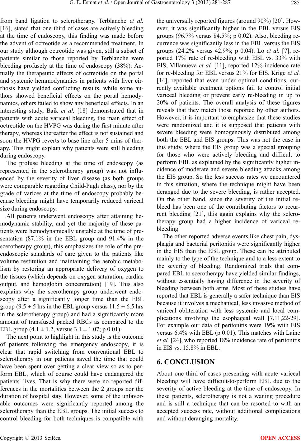 G. E. Esmat et al. / Open Journal of Gastroenterology 3 (2013) 281-287 285 from band ligation to sclerotherapy. Terblanche et al. [16], stated that one third of cases are actively bleeding at the time of endoscopy, this finding was made before the advent of octreotide as a recommended treatment. In our study although octreotide was given, still a subset of patients similar to those reported by Terblanche were bleeding profusely at the time of endoscopy (38%). Ac- tually the therapeutic effects of octreotide on the portal and systemic hemmodynamics in patients with liver cir- rhosis have yielded conflicting results, while some au- thors showed beneficial effects on the portal hemody- namics, others failed to show any beneficial effects. In an interesting study, Baik et al. [18] demonstrated that in patients with acute variceal bleeding, the main effect of octreotide on the HVPG was during the first minute after therapy, whereas thereafter the effect is not sustained and soon the HVPG reverts to base line after 5 mins of ther- apy. This might explain why patients were still bleeding during endoscopy. The profuse bleeding at the time of endoscopy (as represented in the sclerotherapy group) was not influ- enced by the severity of liver disease (as both groups were comparable regarding Child-Pugh class), nor by the grade of varices at the time of endoscopy probably be- cause bleeding might have temporarily reduced variceal size during endoscopy. All patients underwent endoscopy after attaining he- modynamic stability, and yet the majority of these pa- tients were hemodynamically unstable at the time of pre- sentation (87.1% in the EBL group and 91.4% in the scerotherapy group), this emphasizes the role of the pre- endoscopic standards of care given to the patients like volume restitution and maintaining the aerobic metabo- lism by restoring an appropriate delivery of oxygen to the tissues (which depends on oxygen saturation, cardiac output, and hemoglobin concentration) [19]. This also explains why the scerotherapy group underwent endo- scopy after a significantly longer time than the EBL group (9.5 ± 5 hrs in the EBL group versus 11.5 ± 6.5 hrs in the sclerotherapy group) and had a significantly more amount of transfused packed RBCs as compared to the EBL group (4.1 ± 1.2, versus 3.1 ± 1.07; p 0.01). The next point to highlight in this study is the outcome of patients following the emergency endoscopy, it is clear that rapid switching from conventional EBL to sclerotherapy in our patients saved the time that could have been spent over getting a clear view so as to per- form EBL, which of course could have endangered the patients' lives. That is why there were no reported dif- ferences in the mortalities between the 2 groups nor the duration of hospital stay. However, some of the unfavor- able outcomes were significantly reported among the sclerotherapy than the EBL groups. The initial success to control bleeding for both techniques is compatible with the universally reported figures (around 90%) [20]. How- ever, it was significantly higher in the EBL versus EIS groups (96.7% versus 84.5%; p 0.02). Also, bleeding re- currence was significantly less in the EBL versus the EIS groups (24.2% versus 42.9%; p 0.04). Lo et al. [7], re- ported 17% rate of re-bleeding with EBL vs. 33% with EIS, Villanueva et al. [11], reported 12% incidence rate for re-bleeding for EBL versus 21% for EIS. Krige et al. [14], reported that even under optimal conditions, cur- rently available treatment options fail to control initial variceal bleeding or prevent early re-bleeding in up to 20% of patients. The overall analysis of these figures reveals that they match those reported by other authors. However, it is important to emphasize that these studies were randomized and it is supposed that patients with severe bleeding were homogenously distributed among both the EBL and EIS groups. This was not the case in this study, where the EIS group was a special grouping for those who were actively bleeding and difficult to perform EBL as explained by the significantly higher in- cidence of moderate and severe bleeding attacks among the EIS group. So the less success rates we encountered in this situation, where the technique might have been deranged due to the severe bleeding, is rather accepted. On the other hand, since the severity of the initial re- bleed has been one of the contributing factors to recur- rent bleeding [21], this again explains why the sclero- therapy group had a higher incidence of variceal re- bleeding. The other reported adverse events like chest pain, dys- phagia and bacterial peritonitis were significantly higher in the EIS than the EBL group. These can be attributed mainly to the type of the technique and to a less extent to the severity of bleeding. Randomized trials that com- pared EBL to scerotherapy have yielded similar findings, without essentially having difference in the severity of bleeding between both arms. Most of these studies have reported that EBL is generally a safer technique than EIS because it involves a mechanical, less invasive method of variceal obliteration with less systemic and local com- plications involving the esophageal wall [7,11,22-29]. For example our data of peritonitis were 19% with EIS versus 6.4% with EBL (p 0.01). This matches with Laine et al. [24], who reported 18% incidence rate of peritonitis in EIS vs. 15.8% in EBL. 6. CONCLUSION About one third of cases presenting with acute variceal bleeding will have difficult-to-perform EBL due to the severity of active bleeding at the time of endoscopy. In these patients, sclerotherapy is not a waning procedure and is still a technique that can be resorted to with an accepted success rate, without additional complications and without deranging mortality. Copyright © 2013 SciRes. OPEN ACCESS 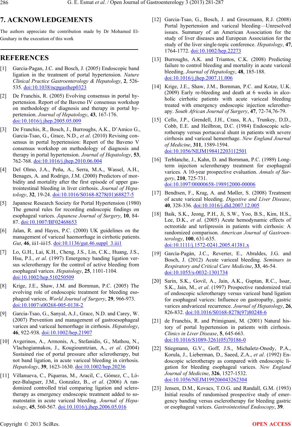 G. E. Esmat et al. / Open Journal of Gastroenterology 3 (2013) 281-287 286 7. ACKNOWLEDGEMENTS The authors appreciate the contribution made by Dr Mohamed El- Gouhary in the execution of this work REFERENCES [1] Garcia-Pagan, J.C. and Bosch, J. (2005) Endoscopic band ligation in the treatment of portal hypertension. Nature Clinical Practice Gastroenterology & Hepatology, 2, 526- 535. doi:10.1038/ncpgasthep0323 [2] De Franchis, R. (2005) Evolving consensus in portal hy- pertension. Report of the Baveno IV consensus workshop on methodology of diagnosis and therapy in portal hy- pertension. Journal of Hepatology, 43, 167-176. doi:10.1016/j.jhep.2005.05.009 [3] De Franchis, R., Bosch, J., Burroughs, A.K., D’Amico G., Garcia-Tsao, G., Grace, N.D., et al. (2010) Revising con- sensus in portal hypertension: Report of the Baveno V consensus workshop on methodology of diagnosis and therapy in portal hypertension. Journal of Hepatology, 53, 762-768. doi:10.1016/j.jhep.2010.06.004 [4] Del Olmo, J.A., Peña, A., Serra, M.A., Wassel, A.H., Benages, A. and Rodrigo, J.M. (2000) Predictors of mor- bidity and mortality after the first episode of upper gas- trointestinal bleeding in liver cirrhosis. Journal of Hepa- tology, 32, 19-24. doi:10.1016/S0168-8278(01)68827-5 [5] Japanese Research Society for Portal Hypertension (1980) The general rules for recording endoscopic findings on esophageal varices. Japanese Journal of Surgery, 10, 84- 87. doi:10.1007/BF02468653 [6] Jalan, R. and Hayes, P.C. (2000) UK guidelines on the management of variceal haemorrhage in cirrhotic patients. Gut, 46, iii1-iii15. doi:10.1136/gut.46.suppl_3.iii1 [7] Lo, G.H., Lai, K.H., Cheng, J.S., Lin, C.K., Huang, J.S., Hsu, P.I., et al. (1997) Emergency banding ligation ver- sus sclerotherapy for the control of active bleeding from esophageal varices. Hepatology, 25, 1101-1104. doi:10.1002/hep.510250509 [8] Krige, J.E., Shaw, J.M. and Bornman, P.C. (2005) The evolving role of endoscopic treatment for bleeding eso- phageal varices. World Journal of Surgery, 29, 966-973. doi:10.1007/s00268-005-0138-2 [9] Garcia-Tsao, G., Sanyal, A.J., Grace, N.D. and Carey, W. (2007) Prevention and management of gastroesophageal varices and variceal hemorrhage in cirrhosis. Hepatology, 46, 922-938. doi:10.1002/hep.21907 [10] Avgerinos, A., Armonis, A., Stefanidis, G., Mathou, N., Vlachogiannakos, J., Kougioumtzian, A., et al. (2004) Sustained rise of portal pressure after sclerotherapy, but not band ligation, in acute variceal bleeding in cirrhosis. Hepatology, 39, 1623-1630. doi:10.1002/hep.20236 [11] Villanueva, C., Piqueras, M., Aracil, C., Gómez, C., Ló- pez-Balaguer, J.M., Gonzalez, B., et al. (2006) A ran- domized controlled trial comparing ligation and sclero- therapy as emergency endoscopic treatment added to so- matostatin in acute variceal bleeding. Journal of Hepa- tology, 45, 560-567. doi:10.1016/j.jhep.2006.05.016 [12] Garcia-Tsao, G., Bosch, J. and Groszmann, R.J. (2008) Portal hypertension and variceal bleeding—Unresolved issues. Summary of an American Association for the study of liver diseases and European Association for the study of the liver single-topic conference. Hepatology, 47, 1764-1772. doi:10.1002/hep.22273 [13] Burroughs, A.K. and Triantos, C.K. (2008) Predicting failure to control bleeding and mortality in acute variceal bleeding. Journal of Hepatology, 48, 185-188. doi:10.1016/j.jhep.2007.11.006 [14] Krige, J.E., Shaw, J.M., Bornman, P.C. and Kotze, U.K. (2009) Early re-bleeding and death at 6 weeks in alco- holic cirrhotic patients with acute variceal bleeding treated with emergency endoscopic injection sclerother- apy. South African Journal of Surgery, 47, 72-74,76-79. [15] Cello, J.P., Grendell, J.H., Crass, R.A., Trunkey, D.D., Cobb, E.E. and Heilbron, D.C. (1984) Endoscopic scle- rotherapy versus portacaval shunt in patients with severe cirrhosis and variceal hemorrhage. New England Journal of Medicine, 311, 1589-1594. doi:10.1056/NEJM198412203112501 [16] Terblanche, J., Kahn, D. and Bornman, P.C. (1989) Long- term injection sclerotherapy treatment for esophageal varices. A 10-year prospective evaluation. Annals of Sur- gery, 210, 725-731. doi:10.1097/00000658-198912000-00006 [17] Bendtsen, F., Krag, A. and Møller, S. (2008) Treatment of acute variceal bleeding. Digestive and Liver Disease, 40, 328-336. doi:10.1016/j.dld.2007.12.005 [18] Baik, S.K., Jeong, P.H., Ji, S.W., Yoo, B.S., Kim, H.S., Lee, D.K., et al. (2005) Acute hemodynamic effects of octreotide and terlipressin in patients with cirrhosis: A randomized comparison. American Journal of Gastroen- terology, 100, 631-635. doi:10.1111/j.1572-0241.2005.41381.x [19] García-Pagán, J.C., Reverter, E., Abraldes, J.G. and Bosch, J. (2012) Acute variceal bleeding. Seminars in Respiratory and Critical Care Medicine, 33, 46-54. doi:10.1055/s-0032-1301734 [20] Sarin, S.K., Govil, A., Jain, A.K., Guptan, R.C., Issar, S.K., Jain, M., et al. (1997) Prospective randomized trial of endoscopic sclerotherapy versus variceal band ligation for esophageal varices: Influence on gastropathy, gastric varices andvariceal recurrence. Journal of Hepatology, 26, 826-832. doi:10.1016/S0168-8278(97)80248-6 [21] de Franchis, R. and Primignani, M. (2001) Natural his- tory of portal hypertension in patients with cirrhosis. Clinics in Liver Disease, 5, 645-663. doi:10.1016/S1089-3261(05)70186-0 [22] Stiegmann, G.V., Goff, J.S., Michaletz-Onody, P.A., Korula, J., Lieberman, D., Saeed, Z.A., et al. (1992) En- doscopic sclerotherapy as compared with endoscopic li- gation for bleeding esophageal varices. New England Journal of Medicine, 326, 1527-1532. doi:10.1056/NEJM199206043262304 [23] Jensen, D.M., Kovacs, T.O.G. and Randall, G.M. (1993) Initial results of randomised prospective study of emer- gency banding versus esclerotherapy for bleeding gastric or esophageal varices. Gastrointestinal Endoscopy, 39. Copyright © 2013 SciRes. OPEN ACCESS  G. E. Esmat et al. / Open Journal of Gastroenterology 3 (2013) 281-287 Copyright © 2013 SciRes. 287 OPEN ACCESS [24] Laine, L., El-Newihi, H.M. and Migikovsky, B. (1993) Endoscopic ligation compared with sclerotherapy for the treatment of bleeding esophageal varices. Annals of In- ternal Medicine, 119, 1-7. doi:10.7326/0003-4819-119-1-199307010-00001 [25] Lo, G.H., Lai, K.H., Chang, C.F., Shen, M.T., Jeng, J.S., Huang, R.L., et al. (1994) Endoscopic injection sclero- therapy vs. endoscopic variceal ligation in arresting acute variceal bleeding for patients with advanced hepatocellu- lar carcinoma. Journal of Hepatology, 21, 1048-1052. doi:10.1016/S0168-8278(05)80616-6 [26] De la Peña, J., Rivero, M., Sanchez, E., Fábrega, E., Cre- spo, J. and Pons-Romero, F. (1999) Variceal ligation compared with endoscopic sclerotherapy for variceal hemorrhage: Prospective randomized trial. Gastrointes- tinal Endoscopy , 49, 417-423. doi:10.1016/S0016-5107(99)70036-2 [27] Patch, D. and Dagher, L. (2001) Acute variceal bleeding: General management. World Journal of Gastroenterology, 7, 466-475. [28] Zargar, S.A., Javid, G., Khan, B.A., Shah, O.J., Yattoo, G.N., Shah, A.H., et al. (2005) Endoscopic ligation vs. sclerotherapy in adults with extrahepatic portal venous obstruction: A prospective randomized study. Gastroin- testinal Endoscopy, 61, 58-66. doi:10.1016/S0016-5107(04)02455-1 [29] Xu, L., Ji, F., Xu, Q.-W. and Zhang, M.-Q. (2011) Risk factors for predicting early variceal re-bleeding after en- doscopic variceal ligation. World Journal of Gastroen- terology, 17, 3347-3352. doi:10.3748/wjg.v17.i28.3347 |