

Paper Menu >>
Journal Menu >>
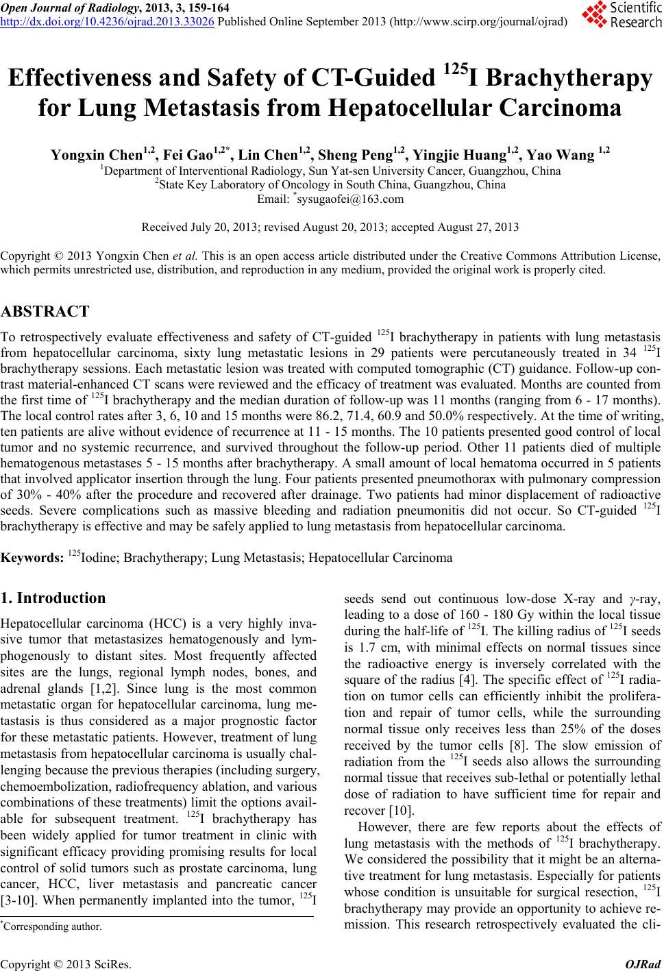 Open Journal of Radiology, 2013, 3, 159-164 http://dx.doi.org/10.4236/ojrad.2013.33026 Published Online September 2013 (http://www.scirp.org/journal/ojrad) Effectiveness and Safety of CT-Guided 125I Brachytherapy for Lung Metastasis from Hepatocellular Carcinoma Yongxin Chen1,2, Fei Gao1,2*, Lin Chen1,2, Sheng Peng1,2, Yingjie Huang1,2, Yao Wang 1,2 1Department of Interventional Radiology, Sun Yat-sen University Cancer, Guangzhou, China 2State Key Laboratory of Oncology in South China, Guangzhou, China Email: *[email protected] Received July 20, 2013; revised August 20, 2013; accepted August 27, 2013 Copyright © 2013 Yongxin Chen et al. This is an open access article distributed under the Creative Commons Attribution License, which permits unrestricted use, distribution, and reproduction in any medium, provided the original work is properly cited. ABSTRACT To retrospectively evaluate effectiveness and safety of CT-guided 125I brachytherapy in patients with lung metastasis from hepatocellular carcinoma, sixty lung metastatic lesions in 29 patients were percutaneously treated in 34 125I brachytherapy sessions. Each metastatic lesion was treated with computed tomographic (CT) guidance. Follow-up con- trast material-enhanced CT scans were reviewed and the efficacy of treatment was evaluated. Months are counted from the first time of 125I brachytherapy and the median duration of follow-up was 11 months (ranging from 6 - 17 months). The local control rates after 3, 6, 10 and 15 months were 86.2, 71.4, 60.9 and 50.0% respectively. At the time of writing, ten patients are alive without evidence of recurrence at 11 - 15 months. The 10 patients presented good control of local tumor and no systemic recurrence, and survived throughout the follow-up period. Other 11 patients died of multiple hematogenous metastases 5 - 15 months after brachytherapy. A small amount of local hematoma occurred in 5 patients that involved applicator insertion through the lung. Four patients presented pneumothorax with pulmonary compression of 30% - 40% after the procedure and recovered after drainage. Two patients had minor displacement of radioactive seeds. Severe complications such as massive bleeding and radiation pneumonitis did not occur. So CT-guided 125I brachytherapy is effective and may be safely applied to lung metastasis from hepatocellular carcinoma. Keywords: 125Iodine; Brachytherapy; Lung Metastasis; Hepatocellular Carcinoma 1. Introduction Hepatocellular carcinoma (HCC) is a very highly inva- sive tumor that metastasizes hematogenously and lym- phogenously to distant sites. Most frequently affected sites are the lungs, regional lymph nodes, bones, and adrenal glands [1,2]. Since lung is the most common metastatic organ for hepatocellular carcinoma, lung me- tastasis is thus considered as a major prognostic factor for these metastatic patients. However, treatment of lung metastasis from hepatocellular carcinoma is usually chal- lenging because the previous therapies (including surgery, chemoembolization, radiofrequency ablation, and various combinations of these treatments) limit the options avail- able for subsequent treatment. 125I brachytherapy has been widely applied for tumor treatment in clinic with significant efficacy providing promising results for local control of solid tumors such as prostate carcinoma, lung cancer, HCC, liver metastasis and pancreatic cancer [3-10]. When permanently implanted into the tumor, 125I seeds send out continuous low-dose X-ray and γ-ray, leading to a dose of 160 - 180 Gy within the local tissue during the half-life of 125I. The killing radius of 125I seeds is 1.7 cm, with minimal effects on normal tissues since the radioactive energy is inversely correlated with the square of the radius [4]. The specific effect of 125I radia- tion on tumor cells can efficiently inhibit the prolifera- tion and repair of tumor cells, while the surrounding normal tissue only receives less than 25% of the doses received by the tumor cells [8]. The slow emission of radiation from the 125I seeds also allows the surrounding normal tissue that receives sub-lethal or potentially lethal dose of radiation to have sufficient time for repair and recover [10]. However, there are few reports about the effects of lung metastasis with the methods of 125I brachytherapy. We considered the possibility that it might be an alterna- tive treatment for lung metastasis. Especially for patients whose condition is unsuitable for surgical resection, 125I brachytherapy may provide an opportunity to achieve re- mission. This research retrospectively evaluated the cli- *Corresponding author. C opyright © 2013 SciRes. OJRad 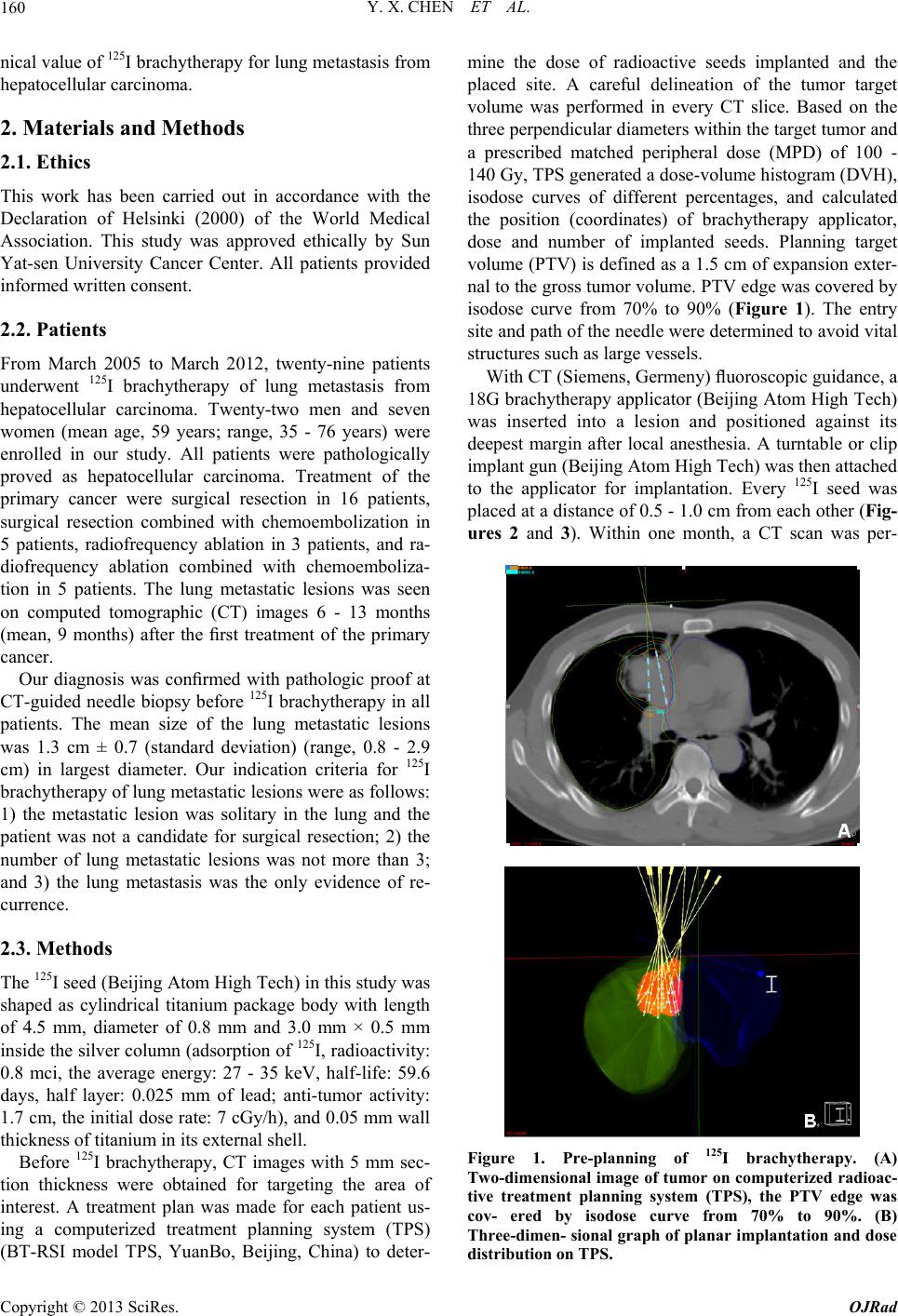 Y. X. CHEN ET AL. 160 nical value of 125I brachytherapy for lung metastasis from hepatocellular carcinoma. 2. Materials and Methods 2.1. Ethics This work has been carried out in accordance with the Declaration of Helsinki (2000) of the World Medical Association. This study was approved ethically by Sun Yat-sen University Cancer Center. All patients provided informed written consent. 2.2. Patients From March 2005 to March 2012, twenty-nine patients underwent 125I brachytherapy of lung metastasis from hepatocellular carcinoma. Twenty-two men and seven women (mean age, 59 years; range, 35 - 76 years) were enrolled in our study. All patients were pathologically proved as hepatocellular carcinoma. Treatment of the primary cancer were surgical resection in 16 patients, surgical resection combined with chemoembolization in 5 patients, radiofrequency ablation in 3 patients, and ra- diofrequency ablation combined with chemoemboliza- tion in 5 patients. The lung metastatic lesions was seen on computed tomographic (CT) images 6 - 13 months (mean, 9 months) after the first treatment of the primary cancer. Our diagnosis was confirmed with pathologic proof at CT-guided needle biopsy before 125I brachytherapy in all patients. The mean size of the lung metastatic lesions was 1.3 cm ± 0.7 (standard deviation) (range, 0.8 - 2.9 cm) in largest diameter. Our indication criteria for 125I brachytherapy of lung metastatic lesions were as follows: 1) the metastatic lesion was solitary in the lung and the patient was not a candidate for surgical resection; 2) the number of lung metastatic lesions was not more than 3; and 3) the lung metastasis was the only evidence of re- currence. 2.3. Methods The 125I seed (Beijing Atom High Tech) in this study was shaped as cylindrical titanium package body with length of 4.5 mm, diameter of 0.8 mm and 3.0 mm × 0.5 mm inside the silver column (adsorption of 125I, radioactivity: 0.8 mci, the average energy: 27 - 35 keV, half-life: 59.6 days, half layer: 0.025 mm of lead; anti-tumor activity: 1.7 cm, the initial dose rate: 7 cGy/h), and 0.05 mm wall thickness of titanium in its external shell. Before 125I brachytherapy, CT images with 5 mm sec- tion thickness were obtained for targeting the area of interest. A treatment plan was made for each patient us- ing a computerized treatment planning system (TPS) (BT-RSI model TPS, YuanBo, Beijing, China) to deter- mine the dose of radioactive seeds implanted and the placed site. A careful delineation of the tumor target volume was performed in every CT slice. Based on the three perpendicular diameters within the target tumor and a prescribed matched peripheral dose (MPD) of 100 - 140 Gy, TPS generated a dose-volume histogram (DVH), isodose curves of different percentages, and calculated the position (coordinates) of brachytherapy applicator, dose and number of implanted seeds. Planning target volume (PTV) is defined as a 1.5 cm of expansion exter- nal to the gross tumor volume. PTV edge was covered by isodose curve from 70% to 90% (Figure 1). The entry site and path of the needle were determined to avoid vital structures such as large vessels. With CT (Siemens, Germeny) fluoroscopic guidance, a 18G brachytherapy applicator (Beijing Atom High Tech) was inserted into a lesion and positioned against its deepest margin after local anesthesia. A turntable or clip implant gun (Beijing Atom High Tech) was then attached to the applicator for implantation. Every 125I seed was placed at a distance of 0.5 - 1.0 cm from each other (Fig- ures 2 and 3). Within one month, a CT scan was per- Figure 1. Pre-planning of 125I brachytherapy. (A) Two-dimensional image of tumor on computerized radioac- tive treatment planning system (TPS), the PTV edge was cov- ered by isodose curve from 70% to 90%. (B) Three-dimen- sional graph of planar implantation and dose distribution on TPS. Copyright © 2013 SciRes. OJRad 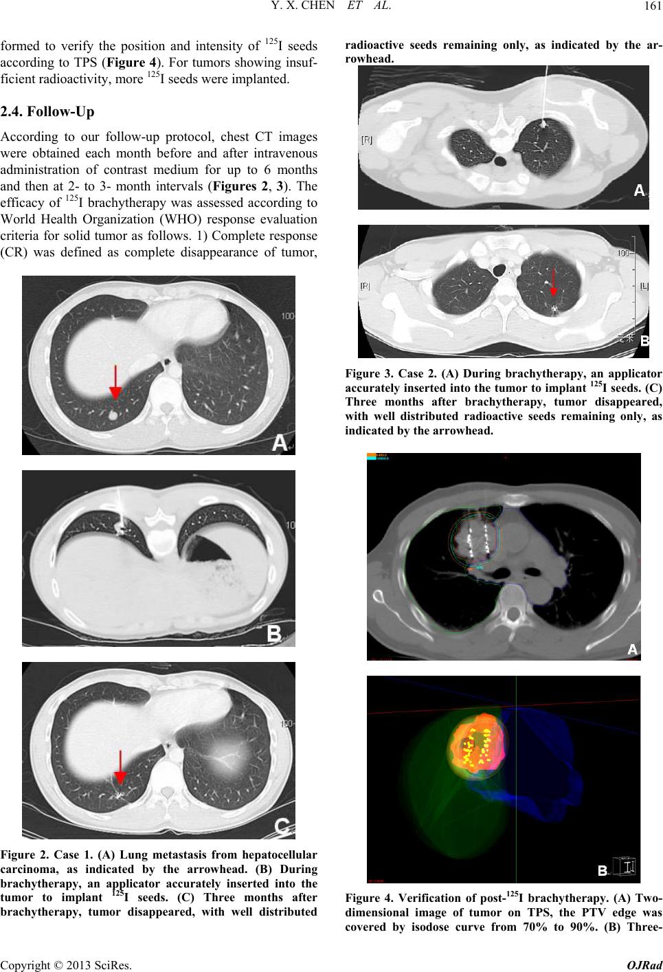 Y. X. CHEN ET AL. 161 formed to verify the position and intensity of 125I seeds according to TPS (Figure 4). For tumors showing insuf- ficient radioactivity, more 125I seeds were implanted. 2.4. Follow-Up According to our follow-up protocol, chest CT images were obtained each month before and after intravenous administration of contrast medium for up to 6 months and then at 2- to 3- month intervals (Figures 2, 3). The efficacy of 125I brachytherapy was assessed according to World Health Organization (WHO) response evaluation criteria for solid tumor as follows. 1) Complete response (CR) was defined as complete disappearance of tumor, Figure 2. Case 1. (A) Lung metastasis from hepatocellular carcinoma, as indicated by the arrowhead. (B) During brachytherapy, an applicator accurately inserted into the tumor to implant 125I seeds. (C) Three months after brachytherapy, tumor disappeared, with well distributed radioactive seeds remaining only, as indicated by the ar- rowhead. Figure 3. Case 2. (A) During brachytherapy, an applicator accurately inserted into the tumor to implant 125I seeds. (C) Three months after brachytherapy, tumor disappeared, with well distributed radioactive seeds remaining only, as indicated by the arrowhead. Figure 4. Verification of post-125I brachytherapy. (A) Two- dimensional image of tumor on TPS, the PTV edge was covered by isodose curve from 70% to 90%. (B) Three- Copyright © 2013 SciRes. OJRad  Y. X. CHEN ET AL. 162 dimensional graph of planar implantation and dose distri- bution on TPS. no tumor or only accumulative metal granule shadow detectable by imaging analysis. 2) Partial response (PR) was defined as the shrinkage of tumor with the P value decreasing by ≥50% compared to the pre-procedure value. 3) No change (NC) was defined as a P value de- crease of <50% or increase of <25% compared to the pre-procedure value. 4) Progression of disease (PD) was defined as a P value increase of ≥25% or the appearance of new tumor foci. Local control rate (LCR) was calcu- lated as cases of (CR + PR)/total × 100%. We com- pared the products (P) of two largest and perpendicular diameters within the tumor under CT examination before and after the procedure. 3. Results 3.1. Implantation of 125I Radioactive Seeds A total of 34 brachytherapies and 60 lung metastatic le- sions were performed on the 29 patients, among which, 29 were successful at the first time, achieving the TPS criteria, and 5 didn’t reach the TPS criteria, followed by additional implantations. The total number of implanted seeds was 493, with an average of 8.2 per tumor. The satisfactory rate of seed implantation was 94.1% (32/34). 3.2. Treatment Efficacy Months are counted from the first time of 125I brachy- therapy and the median duration of follow-up was 11 months (range, 6 - 17 months). The local control rates after 3, 6, 10 and 15 month were 86.2, 71.4, 60.9 and 50.0% respectively. The 15-month survival rate of our study is 48.3% (14/29) excluding 4 patients who were alive without evidence of recurrence at 11, 11, 12 and 14 months. At the time of writing, ten patients are alive without evidence of recurrence at 11 - 15 months. The 10 patients presented good control of local tumor and no systemic recurrence and survived throughout the fol- low-up period. Eleven patients died of multiple hemato- genous metastases 5 - 15 months after brachytherapy. Among the 29 patients, 125I brachytherapy was repeated in 6 patients because of local residue or local progression of the lymph node. The clinical responses of patients to brachytherapy were summarized in Table 1. 3.3. Side Effects Several complications related to the procedure occurred during or after brachytherapy. A small amount of local hematoma occurred in 5 patients that involved applicator insertion through the lung, probably because of injury of small lung vessels. Four patients presented pneumotho- rax with pulmonary compression of 30% - 40% after the procedure and recovered after drainage. During the fol- low-up, two patients had minor displacement of radioac- tive seeds. Severe complications such as massive bleed- ing and radiation pneumonitis did not occur. 4. Discussion Extrahepatic metastases of HCC have been reported in 25 to 65 percent of autopsy series, with lung, bones, and regional lymph nodes as the most commonly involved sites and the most common site of metastasis is lung [2]. The prognosis of patients with extrahepatic metastases remains poor. In two studies, the median survival time was less than 7 months and the 1-year survival rate was as low as 25% [11,12]. Although we recognize that our study was small and had limited follow-up periods, the survival rates seem promising. The 15-month survival rate of our study is higher than the 1-year survival rate of the above two studies. The results of our study seem bet- ter compared with the above reports. At the time of this writing, particularly encouraging is the outcome of 10 patients who are alive without evidence of recurrence for 11 - 15 months after 125I brachytherapy. For these pa- tients, 125I brachytherapy has been curable treatment so far. However, eleven patients died during follow-up. All the 11 patients had multiple hematogenous metastases at the time of follow-up, which resulted in death. In our study, 125I brachytherapy for lung metastases is not indicated if any uncontrollable recurrent foci are recognized at the primary site. In these cases of HCC, however, intrahepatic recurrences often occur and are not regarded to be well controllable by re-resections or other local treatments. In this series, we excluded patients with a history of intrahepatic recurrences as candidates for 125I brachytherapy when the intrahepatic recurrent tumors could not be well controlled. The success of 125I brachytherapy depends on the ac- curate placement of radioactive seeds within a known volume of tumor [13]. Using CT guidance, an image of the implant volume can be seen and the position of each implant needle can be adjusted to ensure proper place- ment. In our study, the positions of the seeds are deter- mined to provide a good dose distribution including a Table 1. Clinical effectiveness of 125I brachytherapy on lung metastasis. Local control efficacy Follow-up time (months) No. of patients CRPR NC PD Local control rate (%) (CR + PR)/Total % 3 29 205 2 2 86.2% 6 28 164 3 5 71.4% 10 23 122 4 5 60.9% 15 14 6 1 1 6 50.0% Copyright © 2013 SciRes. OJRad  Y. X. CHEN ET AL. 163 Note—Months are counted from the time of ablation session. Four patients who were alive without evidence of recurrence at 11, 11, 12 and 14 months at the time of writing were excluded at 15 months of follow-up time. minimum peripheral dose coverage, a uniform dose dis- tribution, and the protection of surrounding tissues since methods for calculating optimal seed locations are avail- able [14,15]. The relative coordinates of the seeds are then used for seed implantation within the lesions. Seeds and spacers are ejected from the needle when the depth of the needle specified by the plan is reached. Achieving this desired seed placement is left to the surgeon, who must take into account tissue deformations during the implant process [16,17]. Also inserting and retracting a needle in soft tissues cause the tissues to move, stretch and deform. This displacement and distortion of the tu- mor during the implantation result in misplaced seeds [17]. During the procedure, with the guidance of CT, the vital blood vessels and organs surrounding the tumor can be clearly displayed, allowing better control of implant- ing applicator and reducing the risk of damaging impor- tant blood vessels. No severe complications such as mas- sive bleeding occurred during procedure, suggesting it is a safe procedure. To avoid radioactive damage on im- portant organs such as vital blood vessels, the distance between implanted seeds should not be less than 10 mm to prevent accumulative radioactivity as a result of two closely positioned seeds. A route with minimal invasion into the lung tissue should be selected to reduce the risk of inducing pneumothorax. After procedure, it is better to keep patient hospitalized for 3 - 5 days and prospectively prescribe antibiotics and hemostatics to reduce the inci- dence of bleeding and infection. Our study also had limitations that we only included 29 patients and followed them up for a maximum of 17 months. More cases and longer follow-up are desired for future survival analysis. We also expect to carry out fur- ther study on combining this local approach with sys- temic approach for tumor treatment. In summary, the application of 125I brachytherapy in treating lung metastasis from hepatocellular carcinoma is still in the developing stage. In this study, we found that 125I brachytherapy has satisfactory short- to mid-term effect on controlling local lung metastasis, without se- vere complications such as massive bleeding or other complications associated with routine radiotherapy such as bone marrow arrest, radiation pneumonitis, digestive response or hair loss. Our study indicates that 125I bra- chytherapy is a promising treatment for lung metastasis from hepato- cellular carcinoma. REFERENCES [1] H. S. Kim, J. W. Shin, G. Y. Kim, et al., “Metastasis of Hepatocellular Carcinoma to the Small Bowel Manifested by Intussusception,” World Journal of Gastroenterology, Vol. 12, No. 12, 2006, pp. 1969-1971. [2] T. Nakashima, K. Okuda, M. Kojiro, et al., “Pathology of Hepatocellular Carcinoma in Japan. 232 Consecutive Cases Autopsied in Ten Years,” Cancer, Vol. 51, 1983, pp. 863-877. doi:10.1002/1097-0142(19830301)51:5<863::AID-CNCR 2820510520>3.0.CO;2-D [3] M. A. Bagshaw, R. S. Cox and J. E. Ramback, “Radiation Therapy for Localized Prostate Cancer. Justification by Long-Term Follow-Up,” Urologic Clinics of North Amer- ica, Vol. 17, No. 4, 1990, pp. 787-802. [4] F.-J. Zhang, P.-H. WU, Y.-K. GU, et al., “CT Guided Radioactive Seed 125I Implantation in Treating Lung Me- tastasis,” Chinese Journal of Radiology, Vol. 38, No. 9, 2004, pp. 906-909. [5] X.-K. Hu, M.-Y. Wang, Z.-G. Yang, et al., “The Study of Curing Central-Type Lung Cancer by CT Guided Percu- taneous Embedding of 125I Seeds,” Chinese Journal of Radiology, Vol. 38, No. 9, 2004, pp. 910-915. [6] M. Johnson, A. Colonias, D. Parda, et al., “Dosimetric and Technical Aspects of Intraoperative I-125 Brachy- therapy for Stage I Non-Small Cell Lung Cancer,” Phys- ics in Medicine and Biology, Vol. 52, No. 2, 2007, pp. 1237-1245. doi:10.1088/0031-9155/52/5/002 [7] T. Peretz, D. Nori, B. Hilaris, et al., “Treatment of Pri- mary Unresectable Carcinoma of the Pancreas with I-125 Implantation,” International Journal of Radiation On- cology, Biology, Physics, Vol. 17, No. 5, 1989, pp. 931- 935. doi:10.1016/0360-3016(89)90138-7 [8] F. J. Zhang, P. H. Wu, M. Zhao, et al., “CT Guided Ra- dioactive Seed 125I Implantation in Treatment of Pancre- atic Cancer,” Zhonghua Yi Xue Za Zhi, Vol. 86, 2006, pp. 223-227. [9] S. Ebara, N. Katayama, R. Tanimoto, et al., “Iodine-125 Seed Implantation (Permanent Brachytherapy) for Clini- cally Localized Prostate Cancer,” Acta Medica Okayama, Vol. 62, No. 1, 2008, pp. 9-13. [10] F.-J. Zhang, C.-X. Li, L. Zhang, et al., “Short- to Mid-Term Evaluation of CT-Guided 125I Brachytherapy on Intra-Hepatic Recurrent Tumors and/or Extra-Hepatic Metastases after Liver Transplantation for Hepatocellular Carcinoma,” Cancer Biology & Therapy, Vol. 8, No. 7, 2008, pp. 585-590. [11] K. Uka, H. Aikata, S. Takaki, et al., “Clinical Features and Prognosis of Patients with Extrahepatic Metastases from Hepatocellular Carcinoma,” World Journal of Gas- troenterology, Vol. 13, 2007, pp. 414-420. [12] M. Natsuizaka, T. Omura, T. Akaike, et al., “Clinical Features of Hepatocellular Carcinoma with Extrahepatic Metastases,” Journal of Gastroenterology and Hepatol- ogy, Vol. 20, 2005, pp. 1781-1787. doi:10.1111/j.1440-1746.2005.03919.x [13] R. Nath, L. L. Anderson, G. Luxton, et al., “Dosimetry of Interstitial Brachytherapy Sources: Recommendations of the AAPM Radiation Committee Task Group No. 43,” Medical Physics, Vol. 22, No. 2, 1995, pp. 209-234. Copyright © 2013 SciRes. OJRad  Y. X. CHEN ET AL. Copyright © 2013 SciRes. OJRad 164 doi:10.1118/1.597458 [14] J. Pouliot, D. Tremblay, J. Roy, et al., “Optimization of Permanent 125I Prostate Implants Using Fast Simulated Annealing,” International Journal of Radiation On- cology, Biology, Physics, Vol. 36, No. 3, 1996, pp. 711- 720. doi:10.1016/S0360-3016(96)00365-3 [15] E. Lee, R. J. Gallagher and M. Zaider, “Planning Implants of Radionuclides for the Treatment of Prostate Cancer: An Application of Mixed Integer Programming,” Optima, Vol. 61, 1999, pp. 1-10. [16] J. E. Dawson, T. Wu, T. Roy, et al., “Dose Effects of Seeds Placement Deviations from Pre-Planned Positions in Ultrasound Guided Prostate Implants,” Radiotherapy and Oncology, Vol. 32, No. 3, 1994, pp. 268-270. doi:10.1016/0167-8140(94)90027-2 [17] P. L. Roberson, V. Narayana, P. L. McShan, et al., “Source Placement Error for Permanent Implant of the Prostate,” Medical Physics, Vol. 24, No. 2, 1997, pp. 251-257. doi:10.1118/1.598058 Abbreviations: Hepatocellular carcinoma (HCC) Computed tomographic (CT) Treatment planning system (TPS) Matched peripheral dose (MPD) Dose-volume histogram (DVH) Planning target volume (PTV) World Health Organization (WHO) Complete response (CR) Partial response (PR) No change (NC) Progression of disease (PD) Local control rate (LCR) Products (P) |