

Paper Menu >>
Journal Menu >>
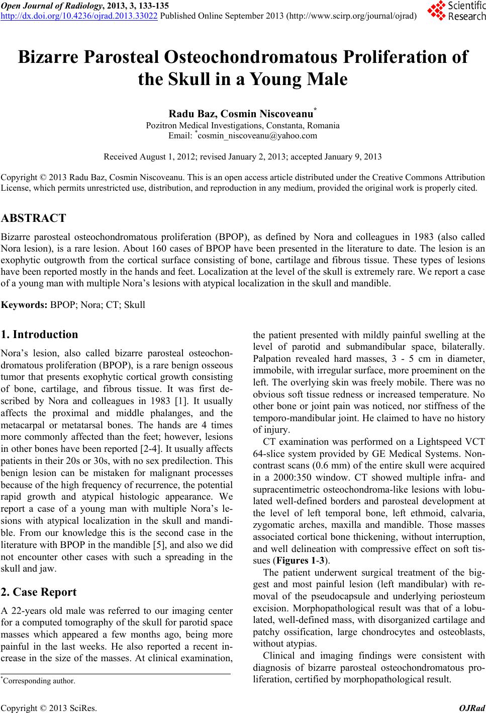 Open Journal of Radiology, 2013, 3, 133-135 http://dx.doi.org/10.4236/ojrad.2013.33022 Published Online September 2013 (http://www.scirp.org/journal/ojrad) Bizarre Parosteal Osteochondromatous Proliferation of the Skull in a Young Male Radu Baz, Cosmin Niscoveanu* Pozitron Medical Investigations, Constanta, Roma nia Email: *[email protected] Received August 1, 2012; revised January 2, 2013; accepted January 9, 2013 Copyright © 2013 Radu Baz, Cosmin Niscoveanu. This is an open access article distributed under the Creative Commons Attribution License, which permits unrestricted use, distribution, and reproduction in any medium, provided the original work is properly cited. ABSTRACT Bizarre parosteal osteochondromatous proliferation (BPOP), as defined by Nora and colleagues in 1983 (also called Nora lesion), is a rare lesion. About 160 cases of BPOP have been presented in the literature to date. The lesion is an exophytic outgrowth from the cortical surface consisting of bone, cartilage and fibrous tissue. These types of lesions have been reported mostly in the hands and feet. Localization at the level of the skull is extremely rare. We report a case of a young man with multiple Nora’s lesions with atypical localization in the skull and mandible. Keywords: BPOP; Nora; CT; Skull 1. Introduction Nora’s lesion, also called bizarre parosteal osteochon- dromatous proliferation (BPOP), is a rare benign osseous tumor that presents exophytic cortical growth consisting of bone, cartilage, and fibrous tissue. It was first de- scribed by Nora and colleagues in 1983 [1]. It usually affects the proximal and middle phalanges, and the metacarpal or metatarsal bones. The hands are 4 times more commonly affected than the feet; however, lesions in other bones have been reported [2-4]. It usually affects patients in their 20s or 30s, with no sex predilection. This benign lesion can be mistaken for malignant processes because of the high frequency of recurrence, the potential rapid growth and atypical histologic appearance. We report a case of a young man with multiple Nora’s le- sions with atypical localization in the skull and mandi- ble. From our knowledge this is the second case in the literature with BPOP in the mandible [5], and also we did not encounter other cases with such a spreading in the skull and jaw. 2. Case Report A 22-years old male was referred to our imaging center for a computed tomography of the skull for paro tid space masses which appeared a few months ago, being more painful in the last weeks. He also reported a recent in- crease in the size of the masses. At clinical examination, the patient presented with mildly painful swelling at the level of parotid and submandibular space, bilaterally. Palpation revealed hard masses, 3 - 5 cm in diameter, immobile, with irregular surface, more proeminent on the left. The overlyin g skin was freely mobile. There was no obvious soft tissue redness or increased temperature. No other bone or joint pain was noticed, nor stiffness of the temporo-mandibular joint. He claimed to have no history of injury. CT examination was performed on a Lightspeed VCT 64-slice system provided by GE Medical Systems. Non- contrast scans (0.6 mm) of the entire skull were acquired in a 2000:350 window. CT showed multiple infra- and supracentimetric osteochondroma-like lesions with lobu- lated well-defined borders and parosteal development at the level of left temporal bone, left ethmoid, calvaria, zygomatic arches, maxilla and mandible. Those masses associated cortical bone thickening, without interruption, and well delineation with compressive effect on soft tis- sues (Figures 1-3). The patient underwent surgical treatment of the big- gest and most painful lesion (left mandibular) with re- moval of the pseudocapsule and underlying periosteum excision. Morphopathological result was that of a lobu- lated, well-defined mass, with disorganized cartilage and patchy ossification, large chondrocytes and osteoblasts, without atypias. Clinical and imaging findings were consistent with diagnosis of bizarre parosteal osteochondromatous pro- liferation, certified by morphopathological result. *Corresponding author. C opyright © 2013 SciRes. OJRad 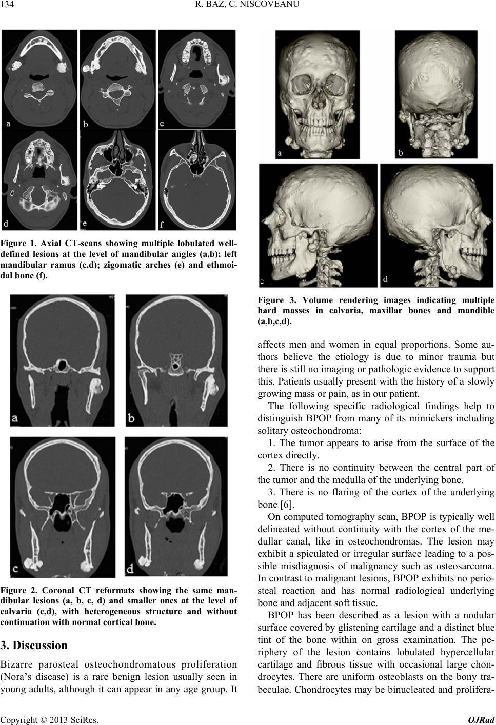 R. BAZ, C. NISCOVEANU 134 Figure 1. Axial CT-scans showing multiple lobulated well- defined lesions at the level of mandibular angles (a,b); left mandibular ramus (c,d); zigomatic arches (e) and ethmoi- dal bone (f). Figure 2. Coronal CT reformats showing the same man- dibular lesions (a, b, c, d) and smaller ones at the level of calvaria (c,d), with heterogeneous structure and without continuation with normal cortical bone. 3. Discussion Bizarre parosteal osteochondromatous proliferation (Nora’s disease) is a rare benign lesion usually seen in young adults, although it can appear in any age group. It Figure 3. Volume rendering images indicating multiple hard masses in calvaria, maxillar bones and mandible (a,b,c,d). affects men and women in equal proportions. Some au- thors believe the etiology is due to minor trauma but there is still no imaging or pathologic evidence to support this. Patients usually present with the history of a slowly growing mass or pain, as in our patient. The following specific radiological findings help to distinguish BPOP from many of its mimickers including solitary osteochondroma: 1. The tumor appears to arise from the surface of the cortex directly. 2. There is no continuity between the central part of the tumor and the medulla of the underlying bone. 3. There is no flaring of the cortex of the underlying bone [6]. On computed tomography scan, BPOP is typically well delineated without continuity with the cortex of the me- dullar canal, like in osteochondromas. The lesion may exhibit a spiculated or irregular surface leading to a pos- sible misdiagnosis of malignancy such as osteosarcoma. In contrast to malignant lesions, BPOP exhibits no perio- steal reaction and has normal radiological underlying bone and adjacent soft tissu e. BPOP has been described as a lesion with a nodular surface covered by glistening cartilage and a distinct blue tint of the bone within on gross examination. The pe- riphery of the lesion contains lobulated hypercellular cartilage and fibrous tissue with occasional large chon- drocytes. There are uniform osteoblasts on the bony tra- beculae. Chondrocytes may be binucleated and prolifera- Copyright © 2013 SciRes. OJRad 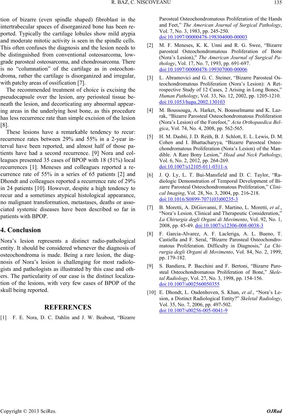 R. BAZ, C. NISCOVEANU Copyright © 2013 SciRes. OJRad 135 tion of bizarre (even spindle shaped) fibroblast in the intertrabecular spaces of disorganized bone has been re- ported. Typically the cartilage lobules show mild atypia and moderate mitotic activity is seen in the spindle cells. This often conf uses the diagnosis and the lesion n eeds to be distinguished from conventional osteosarcoma, low- grade parosteal osteosarcoma, and chondrosarcoma. There is no “columnation” of the cartilage as in osteochon- droma, rather the cartilage is disorganized and irregular, with patc hy areas of ossification [7]. The recommended treatment of choice is excising the pseudocapsule over the lesion, any periosteal tissue be- neath the lesion, and decorticating any abnormal appear- ing areas in the underlying host bone, as this procedure has less recurrence rate than simple excision of th e lesion [8]. These lesions have a remarkable tendency to recur: recurrence rates between 29% and 55% in a 2-year in- terval have been reported, and almost half of those pa- tients have had a second recurrence. [9] Nora and col- leagues presented 35 cases of BPOP with 18 (51%) local recurrences [1]. Meneses and colleagues reported a re- currence rate of 55% in a series of 65 patients [2] and Dhondt and colleagues reported a recurrence rate of 29% in 24 patients [10]. However, despite a high tendency to recur and a sometimes atypical histological appearance, no malignant transformation, metastases, deaths or asso- ciated systemic diseases have been described so far in patients with BPOP. 4. Conclusion Nora’s lesion represents a distinct radio-pathological entity. It should be considered whenever the diagnosis of osteochondroma is made. Being a rare lesion, the diag- nosis of Nora’s lesion is challenging for most radiolo- gists and pathologists as illustrated by this case and oth- ers. The particularity of our case is the distinct localiza- tion of the lesions, with very few cases of BPOP of the skull bein g r ep or t ed. REFERENCES [1] F. E. Nora, D. C. Dahlin and J. W. Beabout, “Bizarre Parosteal Osteochondromatous Proliferation of the Hands and Feet,” The American Journal of Surgical Pathology, Vol. 7, No. 3, 1983, pp. 245-250. doi:10.1097/00000478-198304000-00003 [2] M. F. Meneses, K. K. Unni and R. G. Swee, “Bizarre parosteal Osteochondromatous Proliferation of Bone (Nora’s Lesion),” The American Journal of Surgical Pa- thology, Vol. 17, No. 7, 1993, pp. 691-697. doi:10.1097/00000478-199307000-00006 [3] L. Abramovici and G. C. Steiner, “Bizarre Parosteal Os- teochondromatous Proliferation (Nora’s Lesion): A Ret- rospective Study of 12 Cases, 2 Arising in Long Bones,” Human Pathology, Vol. 33, No. 12, 2002, pp. 1205-1210. doi:10.1053/hupa.2002.130103 [4] M. Boussouga, A. Harket, N. Bousselmame and K. Laz- rak, “Bizarre Parosteal Osteochondromatous Proliferation (Nora’s Lesion) of the Forefoot,” Acta Orthopaedica Bel- gica, Vol. 74, No. 4, 2008, pp. 562-565. [5] H. M. Dashti, J. D. Reith, B. J. Schlott, E. L. Lewis, D. M. Cohen and I. Bhattacharyya, “Bizarre Parosteal Osteo- chondromatous Proliferation (Nora’s Lesion) of the Man- dible. A Rare Bony Lesion,” Head and Neck Pathology, Vol. 6, No. 2, 2012, pp. 264-269. doi:10.1007/s12105-011-0311-x [6] J. Q. Ly, L. T. Bui-Mansfield and D. C. Taylor, “Ra- diologic Demonstration of Temporal Development of Bi- zarre Parosteal Osteochondromatous Proliferation,” Clini- cal Imaging, Vol. 28, No. 3, 2004, pp. 216-218. doi:10.1016/S0899-7071(03)00235-3 [7] B. Moretti, A. DiGiovanni, F. Martino, L. Moretti, et al., “Nora’s Lesion. Clinical and Therapeutic Consideration,” La Chirurgia degli Organi di Movimento, Vol. 92, No. 1, 2008, pp. 45-49. doi:10.1007/s12306-008-0038-3 [8] F. Garcia-Alvarez, A. F. Lacleriga, A. L. Bueno, T. Castiella and F. Seral, “Bizarre Parosteal Osteochondro- matous Proliferation. Difficulty in Diagnosis,” La Chi- rurgia degli Organi di Movimento, Vol. 84, No. 2, 1999, pp. 179-182. [9] S. Bandiera, P. Bacchini and F. Bertoni, “Bizarre Paro- steal Osteochondromatous Proliferation of Bone,” Skele- tal Radiology, Vol. 27, No. 3, 1998, pp. 154-156. doi:10.1007/s002560050355 [10] E. Dhondt, L. Oudenhoven, S. Khan, et al., “Nora’s Le- sion, a Distinct Radiological Entity?” Skeletal Radiology, Vol. 35, No. 7, 2006, pp. 497-502. doi:10.1007/s00256-005-0041-9 |