

Paper Menu >>
Journal Menu >>
 World Journal of AIDS, 2013, 3, 201-206 http://dx.doi.org/10.4236/wja.2013.33027 Published Online September 2013 (http://www.scirp.org/journal/wja) Copyright © 2013 SciRes. WJA 201 Characterization of Candida Species Isolated from Cases of Lower Res piratory Tra ct Infection among HIV/AIDS Patients in Calabar, Nigeria* Ofonime Mark Ogba1, Lydia Nyong Abia-Bassey2, James Epoke2, Baki Idasa Mandor1, Godwin Dickson Iwatt3 1Department of Medical Microbiology/Parasitology, University of Calabar Teaching Hospital, Calabar, Nigeria; 2Department of Medical Laboratory Science, University of Calabar, Calabar, Nigeria; 3Department of Microbiology, University of Calabar, Calabar, Nigeria. Email: [email protected] Received June 2nd, 2013; revised July 2nd, 2013; accepted August 2nd, 2013 Copyright © 2013 Ofonime Mark Ogba et al. This is an open access article distributed under the Creative Commons Attribution Li- cense, which permits unrestricted use, distribution, and reproduction in any medium, provided the original work is properly cited. ABSTRACT This study was designed to identify and characterize the Candida species isolated from lower respiratory tract infections among HIV positive patients and to determine the prevalence rates of Candida infections among these subjects. Two early morning expectorate sputum samples were collected from 272 HIV pos itive subjects visiting the ART clinics and DOTS centre with cases of lower respiratory tract infection, over a period of 14 months from May 2009 to July 2010 in Calabar. Subjects were recruited for this study upon approval by the Ethical Research Committee of the University of Calabar Teaching Hospital and obtaining written informed consent from the patients. Samples were processed by stan- dard methods for isolation of Candida . Speciation was done by a germ tube test, chlamydospore production on corn meal agar and sugar fermentation and assimilation tests using the Microexpr ess Candida identif ication kit (Tulip, India). Out of the 544 sputum samples collected from 272 subjects, Candida species were isolated from 40 (14.7%) and identi- fied after confirming the growth in the second sample. The majority of Candida species among the Candida isolates were Candida albicans (80%) followed by Candida tropicalis 5 (12.5%), Candida dubliniensis 2 (5.0%) and Candida guilliermondii 1 (2.5%). The isolation rate of Candida species from sputum samples was found to be highest among subjects aged 25 - 34 years, followed by those aged 15 - 24 years. Twenty (7.3%) H IV seropositiv e subjec ts had bacte- rial infections, while 4 (1.5%) subjects had mixed fungal and bacterial infections. This study is the first of its kind to be carried out in Calabar and the South-South geopolitical region of Nigeria, and has shown that pulmonary candidiasis is a health problem among HIV positive patients in Calabar. Keywords: Candida Infections; Pulmonary; HIV/A IDS; Characterization 1. Introduction Candida is a polymorphic fungus. It is a Gram positive, oval, budding yeast cell that produces pseudohyphae both in culture and in tissues and exudates [1]. They exhibit fi- lamentous mycellial morphology in the saprophytic phase, but have typical yeast morphology in the parasitic phase, in tissue and when grown at 37˚C in the laboratory [1]. They form pseudohyphae when the buds continue to grow but fail to detach, producing chains of elongated cells that are pinched or constricted at the septations be- tween cells [2]. At temperatures below 26˚C in nutrition- ally poor media such as cornmeal agar, it produces thick- walled resting cells 7 to 17 mm in diameter, called chla- mydospores [3]. It is a member of the normal flora of the mucous membranes in the gastrointestinal, upper respira- tory and female genital tracts [4]. There are about 68 species of Candida, the most path- ogenic is C. albicans [5]. The other non-albicans Candida (NAC) species include C. dubliniensis, C. glabrata, C. guilliermondii, C. tropicalis, C. parapsilosis, C. krusei and C. hisitaniae, C. kefyr, C. lipolytic and C. pelliculos [6,7]. Candida pneumonia is one of the most challenging of all the Candida infections. Primary Candida pneumonia refers to infections limited to the lungs, while secondary Candida pneumonia indicates lung involvement due to * Conflicts of Interests: None declared.  Characterization of Candida Species Isolated from Cases of Lower Respiratory Tracr Infection among HIV/AIDS Patients in Calabar, Nigera Copyright © 2013 SciRes. WJA 202 the movement of invasive Candida from other areas of the body [5]. The criteria for the diagnosis of pulmonary candidiasis are still controversial. The isolation of Can- dida from culture of sputum, endotracheal aspirates, bron - choscopic samples, percutaneous lung needle aspirates and even from lung tissue may only represent coloniza- tion of the trachea-bronchial tree [4]. Despite the debate about the diagnosis of pulmonary candidiasis, the defini- tive diagnosis is still resting on histological demonstra- tion of the yeast in lung tiss ue with associated infla mma- tory changes at autopsy [7,8]. The increase in the number of immunocompromised patients during the last few decades, as a result of che- motherapy or HIV/AIDS has resulted in a parallel in- crease in the number of opportunistic infections, espe- cially those due to Candida species [9,10]. Rapid identification and speciation of Candida species are essential in clinical laboratories. However, no single phenotypic test is highly ef fective in identifying Candida species and the combination of tests is sometimes neces- sary for identification. Molecular techniques have been employed to characterize Candida spp. Although sensi- tive and specific it is not cost effective for routine clini- cal mycology laboratories in a resource constrained setup like ours [11]. 2. Aims and Objectives The aim of the study was to determine the rate of iso- lation of Candida species from sputum samples of HIV positive patients with respiratory symptoms, to identify and characterize the Candida species using various phe- notypic methods such as Germ tube test, Chlamydospore production and identification to specie level with Mi- croxpress Candida identification kit (Tulip diagnostic LTD, India), according to the manufacturer’s instruc- tions. 3. Materials and Methods 3.1. Study Sites and Subjects Selection The present study was carried out in the department of Microbiology/Parasitology, University of Calabar Teach- ing Hospital, Calabar. Subjects were recruited from two tertiary hospitals; the University of Calabar Teaching Hospital and Lawrence Henshaw Hospital located in Ca- labar municipality and Calabar south local Government areas respectively. The cases selected for the study inclu- ded HIV positive subjects with respiratory symptoms who were able to produce sputum. Sputum samples show- ing less than 10 squamous epithelial cells and 25 or more polymorphonuclear leukocytes per low power field (100×) were included in this study. 3.2. Collection and Examination of Samples A total of 272 sputum samples in duplicates were col- lected from May 2009 to July 2010 from the subjects. All sputum sp ecimens were processed by Gram staining, 10% KOH mount, culture, germ tube test, detection of chla- mydospore and sugars fermentation and assimilation tests. A 10% KOH mount of sputum sample showing plenty of Candida pseudohyphae were considered as po- tential pathogens and not as colonizers. The isolates were considered significant by correlation with microscopy and growth of two consecutive cultures. The Candida species were isolated repeatedly in pure culture from two consecutive early mornings expectorate sputum samples [4]. Speciation of Candida was by germ tube test, mor- phology on corn meal agar, sugar fermentation and assi- milation tests using Microexpress Candida id entification kits (Tulip, India) according to the manufacturer’s in- structions [4, 12]. 3.3. Direct Examination of the Specimens 3.3.1. Gram Stain Smears made from the most purulent: or mucopurulent part of the sputum. Smears were examined for the pres- ence of gram positive budding yeast cells with pseudohy- phae. Specimen was considered as acceptable when 25 or more polymorphonuclear leukocytes were seen per low power field (100×) with few (less than 10) squamous epi- thelial cells. 3.3.2. KOH Mount Smears were prepared for each sample by adding a loop full of sputum to a drop of 10% KOH on a clean, grease- free slide and placing a cover slip over it. The prepara- tions were slightly warmed to digest the materials and examined under the microscope with X10 and X40 ob- jective for yeast cells, pseudohyphae [12]. 3.3.3. Culture The most bloody, purulent, or mucus laden portions of sputum samples were inoculated on Sabouraud Dextrose Agar (SDA) with and without chloramphenicol (16 µg∙ml−1) in duplicates incubated at 37˚C and 25˚C. Cul- tures were examined every other day for growth up to 2 weeks before discarding as negative [12,13]. The macro- scopic features of the growth wer e opaque pasty glab rous or membranous texture often with the sweet smell, remi- niscent of ripe apples. The growth was confirmed by ob- serving the characteristic budding with pseudohyphae in the Gram stain [10]. Pure cultures of every isolate were prepared before performing any physiological test. This was done by sub-culturing individual isolates onto fresh SDA plates and incubating at 37˚C and at room tempe- 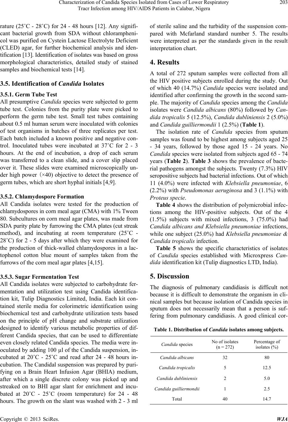 Characterization of Candida Species Isolated from Cases of Lower Respiratory Tracr Infection among HIV/AIDS Patients in Calabar, Nigera Copyright © 2013 SciRes. WJA 203 rature (25˚C - 28˚C) for 24 - 48 hours [12]. Any signifi- cant bacterial growth from SDA without chlorampheni- col was purified on Cystein Lactose Electrolyte D eficient (CLED) agar, for further biochemical analysis and iden- tificatio n [13]. Ide ntification of i s ol a te s wa s ba s e d on gross morphological characteristics, detailed study of stained sampl e s and bi ochemical tests [14]. 3.5. Identification of Candida Isolates 3.5.1. Germ Tube Test All presumptive Candida species were subjected to germ tube test. Colonies from the purity plate were picked to perform the germ tube test. Small test tubes containing about 0.5 ml human serum were inoculated with colonies of test organisms in batches of three replicates per test. Each batch included a known positive and negative con- trol. Inoculated tubes were incubated at 37˚C for 2 - 3 hours. At the end of incubation, a drop of each serum was transferred to a clean slide, and a cover slip placed over it. These slides were examined microscopically un- der high power (×40) objective to detect the presence of germ tubes, which are short hyphal initials [4,9]. 3.5.2. Chlamydospore Formation All Candida isolates were tested for the production of chlamydospores in corn meal agar (CMA) with 1% Tween 80. Subcultures on corn meal agar plates, was made from SDA purity plate by furrowing the CMA plates (cut streak method), and incubating at room temperature (25˚C - 28˚C) for 2 - 5 days after which they were examined for the production of thick-walled chlamydospores in a lac- tophenol cotton blue mount of samples taken from the furrows of the corn meal agar plates [4,15]. 3.5.3. Sugar Fermentation Test All Candida isolates were subjected to carbohydrate fer- mentation and utilization test using Candida identifica- tion kit, Tulip Diagnostics Limited, India. Each kit con- tained sterile media for colorimetric identification using biochemical test and carbohydrate utilization tests based on the principle of pH change and substrate utilization designed to identify various metabolic properties of dif- ferent Candida species, that can be used to differentiate even closely related Candida species. The media were in- oculated by adding 100 µl of the Candida suspension, in- cubated at 20˚C - 25˚C and read after 24 - 48 hours in- cubation. The Candidal suspension was prepared by puri- fying on a Brain Heart Infusion Agar (BHIA) medium, after which a single discrete colony was picked up and streaked on to BHI agar slant for enrichment and incu- bated at 20˚C - 25˚C (room temperature) for 24 - 48 hours. Th e growth on the slant was washed with 2 - 3 ml of sterile saline and the turbidity of the suspension com- pared with Mcfarland standard number 5. The results were interpreted as per the standards given in the result interpretation chart. 4. Results A total of 272 sputum samples were collected from all the HIV positive subjects enrolled during the study. Out of which 40 (14.7%) Candida species were isolated and identified after confirming the growth in the second sam- ple. The majority of Candida species among the Candida isolates were Candida albicans (80%) followed by Can- dida tropicalis 5 (12.5%), Candida dubliniensis 2 (5.0%) and Candida guilliermondii 1 (2.5%) (Table 1 ). The isolation rate of Candida species from sputum samples was found to be highest among subjects aged 25 - 34 years, followed by those aged 15 - 24 years. No Candida species were isolated from subjects aged 65 - 74 years (Table 2). Table 3 shows the prevalence of bacte- rial pathogens amongst the subjects. Twenty (7.3%) HIV seropositive subjects had bacterial infections. Out of which 11 (4.0%) were infected with Klebsiella pneumoniae, 6 (2.2%) with Pseudomonas aeruginosa and 3 (1.1%) with Proteus specie. Table 4 shows the distribution of polymicrobial infec- tions among the HIV-positive subjects. Out of the 4 (1.5%) subjects with mixed infections, 3 (75.0%) had Candida albicans and Klebsiella pneumoniae infections, while one subject (25.0%) had Klebsiella pneumoniae & Candida tropicalis infection. Table 5 shows the specific characteristics of isolates of Candida species established with Microxpress Can- dida identification kit (Tulip diagnostics LTD, India). 5. Discussion The diagnosis of pulmonary candidiasis is difficult not because it is difficult to demonstrate the organism in cli- nical samples but because isolation of Candida species in sputum does not necessarily mean that a person is suf- fering from pulmonary candidiasis. A good clinical cor- Table 1. Distribution of Candida isolates among subjects. Candida species No of isolates (n = 272) Percentage of isolates (%) Candida albicans 32 80 Candida tropical is 5 12.5 Candida dubliniensis 2 5.0 Candida guilliermondii 1 2.5 Total 40 14.7 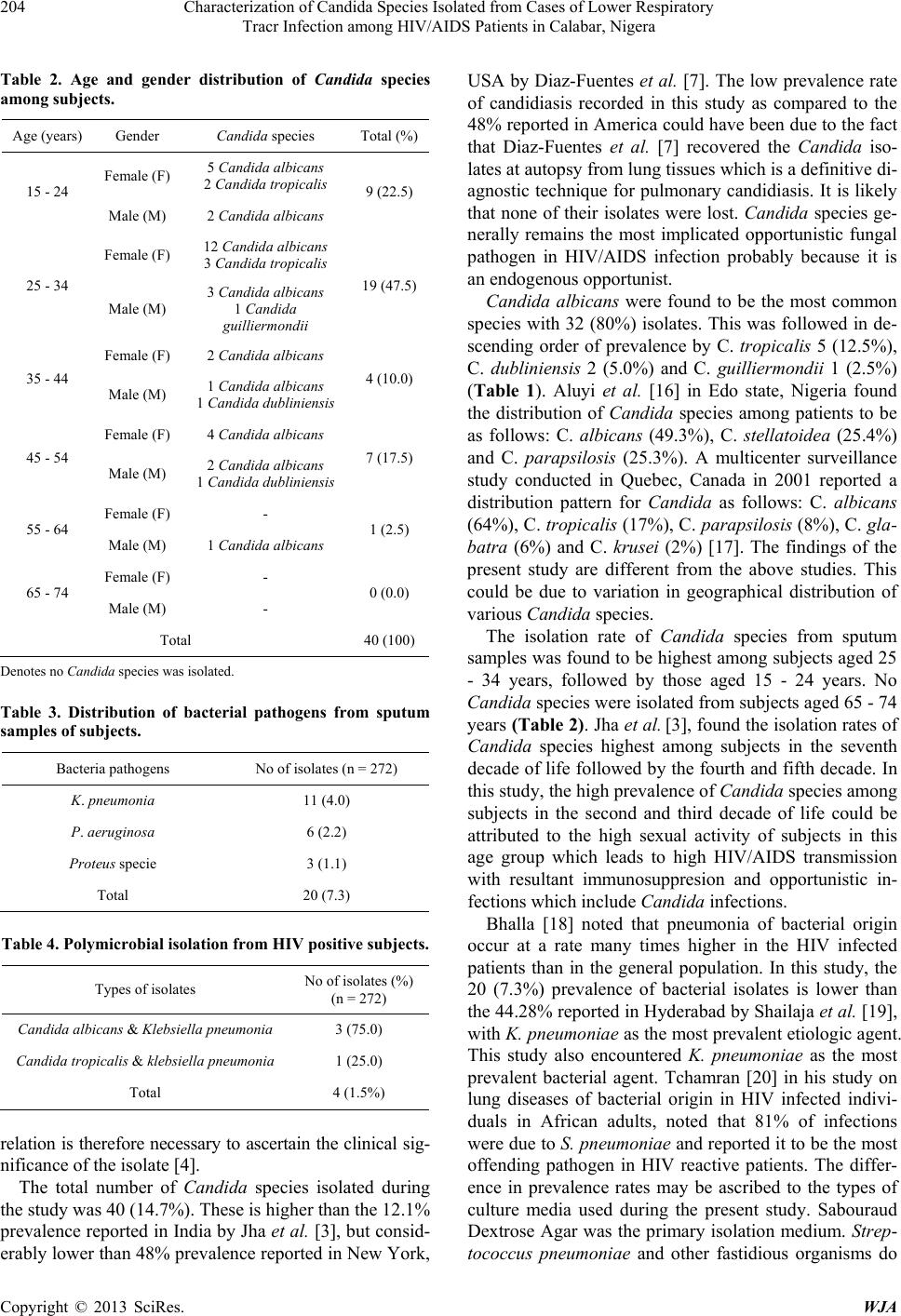 Characterization of Candida Species Isolated from Cases of Lower Respiratory Tracr Infection among HIV/AIDS Patients in Calabar, Nigera Copyright © 2013 SciRes. WJA 204 Table 2. Age and gender distribution of Candida species among subjects. Age (years) Gender Candida species Total (%) 15 - 24 Female (F) 5 Candida albicans 2 Candida tropicalis 9 (22.5) Male (M) 2 Candida albicans 25 - 34 Female (F) 12 Candida albicans 3 Candida tropicalis 19 (47.5) Male (M) 3 Candida albicans 1 Candida guilliermondii 35 - 44 Female (F) 2 Candida albicans 4 (10.0) Male (M) 1 Candida albicans 1 Candida dubliniensis 45 - 54 Female (F) 4 Candida albicans 7 (17.5) Male (M) 2 Candida albicans 1 Candida dubliniensis 55 - 64 Female (F) - 1 (2.5) Male (M) 1 Candida albicans 65 - 74 Female (F) - 0 (0.0) Male (M) - Total 40 (100) Denotes n o Candida species was isolated. Table 3. Distribution of bacterial pathogens from sputum samples of subjects. Bacteria pathogens No of isolates (n = 272) K. pneumonia 11 (4.0) P. aeruginosa 6 (2.2) Proteus specie 3 (1.1) Total 20 (7.3) Table 4. Polymicrobial isolation from HIV positive subjects. Types of isolates No of isolates (%) (n = 272) Candida albicans & Klebsiella pneumonia 3 (75.0) Candida tropical is & klebsiella pneumonia 1 (25.0) Total 4 (1.5%) relation is therefore necessary to ascertain the clinical sig- nificance of the isolate [4]. The total number of Candida species isolated during the study was 40 (14.7%). These is higher than the 12.1% prevalence reported in India by Jha et al. [3], but consid- erably lower than 48% prevalence reported in New York, USA by Diaz-Fuentes et al. [7]. The low prevalence rate of candidiasis recorded in this study as compared to the 48% reported in America could have been due to t he fac t that Diaz-Fu entes et al. [7] recovered the Candida iso- lates at autopsy from lung tissues which is a definitive di- agnostic technique for pulmonary candidiasis. It is likely that none of their isolates were lost. Candida species ge- nerally remains the most implicated opportunistic fungal pathogen in HIV/AIDS infection probably because it is an endogenous op portunist. Candida albicans were found to be the most common species with 32 (80%) isolates. This was followed in de- scending order of prevalence by C. tropicalis 5 (12.5%), C. dubliniensis 2 (5.0%) and C. guilliermondii 1 (2.5%) (Table 1). Aluyi et al. [16] in Edo state, Nigeria found the distribution of Candida species among patients to be as follows: C. albicans (49.3%), C. stellatoidea (25.4%) and C. parapsilosis (25.3%). A multicenter surveillance study conducted in Quebec, Canada in 2001 reported a distribution pattern for Candida as follows: C. albicans (64%), C. tropicalis (17%), C. parapsilosis (8%), C. gla- batra (6%) and C. krusei (2%) [17]. The findings of the present study are different from the above studies. This could be due to variation in geographical distribution of various Candida species. The isolation rate of Candida species from sputum samples was found to be highest among subjects aged 25 - 34 years, followed by those aged 15 - 24 years. No Candida species were isolated from subjects aged 65 - 74 years (Ta ble 2) . Jha et al. [3], found the iso lation rates of Candida species highest among subjects in the seventh decade of life followed by the fourth and fifth decade. In this study, the high prevalence of Candida species among subjects in the second and third decade of life could be attributed to the high sexual activity of subjects in this age group which leads to high HIV/AIDS transmission with resultant immunosuppresion and opportunistic in- fections which i nc l ude Candida infections. Bhalla [18] noted that pneumonia of bacterial origin occur at a rate many times higher in the HIV infected patients than in the general population. In this study, the 20 (7.3%) prevalence of bacterial isolates is lower than the 44.28% reported in Hyderabad by Shailaja et al. [19], with K. pneumoniae as the most prevalent etiologic agent. This study also encountered K. pneumoniae as the most prevalent bacterial agent. Tchamran [20] in his study on lung diseases of bacterial origin in HIV infected indivi- duals in African adults, noted that 81% of infections were due to S. pneumoniae and reported it to be the most offending pathogen in HIV reactive patients. The differ- ence in prevalence rates may be ascribed to the types of culture media used during the present study. Sabouraud Dextrose Agar was the primary isolation medium. Strep- tococcus pneumoniae and other fastidious organisms do  Characterization of Candida Species Isolated from Cases of Lower Respiratory Tracr Infection among HIV/AIDS Patients in Calabar, Nigera Copyright © 2013 SciRes. WJA 205 Table 5. Characterization of Candida species with Microxpress Candida identifi cation kit. SPECIES Urease utilisation Melibose utilisation Lactose utilization Maltose utilization Sucrose utilization Galactose utilization Cellobiose utilisation Inositol utilization Xylose utilization Dulcitol utilization Raffinose utilization C. albicans − − − + − + − − + − − C. dubliniensis − − − + − + − − V − − C. tropicalis − − − + + V + − + − − C. guilliemondii − + − − + V + − + + + Key: + = positive; − = negative; v = variable. not grow on this media, thus such organisms were not isolated. Also the polymicrobial etiology (mixed Candida and bacterial infections) in 4 (1.5%) of the HIV reactive sub- jects indicates the severity and the effect of immunosup- presion in this group. 6. Conclusion Pulmonary candidiasis is a health problem among HIV positive patients in Calabar, Nigeria. Microexpress can- dida identification kit has been found to be useful in the identification of Candida to species level, as PCR is ex- pensive and not easily assessable. Periodic study of pul- monary Candida infections among HIV infected patients is the present day’s need, to know the changing pattern in incidence of Candida species. REFERENCES [1] A. Chakrabati and M. R. Shivaprakash, “Microbiology of Systemic Fungal Infections,” Journal of Postgraduate Medicine, Vol. 51, No. 5, 2005, pp. 16-20 . [2] R. A. Calderone and W. A. Fronzi, “Virulence of Candi- da albicans,” Trends in Microbiology, Vol. 9, No. 7, 2001, p. 327. doi:10.1016/S0966-842X(01)02094-7 [3] B. K. Jha, S. Dey, M. D. Tamang, M. E. Joshy, P. G. Shi- vananda and K. N. Brahmadatan, “Characterization of Candida Species Isolated from Cases of Lower Respirato- ry Tract Infection,” Kathmandu University Medical Jour- nal, Vol. 4, No. 3, 2006, pp. 290-294. [4] E. Segal and D. Elad, “Candidiasis,” In: W. M. Scheld, D. C. Hooper and J. M. Hughes, Eds., Topley and Wilson’s Microbiology and Mic robial Infections, 10th Edition, Hod- der Arnold ASM Press, Washington DC, 2005, pp. 479- 623. [5] D. R. Soll, “Candida and Virulence: The Evolution of Phe- notypic Plasticity,” Acta Tropica, Vol. 81, No. 2, 2002, p. 101. doi:10.1016/S0001-706X(01)00200-5 [6] Z. U. Khan, S. Ahmed, E. Mokaddas and R. Chandy, “To- bacco Agar, a New Medium for Differentiating Candida dubliniensis from Candida albicans,” Journal of Clinical Microbiology, Vol. 42, No. 10, 2004, pp. 4796-4798. doi:10.1128/JCM.42.10.4796-4798.2004 [7] G. Diaz-Fuentes, C. Shin, E. R. Sy, M. Niazi and L. Me- non, “Pulmonary Fungal Involvement in HIV-Positive Pa- tients in an Inner City Hospital in New York,” The Inter- net Journal of Pulmonary Medicine, Vol. 7, No. 2, 2007, pp. 1531-2984. [8] A. Usharani, M. Bharathi and C. Sandhya, “Isolation and Characterization of Candida Species from Oropharyngeal Secretions of HIV Positive Individuals,” Nasza Derma- tology Online, Vol. 2, No. 3, 2011, pp. 119-124. [9] L. Y. Binesh and M. Kalyani, “Phenotypic Characteriza- tion of Candida Species and Their Antifungal Suscepti- bility from a Tertiary Care Centre,” Journal of Pharma- ceutical and Biomedical Sciences, Vol. 11, No. 12, 2011. [10] C. R. Juliana, “Phenotypic and Genotypic Identification of Candida spp. Isolated from Hospitalized Patients,” Re- vista Iberoamericana de Micología, Vol. 21, 2004, pp. 24-26. [11] E. W. Koneman, S. D. Allen, W. M. Janda, P. C . Schrec- kenberger and W. C. Winn, “The Role of Microbiology Laboratory in the Diagnosis of Infectious Disease: Guide- lines to Practice and Management,” In: R. P. Pfeiffer and B. C. Magnus, Eds., Color Atlas and Text Book of Diag- nostic Microbiology, 5th Edition, Lippincot Press, New York, 1997, p. 69. [12] B. A. Forbes, D. F. Sahm and A. S. Weissfeld, “Laborato- ry Methods in Basic Mycology,” In: Bailey and Scott’s Diagnostic Microbiology, 11th Edition, Mosby, St. L ouis, 2002, pp. 711-798. [13] L. S. Garcia, G. W. Procop, G. D. Roberi a nd R. B. Thom- pson, “Overview of Conventional Methods for Bacterial Identification, Chapter 13,” In: B. A. Forbes, D. F. Sahm and A. S. Weissfeld, Eds., Bailey and Scott’s Diagnostic Microbialogy, 10th Edition, Mosby, 1998, pp. 167-181. [14] M. Cheesbrough, “Medical Laboratory Manual for Tropi- cal Countries: Microbiology Vol. 2. Butterworth-Heine- mann Ltd.,” University Press, Cambridge, 1995, pp. 248- 273. [15] G. S. De Hoog, J. Guarro and M. E. Grauer, “Atlas of Cli- nical Fungi,” 2nd Edition, Centraalbureauvoor Schimmel- cultures, Baarn, 2000. [16] H. S. A. Aluyi, F. D. Otajev wo and O. Iweriebor, “Incidence of Pulmonary Mycoses in Patients with Acquired Immu- nodeficiency Diseases,” Nigerian Journal of Clinical  Characterization of Candida Species Isolated from Cases of Lower Respiratory Tracr Infection among HIV/AIDS Patients in Calabar, Nigera Copyright © 2013 SciRes. WJA 206 Practice, Vol. 13, No. 1, 2010, pp. 78-83. [17] G. St. Germain, M. Laverdiere and R. Pelletier, “Preva- lence and Antifungal Susceptibility of 442 Candida Iso- lates from Blood and Other Normally Sterile Sites: Re- sults of a 2 Year (1996-1998) Multicentre Surveillance Study in Quebec, Canada,” Journal of Clinical Microbi- ology, Vol. 39, No. 3, 2001, pp. 949-953. doi:10.1128/JCM.39.3.949-953.2001 [18] P. Bhalla, “Non Mycobac teri al and Bacterial Infections in HIV/AIDS,” In: U. K. Baveja and J. Sokhey, Eds., Man- ual on Laboratory Diagnosis of Common Opportunistic Infections Associated with HIV/AIDS, National Institute of Communicable Diseases, New Delhi, 2002, pp. 100- 118. [19] L. A. Shailaja, L. A. Pai, D. R. Mathur and V. Lakshmi, “Prevalence of Bacterial and Fungal Agents Causing Lo- wer Respiratory Tract Infections in Patients wi th HIV In- fection,” Indian Journal of Medical Microbiology, Vol. 22, No. 1, 2004, pp. 28-33. [20] N. Koffi, A. Ngom, B. Kouassi and M. Tchamran, “Bac- terial Lung Disease from Common Bacteria during HIV Infection in African Adults Hospitalized in Abidjan, Cote d’Ivoire,” Bulletin de la Societe de Pathologie Exotique, Vol. 90, No. 5, 1997, pp. 370-372. List of Abbreviations Abbreviation/Symbol Meaning HIV: Human immunodeficiency syndrome AIDS: Acquired immune deficiency syndrome ART: Antiretroviral therap y DOTS: Directly Observed Treatment Short Course KOH: Potassium hydroxide SDA: Sabouraud Dextrose Agar CMA: Corn meal agar BHIA: Brain Heart Infusion Agar ˚C: Degree Celsius χ2: Chi Square mg: Milligramme ml: Milliliter L: Litre µl: Microliter g: Gram %: Percent No: Number TB: Tuberculosis |